First Anatomy Quiz - Cognitive Neuro
 Call Kai
 Learn
Learn Practice Test
 Spaced Repetition
 Match
Match Flashcards
 Knowt Play
1/15

There's no tags or description
Looks like no tags are added yet.
Name | Mastery | Learn | Test | Matching | Spaced | Call with Kai | Chat |
|---|
No analytics yet
Send a link to your students to track their progress
16 Terms
Directional Terms: General
Superior - inferior: top-bottom/above-below
Anterior - posterior: front-behind
Medial (middle) - lateral (away from middle)
Directional terms: brain-oriented
Rostral - caudal: towards the front (nose, beak) - caudal (towards the back, tail)
Dorsal - ventral: top of brain - bottom of brain
Directional Terms: Body-oriented
Rostral - caudal: towards head - tail
Ventral - dorsal: stomach - spinal cord axis (back)
Planes of section & directional terms
Horizontal/axial/transverse plane: divides brain into upper & lower planes
Sagittal (sagittus: parallel) plane: left and right halves
Coronal plane: front and back halves
Major lobes
Frontal lobe
Parietal lobe
Occipital lobe
Temporal lobe
Major Fissures:
Longitudinal fissures: separates left & right hemispheres
Central fissure: separates frontal and parietal lobes
Run from longitudinal fissure down to lateral fissures
Lateral fissure/sylvian: separates frontal & parietal lobe from the temporal lobe.

Cerebral Commissures
Tracts that span the longitudinal fissure:
Corpus callosum: largest cerebral commissure
Anterior commissure: inferior to anterior tip of corpus callosum —> left and right temporal lobe communication
Massa intermedia: located in the middle of the third ventricle ==> communication between left & right diencephalon

Major Gyri of Cerebral Hemisphere
Tend to be adjacent to fissures —> bumps
Frontal lobe gyri:
Precentral gyrus: anterior to central fissure = primary MOTOR cortex
Superior frontal gyrus: Runs horizontally along the lobe
Middle frontal gyrus: located between superior & inferior frontal gyri
Inferior frontal gyri
Temporal gyri:
Superior (inferior to lateral fissure), middle, inferior
Parietal lobe:
Post-central gyrus: posterior to central fissure = primary somatosensory cortex
Angular gyrus: located on borther with parietal lobe, temporal and occipital

Cereral cortex
Neocortex: layers 1-6
Allocortex: hippocampus & adjacent cortex
Divisions of Frontal Lobe
Prefrontal cortex:
Orbitofrontal cortex
Dorsolateral prefrontal
Ventrolateral prefrontal (closer to the sylvian fissure)
Motor cortex (pre-central gyrus)
Somatosensory cortex (post-central gyrus.
Sagittal view:
Medial prefrontal cortex: located on the inner surface of the brain
Anterior cingulate cortex (ACC) is located posterior to mPFC
Supplementary motor area: inner surface

Five Divisions of Mature Brain
Telencephalon: Largest and highest region of the human brain that contains:
Cerebral cortex
Basal ganglia
Limbic structures
White matter tracts: fiber bundles (e.g. corpus callosum) that connect different brain areas.
Diencephalon: contains the thalamus and hypothalamus.
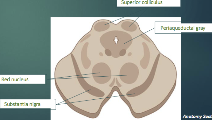
Mesencephalon: midbrain → most superior part of brainstem. ⇒ tectum (dorsal) and tegmentum (ventral)
Where a lot of neurotransmitters are produced: superior colliculus, periaqueductal gray, red nucleus, substantia nigra.
Metencephalon: pons (brainstem) and cerebellum (which is NOT part of the brainstem)
Myelencephalon: contains medulla oblongata (brainstem)
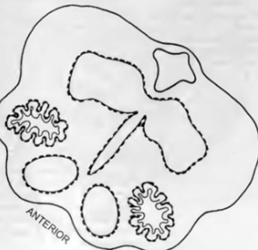
Major structures of Myelencephalon
Inferior part of brainstem containing the medulla oblongata
Olives (olivary nucleus): on lateral sides of medulla —> connect to cerebellum
Pyramids (protrusions): ventral side of medulla —> voluntary motor signals from brain travel down these tracts to spinal cord; decussation site.
Reticular formation (posterior) to other structures. —> core of brainstem
Metencephalon
Contains:
Pons (part of brainstem)
Bulbous section in the middle of the brainstem
Cerebellum (NOT part of brainstem)
Dorsal to pons
Contains the superior, middle, and inferior cerebellar peduncles.
Inferior cerebellar penduncles connect cerebellum to brainstem at olives of medulla.

Mesencephalon
Superior part of brainstem (midbrain)
Superior portion of midbrain:
Tectum: dorsal (top)
Superior colliculi (& inferior)
Tegmentum: Ventral
Periaqueductal gray: pain modulation
Red nucleus
Substantia nigra
Diencephalon: Thalamus
Has two lobes (bilateral) connected by massa intermedia.
Medial geniculate nucleus (MGN): auditory information => furthest from midbrain connection (towards end of thalamus)
Lateral geniculate nucleus (LGN): visual information
Ventral posterior nucleus (VPN): tactile information => closest to midbrain connection
Also has internal lamina: Y-shaped tracts of white matter.
Diencephalon: Hypothalamus
Inferior to thalamus on its anterior side.
Preoptic nucleus: sexual behavior
Suprachiasmatic nucleus: circadian rhythm
Mammillary body: emotion (bulge in the back)
Ventromedial nucleus: blood glucose —> fat