Pathology Test 4 Picture
 Call Kai
 Learn
Learn Practice Test
 Spaced Repetition
 Match
Match Flashcards
 Knowt Play
1/46

There's no tags or description
Looks like no tags are added yet.
Name | Mastery | Learn | Test | Matching | Spaced | Call with Kai | Chat |
|---|
No analytics yet
Send a link to your students to track their progress
47 Terms

1. Red Arrow
2. What is Happening?
1. Red Arrow - Atrophy
2. brain - hydrocephalus (cause - FIP)

What is Happening
Atrophy

1. Red Arrow
2. Morph. dg
1. Red Arrow - Atrophy
2. Morph. dg.: heart - epicardial serous atrophy of fat, diffuse, severe
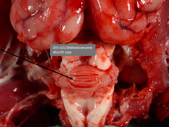
Morph. Dg and Cause
Morph. dg.: cerebellum - hypoplasia, diffuse, severe
Cause: intrauterine infection with panleukopenia

Morph Dg and Name od disease
Morf. dg.: Cardiac -left ventricular hypertrophy, severe, chronic
Name of the disease: hypertrophic cardiomyopathy
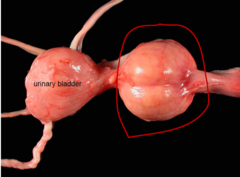
Morph. Dg
M. dg.: prostate -hyperplasia, bilateral, severe, chronic

Morph. Dg
M. dg.: pancreas - nodular hyperplasia, multifocal, moderate, chronic

Morph. Dg
M. dg.: liver - nodular hyperplasia, multifocal, severe, chronic
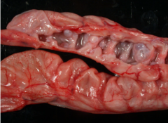
Morph. Dg
M. dg.: uterus - cystic endometrial hyperplasia, diffuse, severe, chronic

What?
Parakeet - squamous cell metaplasia in the esophageal glands

Right and Left
Arrows
1. Right Hydropic degeneration. Left Normal Hepatocytes
2. Thin Black : Central vein
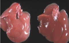
Right and Left
Right: Normal
Left: Degeneration

What?
Cutaneous interface dermatitis in a dog (discoid lupus erythematosus

What is this?
Dog-haired skin with epidermal hyperplasia and hydropic degeneration of keratinocytes in the basal aspect of the epidermis
apoptosis
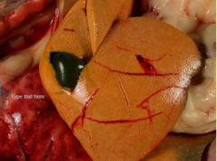
What is it
Lipidosis (steatosis / fatty degeneration
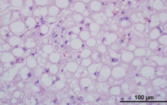
Liver with macrovesicular lipidosis
Liver with microvesicular lipidosis

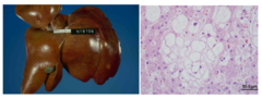
steroid hepatopathy
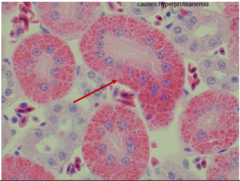
Glomerular disease - proteins in urinary space → hyaline droplets in tubular epithelial cells

Constipated» plasma cell (Mott cell; Russel bodies)

Viral inclusion bodies in the biliary epithelium of a dog (canine distemper virus)

Dystrophic mineralization

Yellow - Endocaedial surface
Red - Mineralization
Green- Viable myocardium

Lakes of mineral
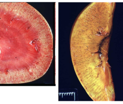
Amyloidosis
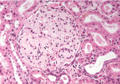
Kidney, glomerulus

Liver

Gout
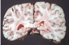
Carboxyhemoglobin

Methemoglobin

Intravascular hemolysis and hemoglobinuria
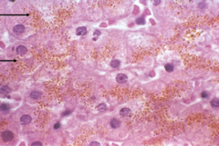
Liver with intracellular hemosiderin (brown granular material) in the cytoplasm of hepatocytes
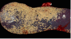
Sidero-fibrotic plaque - very common change in dog spleen

Dog, lungs -chronic left-side heart failure → chronic congestion in the lungs → hemosiderophages in the alveoli

Heart -lipofuscin in cardiomyocytes -perinuclear yellow granularpigment deposits
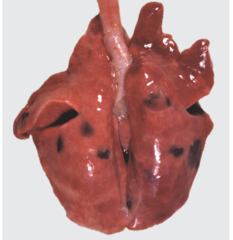
pig
Pig: pulmonary melanosis -multifocal black pigment deposits

Sheep
Sheep: melanosis of the meninges(black pigment)

Lungs - pulmonary anthracosis

Lungs -anthracosis(carbon particles in the peribronchialarea, in the cytoplasm of macrophages)
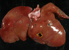
liver, cystic biliary hyperplasia, multifocal

liver, cystic biliary hyperplasia, multifocal.

what happneed and etiology
Dog: Musculus frontoscutularis, musculus temporalis atrophy, unilateral.
Etiology: denervation

Gland with cavity, dilated → glandular epithelium has
transformed into squamous epithelium [metaplasia]

Ruptured cyst with inflammation surrounding the ruptured region

Neutrophils
(blue arrows)
Lymphocytes Green
Plasma cell
(yellow arrows)
fibroblast balck
may be wrong
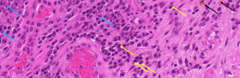
Neutrophils
(blue arrows)
Siderophage
(black arrow)
Plasma cell
(yellow arrows)
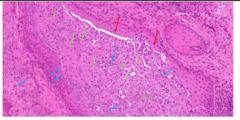
Gland cavity - dilated, lined with squamous epithelium(red) [metaplasia] and in the lumen there are exfoliated acantholytic epithelial cells(green), debri, neutrophils(blue). Neutrophils also in the interstitium