Neuro Quiz 1
 Call Kai
 Learn
Learn Practice Test
 Spaced Repetition
 Match
Match Flashcards
 Knowt Play
1/133

There's no tags or description
Looks like no tags are added yet.
Name | Mastery | Learn | Test | Matching | Spaced | Call with Kai | Chat |
|---|
No analytics yet
Send a link to your students to track their progress
134 Terms
What years were declared the “Decade of the Brain”?
1990-2000
Enhance public awareness of the benefits of brain research
Presidential Proclamation: George H. W. Bush on July 17,1990
BRAIN Initiative - What does it stand for, when, funding, purpose
Brain Research through Advancing Innovative Neurotechnologies
April 2, 2013
$100 million - initial stated funding level
→ purpose: revolutionize understanding of human brain by accelerating the development and application of innovative technologies to improve treatment, prevention, and cures for brain disorders
Example: mouse and human brain cell atlases
What 2 broad tasks - demonstrate complexity of nervous system and tennis?
Sensing and movement control
What does the visual cortex do during the phase of catching / hitting the ball?
Judging the ball
What does the PREMOTOR CORTEX do during the recognition phase - tennis?
Develops a motor program
What role does the AMYGDALA play in the recognition phase - tennis?
Influences heart rate and respiration
What structures execute movement during the movement phase - tennis?
Motor cortex and basal ganglia
What structure fine-tunes movement - tennis?
Cerebellum
Major point of the Einstein example
Cognitive functions involve several cortical areas
What differences were reported between Einstein’s brain and other brains?
Above average # of GLIAL cells
Possibly MISSING the PARIETAL OPERCULUM
Parietal lobe - 15% wider than normal
Incomplete SYLVIAN FISSURE
What function was associated with the englarged parietal lobe - Einstein slide?
Mathematical reasoning
What is the other name for the SYLVIAN FISSURE?
Lateral sulcus
What is the parietal operculum associated with?
Secondary somatosensory complex
Approx. how many NEURONS are in the human brain?
10^11 neurons
Approx. how many NEURONAL CONNECTIONS are in the human brain?
10^14
Two major methods - create high-resolution mouse brain cell atlas described on the slide?
Single-cell RNA sequencing and MERFISH
*MERFISH= multiplexed error-robust fluorescence in situ hybridization
What neurological disorder listed has the greatest # of US cases?
Chronic pain - 100 million cases
$300 billion
Highest annual economic cost disorder
Alzheimer’s disease - $780 billion / year
About 7.2 million cases
Two main cell types in the nervous system
Neurons and Glial cells
Primary function of NEURONS
Signaling / Communication
Primary role of GLIAL CELLS
Maintenance and Support
What functions can the nervous system control?
Simple reflexes to complex functions including…
-Vision
-Hearing
-Speaking
-Thinking
-Learning
-Emotions
Main Signaling Unit of the Nervous System
The Neuron
Do neurons contain the same basic organelles as other cells?
Yes, but they are highly specialized for intercellular communication.
What does neuronal polarization mean?
Different parts are structurally and functionally different, it does NOT mean electrical charge.
Are mature neurons generally MIOTIC?
NO, mature neurons are POSTmiotic and can live for many years
What are the basic visible parts of a typical neuron?
Cell body/soma, dendrites, axon, pre-synaptic terminal
What are the 4 morphologically and functionally defined regions of a typical neuron?
Cell body (nucleus, ER, Golgi)
Dendrites (receive information)
Axon (conducts information)
Pre-synaptic neuron (conversion of electrical to chemical signal)
*NEURONS ARE POLARIZED CELLS
Primary function of DENDRITES
Receive information
Primary function of the AXON
Conduction / conduct information
What happens at the pre-synaptic terminal?
Electrical signal is converted to a chemical signal
What are the 3 components of a SYNAPSE?
Presynaptic terminal
Synaptic cleft
Postsynaptic membrane
Average # of NEURONS in the human brain
100 billion, chain of 600 miles
Ramon y Cajal
“Father of Neuroscience”
-Used silver staining to show neurons are single cells (dont form a syncytium)
Top 4 Major Ramon y Cajal discoveries:
1) Neuron Doctrine - single unit is a distinct cell
2) Principle of Polarization - signal travels in one direction
3) Principle of connectional specificity
4) Neurons have Distinct Shapes - concept of synapse; Cajal identified the GROWTH CONE for the first time
Neurons are…
Extremely polarized, very asymmetrical cells, with fine long processes
Shapes of Neurons:
Unipolar, Bipolar, Pseudo-Unipolar, Multipolar

What does the shape of Neurons reflect?
More elaborate branching of the dendritic branches = the more complex functions are mediated by the neuron
→ PURKINJE CELL (cerebellum)
Techniques - Identifying Neurons and Connections
Golgi Silver Staining
Injection - fluorescent markers (Lucifer yellow, fluorescent dextran)
GFP expression
Labeling specific neurons - immunocytochemistry
Functional Classification of Neurons
Sensory - receivers of information from periphery
Motor - senders of information to periphery
Interneurons - make up the bulk of the brain
A) projection neurons: long axons, connect to parts far away
B) local neurons: short axons, connect close to brain
4 Signaling Components of Neurons
Input Component = dendrites, cell body
Trigger Component = axon hillock, action potential (beginning)
Conduction Component = axon
Output Component = pre-synaptic terminal, most synapses
Stimulus INTENSITY determines…
FREQUENCY of action potentials
Increased intensity = faster (more frequent) APs
Stimulus DURATION determines…
The NUMBER of action potentials
Increased duration = increased # of APs
Electrical signals are used for
LONG distances (along the neuron)
CHEMICAL signals (transmitters) are used for…
SHORT distances (between neurons)
Universality of electrical signaling
The nature of electrical signals is identical among different neurons and among species
Information specificity of electrical signals
It is given by the connection and the transmitter type
The basic mechanism of signaling is…
The SAME in all neurons
Where do neurons differ?
At the MOLECULAR level (ion channels, receptors, transmitters)
The specificity of information (visual, pain, motor) is given by…
Specific circuitry along which the signal travels
Glial Cell Functions
1) Development: acts as GUIDES for migrating neurons and axons
-Radial glial cell
2) Maintenance: homeostasis, regulation of the blood brain barrier (BBB)
-Astrocytes
3) Structural Support (Glue)
-Astrocytes
4) Providing Nutrients to Neurons
-Astrocytes (CNS), Schwann Cells (PNS)
5) Immune Function: removing debris
-Microglia
6) Myelination: insulation of axon
-Oligodendrocyte (CNS), Schwann Cell (PNS)
7) Production of Growth: factors and GCM
-Astrocytes
8) Secretion of Cerebralspinal fluid (CSF)
-Ependymal cell
Microglia - CNS only
Act as macrophages (immune defense cells), remove neuronal debris after injury, infection, disease via PHAGOCYTOSIS
Activated by multiple sclerosis, Parkinson’s, Alzheimer’s
Astrocytes
Astrology → keeps everything in check → homeostasis
-most ABUNDANT glial cell type in the CNS
-GFAP is an astrocyte protein marker
-Structural support
-Control chemical environment via spatial buffering → highly permeable to K+
-Supply neurons with nutrients (lactate from glycogen)
-Glial end feet: regulate development of BBB
-do not have direct connections with neurons, form gap junctions among themselves
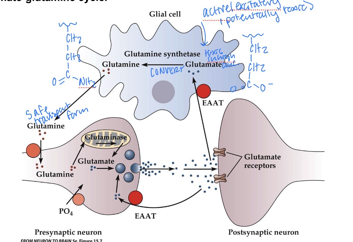
Astrocytes - Glutamate-Glutamine Cycle
Recycles Glutamate at the synapse
-Glutamine synthetase converts glutamate → glutamine
-Glutamine is released and taken back up by the pre-synaptic neuron, where glutaminase converts glutamine → glutamate for reuse.
*This process happens in the astrocyte
Oligodendrocytes
-Mostly in white matter
-Insulates axons with myelin in CNS, one oligodendrocyte can wrap several axons
Myelin Difference
Myelin in Oligodendrocytes DOES NOT = Myelin in Schwann Cells
→ they have different composition
Radial Glia
Development and Positioning
→ postmiotic neurons migrate to the appropriate layer along radial glial cells during development
Schwann Cells
Insulate axons with myelin in the PNS, 1:1 ratio of Schwann cell to axon
-Non-Myelinated spaces are the Nodes of Ranvier → sodium channels are highly concentrated (action potential jumps from one to another)
Key Features of Fast and Directional Signaling - Neurons
Electrical Excitability
Polarization (polarity)
What organisms are used to study cell polarity typically?
Drosophilia, C. Elegans, Yeast
Establishment of Polarity in Hippocampal Neurons:
Lamellipodia → Immature neurites → axon formation → dendrite formation → further maturation
Neuronal Polarity - how is it established and maintained?
Selective Delivery of Protein: need voltage gated/activated ion channel protein channels, need neurotransmitter receptor proteins
Selective Fusion of Membranes: SNARE proteins = mediate vesicle fusion with plasma membrane
Selective Retention of Proteins: diffusion barrier of axon initial segment restricts unwanted mixing of membrane potential
Differences between Axon and Dendrites
AXON:
-1 axon
-Generally long
-Intital segment
-Can be myelinated
-Na+/K+ channels
-Not all organelles
-tau
DENDRITE:
-Less than or equal to 1 dendrite
-Generally shorter, branched
-Continuous w/ cell body
-Not myelinated
-Neurotransmitter receptors
-Most organelles
-Map 2
Plasma Membrane Components - Neuron
Asymmetric Lipid Bilayer w/ Proteins
Hydrophobic Barrier Impermeable to Most Water-Soluable Molecules
*Very important for maintains specific intra and extra cellular concentrations of ions, metabolites, and transmitters → due to pumps, transporters, channels
Cytosol - Neuron
Consists soluble proteins, enzymes with metabolic functions, signaling proteins concentrated in the cytoskeletal-membrane interface
Cytoskeleton - Neuron
Function - cell division, shape determination, motility, organelle transport
Contains micotubules, actin filaments, neurofilaments
Accounts for 25% of the neuronal protein, tubules makes up 10% of total brain protein
Microtubules (MTs)
Made of Alpha and Beta Dimers
Plus ends toward the Beta subunit
GTP or GDP Binding for B tubulin, GTP binding for A tubulin
-Rapid changes of growing / shrinking regulated by free dimer concentration, rate of GTP hydrolysis, MT associated proteins
-GTP cap favors microtubule growth
-MAPs can stabilize MTs, marker proteins
→ tau is axonal marker
→ MAP2 is specfic to dendrites
Growing Microtubule is…
Rate of subunit addition EXCEEDS rate of GTP hydrolysis
Shrinking Microtubule =
Rate of GTP hydrolysis EXCEEDS rate of subunit addition
Microtubule Function in Neurons
-Axonal growth and balance
-Provide tracks for long-range protein and organelle transport
-Kinesin motors: mediate either plus or minus end oriented transport
-Dynein motors: minus end oriented transport
Microtubule polarity in neurons
Axons: all MT plus ends are oriented towards growth cone
Dendrites: mixed MT polarity in higher organisms (vertebrates)
F-Actin
-Very conserved and abundant
-Two parallel protofilaments made from a globular monomer, forms a right handed helix
-Plus end is the growing end
-ATP/ADP binding
F-Actin Functions in Neurons
Structural support, growth cone motility and guidance
F-Actin Dynamics
-Free monomer concentration and actin associated proteins
ATP-Bound → ASSEMBLE ACTIN
ADP-Bound → DISASSEMBLE ACTIN
Neurofilaments
-Tetramer is an organizing unit, fiber is not polarized
-Most abundant fibrillation components in axon that provide strength
-Dont have a plus or minus end, not as dynamic
Where is genetic information stored in neurons?
Nucleus, some in mitochondria (mito tRNA, mito rRNA, a few mito proteins)
Where does transcription and RNA processing occur?
Nucleus
Where does translation occur?
In the cell body and dendrites
→ there is also evidence for protein synthesis in the axon and nerve endings
Cytosolic proteins are translated …
On free ribosomes
Secretory and membrane proteins are synthesized by…
Ribosomes bound to the rough ER (RER)
Proteins with special destinations have…
Signal sequences
→ mitochondria, nucleus, peroxisomes, ER
→ the classic ER retention signal is the C-terminal KDEL sequence
What are the post-translational modifications?
1) Glycosylation
2) Phosphorylation / Dephosphorylation
3) N -acylation
4) Isoprenylation
5) Ubiquitination
Glycosylation
Attaching a sugar, carbohydrate modification
→ important for outer membrane proteins for molecular recognition
→ N-linked, O-linked
Phosphorylation / Dephosphorylation
Kinases, Phosphatases
→ affects signal transduction, protein function/activity
N-acylation
Attached carbon chain to protein
→ membrane association
→ can be diff lengths
Isoprenylation
Membrane association
→ prenyl-group
→ Ras-GTPase
Ubiquitination
Ubiquitin → 76 a.a.
→ protein degradation
→ proteasome
2 major functions of the Golgi Complex
1) Modifications of proteins: Glycosylation and Phosphorylation
2) Sorting and targeting of proteins into different vesicles
Why is ENDOCYTOSIS important in neurons?
1) Recycling of synaptic vesicles (membrane)
2) Keeping the neuron at the same size
3) Regulation of the amount of membrane receptors
*Clathrin mediates endocytosis
Proteins and Axonal transport
Most proteins are synthesized in the cell body → need to be transported along the axon
-Paul Weiss tied off sciatic nerve
-Radioactive labeling of neuronal proteins
-Video enhanced DIC light microscopy
-Fluorescent labeling techniques combined with photobleaching and photoactivation
Kinesin moves…
Minus end to Plus end (anterograde)
Dynein moves…
Plus end to Minus end (retrograde)
Central Nervous System is composed of…
Brain, Spinal Cord
Peripheral Nervous System (PNS) is composed of…
Somatic Division (Sensory neurons) and Autonomic division (motor)
Parasympathetic
Rest and Digest
Sympathetic
Fight or Flight
Cephalic Flexure
Remains after development causing the tilt between forebrain and spinal cord axes
Spinal Cord
Processes sensory information from limbs and trunk and many internal organs
Controls body movements
Transmits sensory information in ascending tracts and motor information in descending tracts
Amount of Segments (spinal cord)
Cervical = 8
Thoracic = 12
Lumbar = 5
Sacral = 5
White matter is most abundant…
At the cervical level
→ more ascending and descending axons