Seasons Structure Week 4
 Call Kai
 Learn
Learn Practice Test
 Spaced Repetition
 Match
Match Flashcards
 Knowt Play
1/137

Earn XP
Description and Tags
Aorta, Thoracic Vasculature, Nervous System
Name | Mastery | Learn | Test | Matching | Spaced | Call with Kai | Chat |
|---|
No analytics yet
Send a link to your students to track their progress
138 Terms
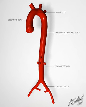
What are the three anatomical segments of the thoracic aorta?
Answer: Ascending aorta → aortic arch → descending thoracic aorta.
Extra Information:
The ascending aorta is the most proximal segment.
The descending thoracic aorta continues inferiorly through the posterior mediastinum.
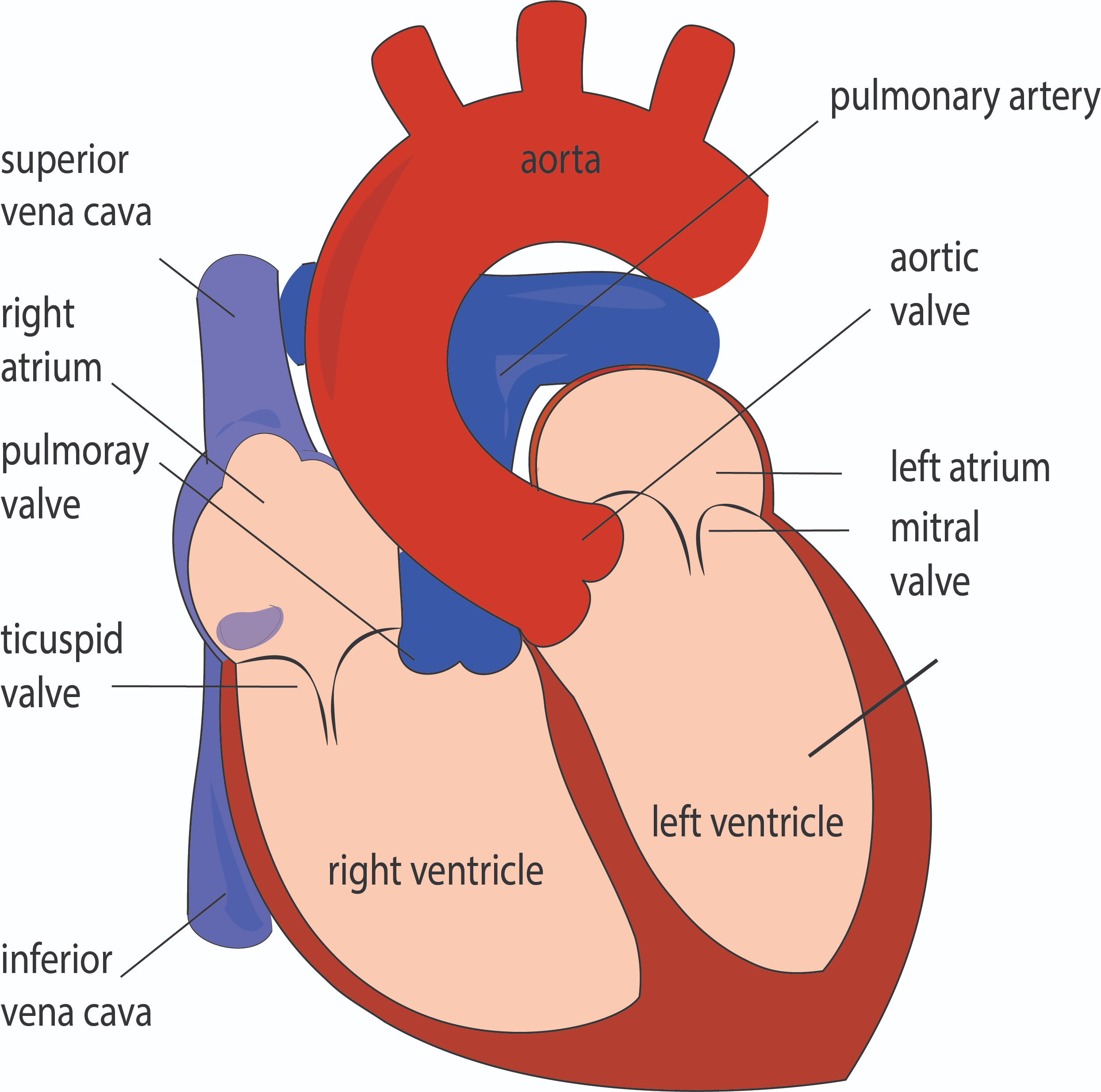
Where does the ascending aorta originate?
Answer: The left ventricle.
Extra Information:
It is the most proximal portion of the thoracic aorta.
The aortic valve is located at its base.
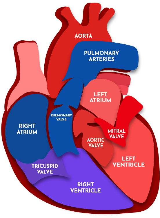
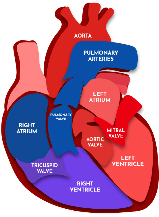
What valve is located at the base of the ascending aorta?
Answer: The aortic valve.
Extra Information:
The ascending aorta emerges directly from the left ventricle.
At what vertebral level does the aortic arch begin and end?
Answer: T4/T5 at the level of the sternal angle.
Extra Information:
- The transition to the descending thoracic aorta also occurs around this level.
- The sternal angle is therefore an important landmark for identifying the aortic arch.
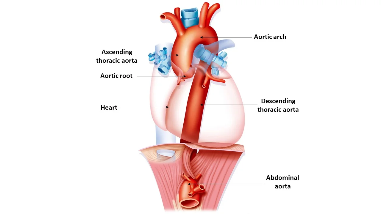
In what direction does the aortic arch travel?
Answer: Posteriorly and to the left.
Extra Information:
It connects the ascending aorta to the descending thoracic aorta.
What important structures does the aortic arch cross over?
Answer: The right pulmonary artery and left main bronchus.
Extra Information:
These relationships help anatomically identify the arch in the thorax.
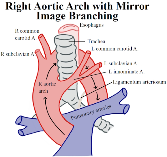
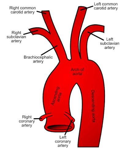
What are the three major branches of the aortic arch from proximal to distal?
Answer: Brachiocephalic trunk → left common carotid artery → left subclavian artery.
Extra Information:
The branches arise in this order from the arch.
Memory: ABC's of the Arch = Brachiocephalic, Carotid, Subclavian.
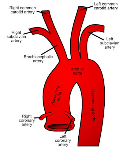
What is the first and largest branch of the aortic arch?
Answer: The brachiocephalic trunk.
Extra Information:
It subsequently divides into the right common carotid and right subclavian arteries.
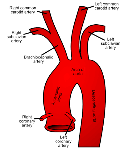
Into which arteries does the brachiocephalic trunk divide?
Answer: Right common carotid artery and right subclavian artery.
Extra Information:
- This explains why there is a brachiocephalic arterial trunk on the right rather than an equivalent left-sided trunk in the normal arch pattern.
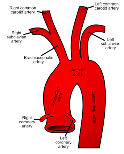
What is the second major branch of the aortic arch?
Answer: The left common carotid artery.
Extra Information:
It supplies the left side of the head and neck.
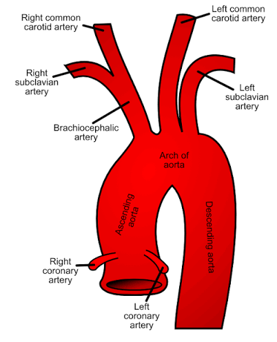
What is the third major branch of the aortic arch?
Answer: The left subclavian artery.
Extra Information:
It supplies the left upper limb.
Trace the major arterial pathway through the thoracic aorta from the heart.
Answer: Left ventricle → ascending aorta → aortic arch → descending thoracic aorta.
Extra Information:
The arch begins and ends around the T4/T5 sternal-angle level.
The descending thoracic aorta eventually passes through the diaphragm.
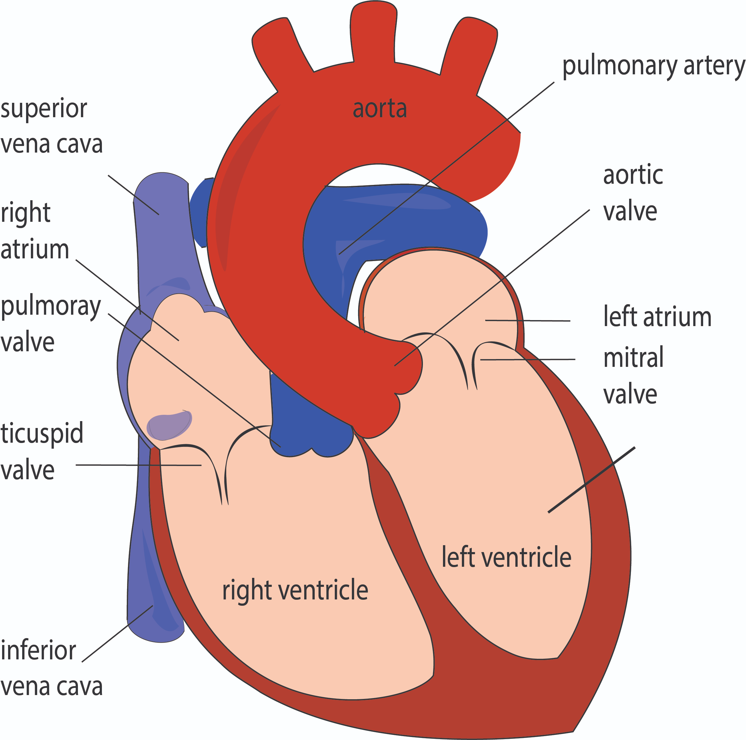
Where does the descending thoracic aorta begin?
Answer: Around T4/T5.
Extra Information:
It begins after the aortic arch.
It descends through the posterior mediastinum.
Through which part of the mediastinum does the descending thoracic aorta travel?
Answer: The posterior mediastinum.
Extra Information:
It courses inferiorly toward the diaphragm.
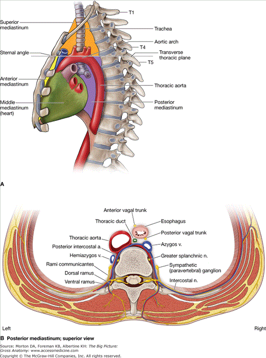
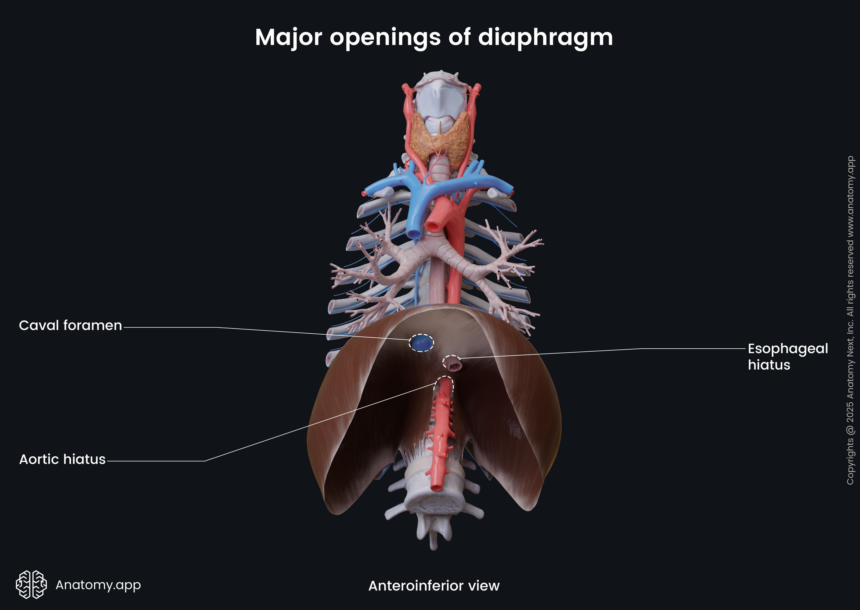
At what vertebral level does the aorta pass through the diaphragm?
Answer: T12.
Extra Information:
After passing through the diaphragm, it becomes the abdominal aorta.
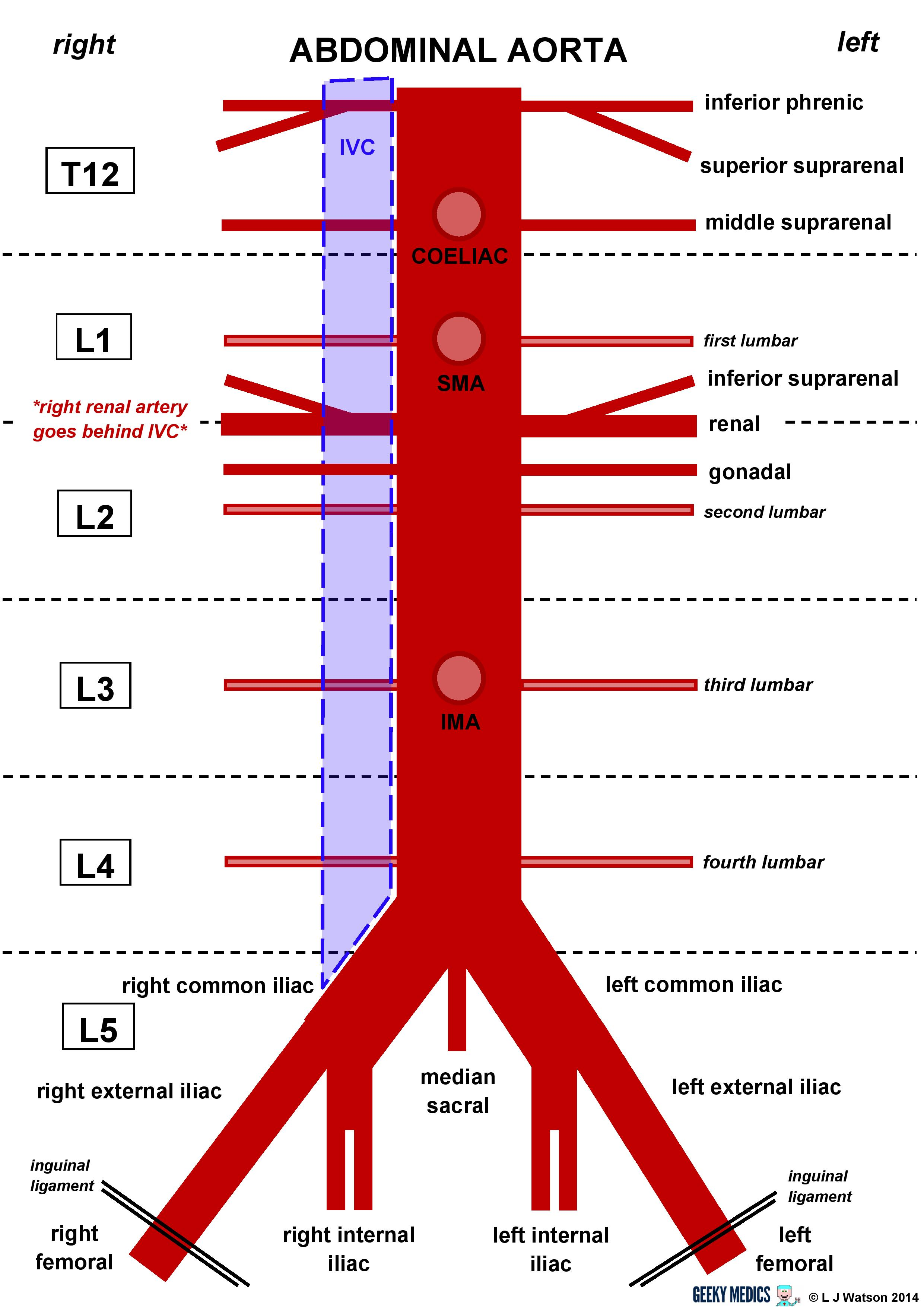
What does the descending thoracic aorta become after passing through the diaphragm?
Answer: The abdominal aorta.
Extra Information:
The transition occurs at the T12 aortic hiatus.
What are the visceral branches of the descending thoracic aorta listed in the course guide?
Answer: Bronchial, esophageal, mediastinal, and pericardial branches.
Extra Information:
These branches supply thoracic viscera and associated mediastinal structures.
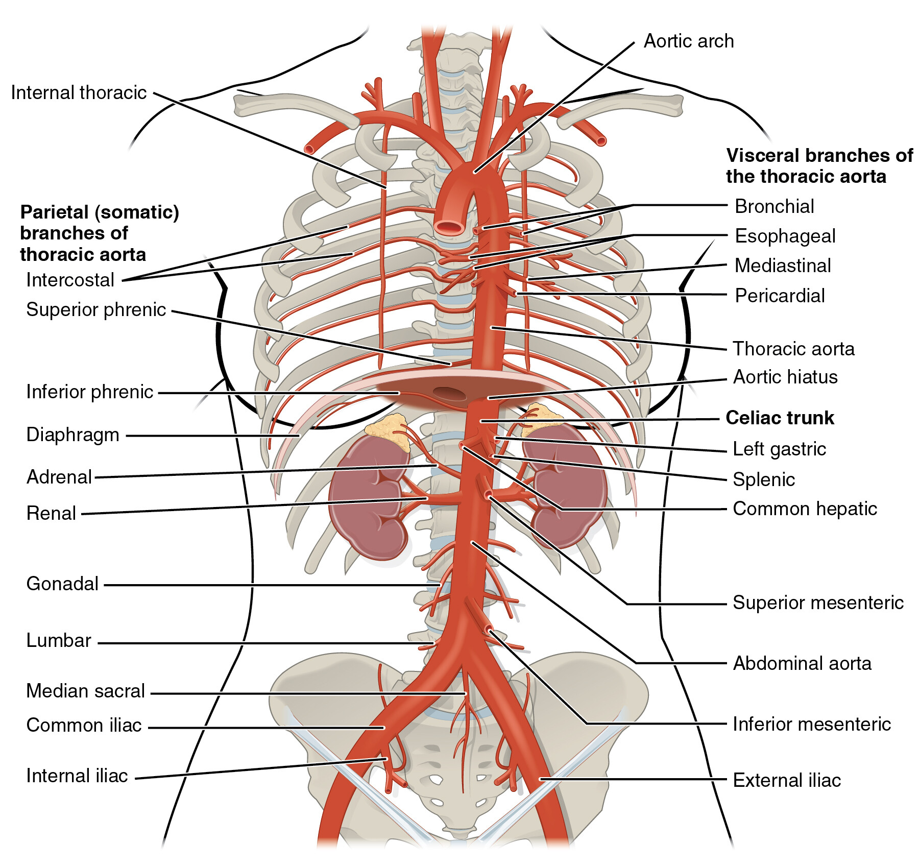
What are the parietal branches of the descending thoracic aorta listed in the course guide?
Answer: Posterior intercostal and phrenic branches.
Extra Information:
Parietal branches supply structures associated with the thoracic body wall.
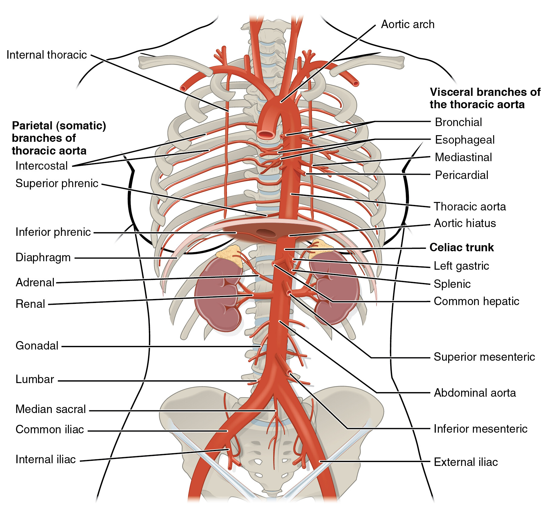
Which arteries contribute to the blood supply of the thoracic wall?
Answer: Posterior intercostal arteries and anterior intercostal arteries.
Extra Information:
The prework diagrams show posterior intercostal arteries associated with the thoracic aorta.
Anterior intercostal arteries are associated with the internal thoracic arterial system.
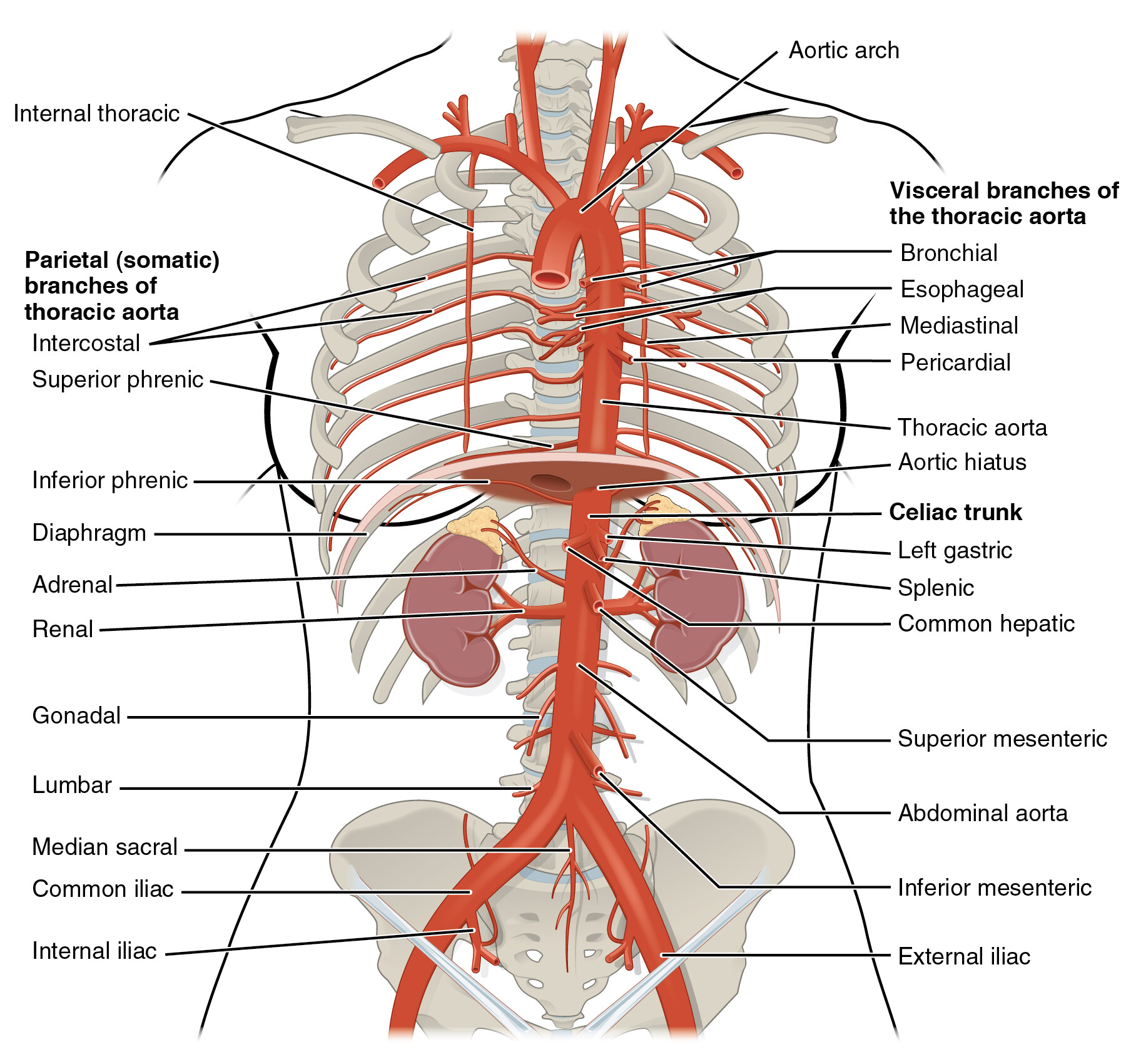
The internal thoracic arteries are branches of the ______.
Answer: Subclavian arteries.
Extra Information:
They descend along the internal surface of the anterior thoracic wall.
The prework notes their use in coronary artery bypass grafting.
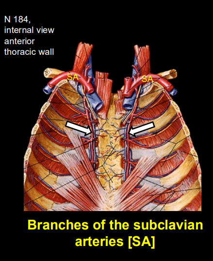
How many anterior intercostal arteries are shown per intercostal space in the prework?
Answer: Two per intercostal space.
Extra Information:
They contribute to the arterial supply of the anterior thoracic wall.
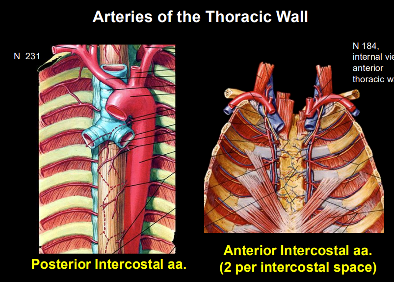
What is an aortic dissection?
Answer: A tear in the tunica intima that allows blood to track into a false lumen within the tunica media.
Extra Information:
It is a life-threatening vascular emergency.
The false lumen can interfere with normal blood flow to vital organs.
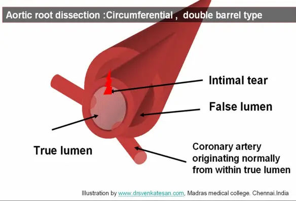
What is the key anatomical sequence to remember for the thoracic aorta?
Answer: Left ventricle → ascending aorta → arch at T4/T5 → descending thoracic aorta → diaphragm at T12 → abdominal aorta.
Extra Information:
- This integrates the major anatomical landmarks of S4S.1.
What is the major venous destination for blood returning from structures superior to the diaphragm?
Answer: The superior vena cava.
Extra Information:
- The course excludes venous return from the heart and lungs from this general description.
- The superior vena cava empties into the right atrium.
How is the superior vena cava formed?
Answer: By union of the right and left brachiocephalic veins.
Extra Information:
- The brachiocephalic veins collect venous return from structures including the head, neck, and upper limbs.
How is each brachiocephalic vein formed?
Answer: By the junction of the internal jugular vein and subclavian vein.
Extra Information:
- There is a right and a left brachiocephalic vein.
- Both ultimately contribute to formation of the superior vena cava.
Why is the left brachiocephalic vein longer than the right?
Answer: It must cross the midline to join the right brachiocephalic vein.
Extra Information:
- Their union forms the superior vena cava.
Trace venous return through a brachiocephalic vein to the heart.
Answer: Internal jugular or subclavian vein → brachiocephalic vein → superior vena cava → right atrium.
Extra Information:
- The right and left brachiocephalic veins unite to form the superior vena cava.
What is the primary function of the azygos venous system?
Answer: It drains the thoracic wall and posterior mediastinum and provides an important collateral venous pathway.
Extra Information:
- It ultimately drains into the superior vena cava.
- It can provide an alternative route when major venous flow is obstructed.
On which side of the thorax is the azygos vein located?
Answer: The right side.
Extra Information:
- Memory: Azygos is Always on the Right, mostly.
What does the azygos vein arch over before entering the superior vena cava?
Answer: The root of the right lung.
Extra Information:
- The azygos arch then empties into the superior vena cava.
Where does the azygos vein ultimately drain?
Answer: The superior vena cava.
Extra Information:
- It is an important component of thoracic venous return.
On which side are the hemiazygos and accessory hemiazygos veins located?
Answer: The left side.
Extra Information:
- Both ultimately cross the midline to drain into the azygos vein.
Where does the hemiazygos vein ultimately drain?
Answer: Into the azygos vein after crossing the midline.
Extra Information:
- It forms part of the left-sided component of the azygos system.
Where does the accessory hemiazygos vein ultimately drain?
Answer: Into the azygos vein after crossing the midline.
Extra Information:
- Like the hemiazygos vein, it is primarily located on the left.
Trace the azygos venous pathway to the heart.
Answer: Thoracic veins → azygos system → azygos vein → superior vena cava → right atrium.
Extra Information:
- Left-sided hemiazygos veins first cross to the azygos vein.
Where do the internal thoracic veins typically drain?
Answer: Into the brachiocephalic veins.
Extra Information:
- The brachiocephalic veins then drain into the superior vena cava.
- The internal thoracic veins accompany the internal thoracic arteries.
Into which two major systems do the intercostal veins drain?
Answer: The azygos system and the internal thoracic veins.
Extra Information:
- The azygos system drains toward the superior vena cava.
- Internal thoracic veins drain toward the brachiocephalic veins and then the superior vena cava.
Trace anterior thoracic wall venous return through the internal thoracic veins.
Answer: Intercostal veins → internal thoracic veins → brachiocephalic veins → superior vena cava → right atrium.
Extra Information:
- This pathway complements drainage through the azygos system.
How does the azygos system function as a collateral pathway?
Answer: It provides an alternative venous route between major venous territories when normal flow is obstructed.
Extra Information:
- The course specifically emphasizes its importance as a collateral pathway between the inferior and superior vena caval systems.
What can occur clinically when the superior vena cava is obstructed?
Answer: Facial swelling and visible collateral veins on the chest wall.
Extra Information:
- Blood can be redirected through collateral pathways such as the azygos system.
- This clinical pattern is associated with superior vena cava syndrome.
What is the major thoracic venous flow pattern to remember?
Answer: Thoracic venous blood converges toward the superior vena cava through the brachiocephalic veins and azygos system.
Extra Information:
- The azygos vein is right-sided.
- The hemiazygos and accessory hemiazygos veins are primarily left-sided.
What is the major functional distinction between somatic and visceral innervation in the thorax?
Answer: Somatic nerves primarily innervate the body wall and skeletal muscle, while visceral nerves primarily innervate internal organs and associated smooth or cardiac muscle.
Extra Information:
- Somatic functions are associated with conscious sensation and voluntary skeletal muscle control.
- Visceral functions are associated with autonomic and organ regulation.
Which thoracic structures are identified as somatically innervated in the course guide?
Answer: Thoracic wall, skin, ribs, intercostal muscles, diaphragm, and larynx.
Extra Information:
- The guide emphasizes somatic innervation of the wall and skeletal muscle.
Which thoracic structures are identified as viscerally innervated?
Answer: Heart, lungs, esophagus, and blood vessels.
Extra Information:
- These are internal thoracic viscera or structures regulated by autonomic pathways.
What type of innervation supplies the intercostal muscles?
Answer: Somatic innervation.
Extra Information:
- Intercostal muscles are skeletal muscles of the thoracic wall.
What type of innervation supplies the thoracic skin?
Answer: Somatic sensory innervation.
Extra Information:
- Somatic sensory information includes conscious and well-localized sensations.
What type of innervation supplies the heart?
Answer: Visceral innervation.
Extra Information:
- The heart receives autonomic sympathetic and parasympathetic efferents.
- It also sends visceral afferent information back toward the CNS.
What type of innervation supplies the lungs?
Answer: Visceral innervation.
Extra Information:
- Pulmonary autonomic pathways contribute to involuntary regulation.
- Visceral afferent information also travels from the lungs.
What type of innervation supplies the esophagus?
Answer: Visceral innervation.
Extra Information:
- The course diagrams show an esophageal autonomic plexus.
What type of innervation regulates blood vessels?
Answer: Visceral autonomic innervation.
Extra Information:
- The histology material identifies unmyelinated autonomic nerve fibers in vascular walls.
- These fibers can release norepinephrine to produce vasoconstriction.
What type of innervation supplies the diaphragm according to the course guide?
Answer: Somatic innervation.
Extra Information:
- The phrenic nerve is important for both diaphragm motor innervation and somatosensory pathways from parietal serous membranes.
What clinical finding can result from left recurrent laryngeal nerve damage caused by an aortic arch aneurysm?
Answer: Hoarseness.
Extra Information:
- The course relates this to paralysis of laryngeal muscles.
- The example demonstrates the close anatomical relationship between thoracic structures and neural pathways.
What is the basic memory distinction between somatic and visceral thoracic innervation?
Answer: Somatic = skin and skeletal structures, visceral = viscera and organs.
Extra Information:
- This distinction helps organize the sensory and motor pathways in the following LOs.
What information do visceral afferent fibers carry?
Answer: Sensory information from internal organs toward the CNS.
Extra Information:
- This can include visceral pain or physiological reflex information.
- Afferent means traveling toward the CNS.
From which supra-diaphragmatic organs can visceral afferent information arise in these materials?
Answer: Thoracic organs such as the heart, lungs, and esophagus.
Extra Information:
- Sensory endings are associated with visceral structures and autonomic plexuses.
How do visceral afferent fibers generally travel back toward the CNS?
Answer: They travel retrograde alongside autonomic pathways.
Extra Information:
- Pain information typically follows sympathetic pathways.
- Reflex information typically follows parasympathetic pathways through the vagus nerve.
Which autonomic pathway do visceral pain afferents typically accompany?
Answer: Sympathetic pathways.
Extra Information:
- For thoracic visceral pain, the course emphasizes upper thoracic spinal levels.
Where are the cell bodies of visceral pain afferent neurons located?
Answer: Dorsal root ganglia.
Extra Information:
- For the thoracic visceral pathways emphasized in the guide, these are associated with approximately T1–T4/5.
Which spinal levels are emphasized for visceral pain afferents from the heart and related thoracic viscera?
Answer: Approximately T1–T4/5.
Extra Information:
- Their sensory neuron cell bodies are located in the dorsal root ganglia.
Through which root do visceral pain afferents enter the spinal cord?
Answer: The dorsal root.
Extra Information:
- After entering, they can synapse in the dorsal horn.
Where do visceral pain afferents synapse after entering the spinal cord?
Answer: The dorsal horn.
Extra Information:
- Their first-order neuron cell bodies are located in the dorsal root ganglia.
Trace the visceral pain afferent pathway from a supra-diaphragmatic organ to the spinal cord.
Answer: Visceral receptor → travels with sympathetic pathway → spinal nerve pathway → dorsal root ganglion → dorsal root → dorsal horn.
Extra Information:
- The dorsal root ganglion contains the sensory neuron cell body.
- Visceral pain generally accompanies sympathetic pathways.
Which pathway carries visceral reflex information from thoracic organs?
Answer: Parasympathetic afferents traveling with the vagus nerve.
Extra Information:
- This differs from visceral pain, which generally accompanies sympathetic pathways.
Where are the sensory neuron cell bodies for vagal visceral reflex afferents located?
Answer: In sensory ganglia of the vagus nerve.
Extra Information:
- These afferents carry physiological reflex information toward the brainstem.
Where do vagal visceral reflex afferents ultimately enter the CNS?
Answer: The brainstem.
Extra Information:
- Their pathway differs from sympathetic visceral pain fibers entering the spinal cord through dorsal roots.
Trace visceral reflex afferent information from a thoracic organ toward the CNS.
Answer: Visceral receptor → vagal afferent fiber → vagal sensory ganglion → vagus nerve pathway → brainstem.
Extra Information:
- This pathway primarily carries visceral reflex information.
- The sensory neuron cell body lies in a vagal sensory ganglion.
What is the major difference between visceral pain and visceral reflex afferent pathways?
Answer: Visceral pain generally travels with sympathetic pathways to dorsal root ganglia and the spinal cord, while visceral reflex information generally travels with the vagus nerve to vagal sensory ganglia and the brainstem.
Extra Information:
- Pain = sympathetic-associated.
- Reflex information = parasympathetic or vagal-associated.
What system provides visceral efferent innervation to thoracic organs?
Answer: The autonomic nervous system.
Extra Information:
- Visceral efferents provide motor control to structures such as the heart and thoracic smooth muscle.
- Both sympathetic and parasympathetic pathways are involved.
Where are sympathetic preganglionic neuron cell bodies for the thoracic visceral pathway located?
Answer: In the lateral horn of the upper thoracic spinal cord.
Extra Information:
- The course guide emphasizes approximately T1–T4/5 for supra-diaphragmatic thoracic viscera.
What is the first structure traveled through by a sympathetic preganglionic axon after leaving the spinal cord?
Answer: The ventral root.
Extra Information:
- The ventral root carries efferent fibers away from the spinal cord.
Trace the sympathetic preganglionic pathway from the spinal cord to the sympathetic trunk.
Answer: Lateral horn → ventral root → spinal nerve → ventral ramus → white ramus communicans → sympathetic trunk.
Extra Information:
- The preganglionic neuron cell body is located in the lateral horn.
- The white ramus carries the preganglionic fiber into the sympathetic chain.
What does the white ramus communicans carry in the sympathetic pathway?
Answer: Preganglionic sympathetic fibers into the sympathetic trunk.
Extra Information:
- It is part of the route from the spinal nerve to the paravertebral sympathetic chain.
Where are sympathetic postganglionic neuron cell bodies for supra-diaphragmatic thoracic viscera located?
Answer: In cervical or upper thoracic paravertebral ganglia of the sympathetic chain.
Extra Information:
- Preganglionic fibers synapse on these neurons.
- Postganglionic fibers then travel toward thoracic organs.
What happens after a sympathetic preganglionic fiber reaches the appropriate paravertebral ganglion?
Answer: It synapses with a postganglionic sympathetic neuron.
Extra Information:
- The postganglionic neuron's cell body is located within the sympathetic-chain ganglion.
What type of sympathetic fibers travel from paravertebral ganglia toward the heart and lungs?
Answer: Postganglionic sympathetic fibers.
Extra Information:
- The course gives cardiac nerves as an example.
- Cardiopulmonary splanchnic nerves are postganglionic sympathetic pathways.
Trace the complete sympathetic visceral efferent pathway to a supra-diaphragmatic thoracic organ.
Answer: Lateral horn preganglionic cell body → ventral root → spinal nerve → ventral ramus → white ramus communicans → sympathetic trunk → cervical or upper thoracic paravertebral ganglion → postganglionic fiber → autonomic plexus → target organ.
Extra Information:
- The preganglionic neuron cell body is in the spinal cord.
- The postganglionic neuron cell body is in a paravertebral ganglion.
What is the typical fiber-length pattern for sympathetic visceral efferents?
Answer: Short preganglionic fibers and long postganglionic fibers.
Extra Information:
- Sympathetic ganglia are relatively close to the spinal cord.
Where are parasympathetic preganglionic neuron cell bodies for thoracic visceral innervation located?
Answer: In the brainstem, particularly the medulla for the vagal pathways emphasized in the course.
Extra Information:
- Their axons travel through the vagus nerve.
Which cranial nerve carries parasympathetic efferent fibers to thoracic viscera?
Answer: The vagus nerve, cranial nerve X.
Extra Information:
- It travels through the mediastinum toward thoracic autonomic plexuses and organs.
Where are parasympathetic postganglionic neuron cell bodies for thoracic organs located?
Answer: In microscopic ganglia within or very near the target organ wall.
Extra Information:
- This arrangement produces very short postganglionic fibers.
What is the typical fiber-length pattern for parasympathetic visceral efferents?
Answer: Long preganglionic fibers and very short postganglionic fibers.
Extra Information:
- The ganglia are located near or within the target organ.
- This contrasts with the sympathetic short-pre and long-post arrangement.
Trace the parasympathetic visceral efferent pathway to a thoracic organ.
Answer: Brainstem preganglionic cell body → vagus nerve → thoracic autonomic plexus → microscopic ganglion in or near the target organ → short postganglionic fiber → target tissue.
Extra Information:
- The preganglionic neuron cell body is in the brainstem.
- The postganglionic neuron cell body is located in or near the organ wall.
What is the major ganglion-location difference between sympathetic and parasympathetic pathways to thoracic viscera?
Answer: Sympathetic neurons synapse in paravertebral ganglia, while parasympathetic neurons synapse in ganglia within or near the target organ.
Extra Information:
- This difference explains their characteristic preganglionic and postganglionic fiber lengths.
Compare the complete sympathetic and parasympathetic visceral efferent organization.
Answer: Sympathetic pathways begin in the thoracic spinal cord and synapse in paravertebral ganglia, while parasympathetic pathways begin in the brainstem, travel through the vagus nerve, and synapse in or near the target organ.
Extra Information:
- Sympathetic = short preganglionic and long postganglionic.
- Parasympathetic = long preganglionic and short postganglionic.
What structures provide the somatosensory information emphasized in S4S.6?
Answer: The thoracic wall and parietal serous membranes such as parietal pleura and parietal pericardium.
Extra Information:
- The course notes that true viscera generally use visceral sensory pathways.
- Somatosensory pain is sharp and well localized.
Where are somatosensory receptors located in the thoracic pathways emphasized by the guide?
Answer: In structures such as thoracic skin and parietal serous membranes.
Extra Information:
- These receptors provide conscious and localized sensory information.
Which nerves carry somatosensory information from the thoracic wall?
Answer: Intercostal nerves.
Extra Information:
- Intercostal nerves are ventral rami of thoracic spinal nerves.
What are intercostal nerves anatomically?
Answer: Ventral rami of thoracic spinal nerves.
Extra Information:
- They carry somatic information associated with the thoracic wall.
Which nerve carries somatosensory information from important parietal serous membranes in the thorax?
Answer: The phrenic nerve.
Extra Information:
- The guide includes the phrenic nerve as a somatosensory pathway from parietal membranes.
Where are the cell bodies of thoracic somatosensory afferent neurons located?
Answer: In the dorsal root ganglia.
Extra Information:
- These are first-order sensory neuron cell bodies.
Through which root do somatosensory afferents enter the spinal cord?
Answer: The dorsal root.
Extra Information:
- They subsequently synapse in the dorsal horn.
Where do thoracic somatosensory afferents synapse after entering the spinal cord?
Answer: The dorsal horn.
Extra Information:
- Their cell bodies are located in the dorsal root ganglion before entering the spinal cord.
Trace somatosensory information from the thoracic wall to the spinal cord.
Answer: Somatic receptor → intercostal nerve → spinal nerve pathway → dorsal root ganglion → dorsal root → dorsal horn.
Extra Information:
- The sensory neuron cell body is in the dorsal root ganglion.
- This pathway carries sharp, localized sensation.
Trace the somatosensory pathway emphasized for parietal thoracic serous membranes.
Answer: Parietal sensory receptor → phrenic or appropriate somatic sensory nerve → dorsal root ganglion → dorsal root → dorsal horn.
Extra Information:
- The course contrasts this well-localized somatic sensation with poorly localized visceral pain.
How does somatic pain differ from visceral pain?
Answer: Somatic pain is typically sharp and well localized, while visceral pain is typically dull and poorly localized.
Extra Information:
- Somatosensory neuron cell bodies are in dorsal root ganglia.
- Visceral pain afferents also use dorsal root ganglia but travel back alongside sympathetic pathways.
What are the three tunics found in the walls of large blood vessels?
Answer: Tunica intima, tunica media, and tunica adventitia.
Extra Information:
- These layers are present in larger arteries and veins.
- Their relative thickness and composition differ between vessel types.
What structures make up the tunica intima?
Answer: Endothelium and a thin subendothelial layer of loose connective tissue.
Extra Information:
- The subendothelial layer can contain smooth muscle fibers.
- Arterial intima also includes an internal elastic lamina.
What elastic structure is associated with the arterial tunica intima?
Answer: The internal elastic lamina.
Extra Information:
- It is composed of elastin.
- It helps distinguish arterial wall organization histologically.
What is the general composition of the tunica media?
Answer: Concentric layers of helically arranged smooth muscle with variable elastic fibers, elastic lamellae, reticular fibers, and proteoglycans.
Extra Information:
- The smooth muscle cells produce many of the extracellular components of the media.
- Arteries may also have an external elastic lamina.