NEPHROLOGY
 Call Kai
 Learn
Learn Practice Test
 Spaced Repetition
 Match
Match Flashcards
 Knowt Play
1/43

There's no tags or description
Looks like no tags are added yet.
Name | Mastery | Learn | Test | Matching | Spaced | Call with Kai | Chat |
|---|
No analytics yet
Send a link to your students to track their progress
44 Terms
Normal range in ABG
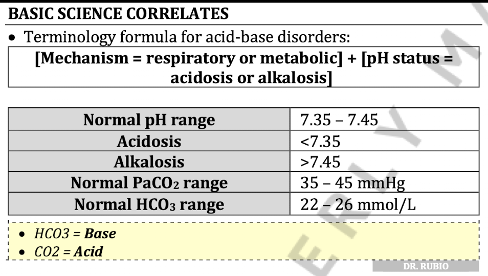
When does Kussmaul breathing occur
Metabolic acidosis
Decreased HCO3 » Body compensates and tries to decrease pH by decreasing CO2 » increase RR
When must anion gap be computed
Metabolic Acidosis
measured to find out what is causing acid buildup
Differentiate between high and normal anion gap
Formula for Anion Gap
Normal: 8-12 mEqs/L
High": >12mEqs/L
consider presence of organic acids that would cause increase in the anion gap

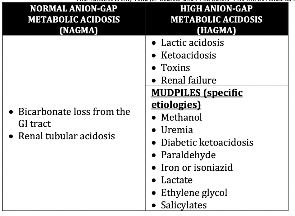
Causes of Normal vs High Anion Gap
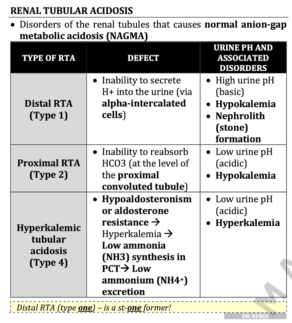
Three Disorders of Renal Tubular Acidosis
RTA is NAGMA type of metabolic acidosis
Normal plasma K+
What level is considered severe hyperkalemia
Where is K+ excreted
N: 3.5-5.5 mM
Severe: >6 mM
90% urine 10% stool
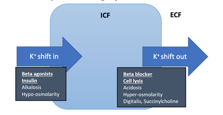
4 Mechanisms that can cause hyperkalemia
Decrease in renal K+ excretion (due to decrease in aldosterone)
RAAS inhibitors (Inhibition of ACE and Angiotensin II pathway » decrease in aldosterone)
Hypoaldosteronism
Adrenal insufficiency (Addison disease)
Redistribution and reduced tissue uptake (beta blocker)
Excessive potassium intake (only if with renal failure)
Increase potassium release from tissues (tumor lysis syndrome, rhabdomyolysis)
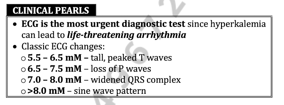
Describe ECG changes based on K+ level
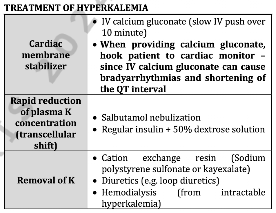
Treatment of hyperkalemia
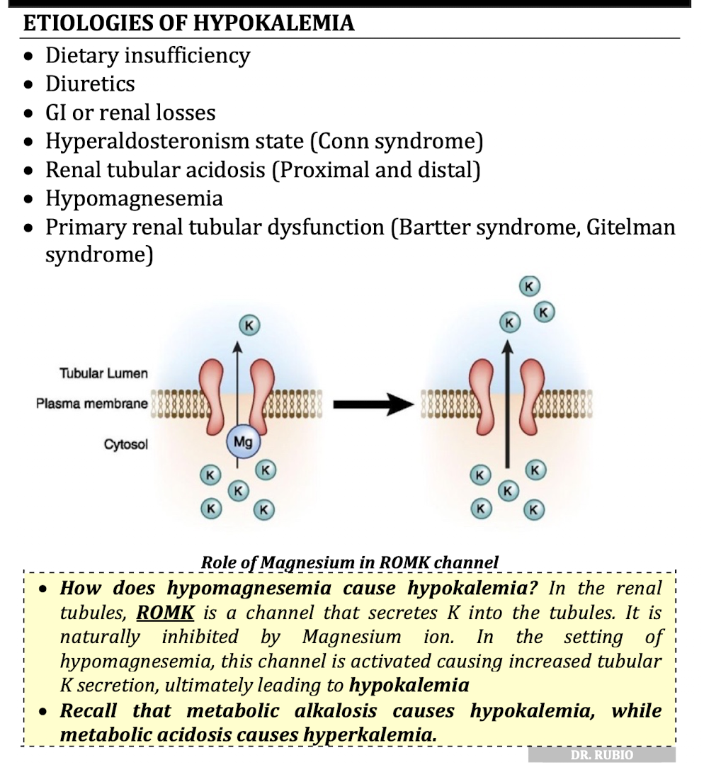
7 Etiologies of hypokalemia
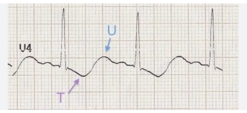
ECG change in hypokalemia
U waves
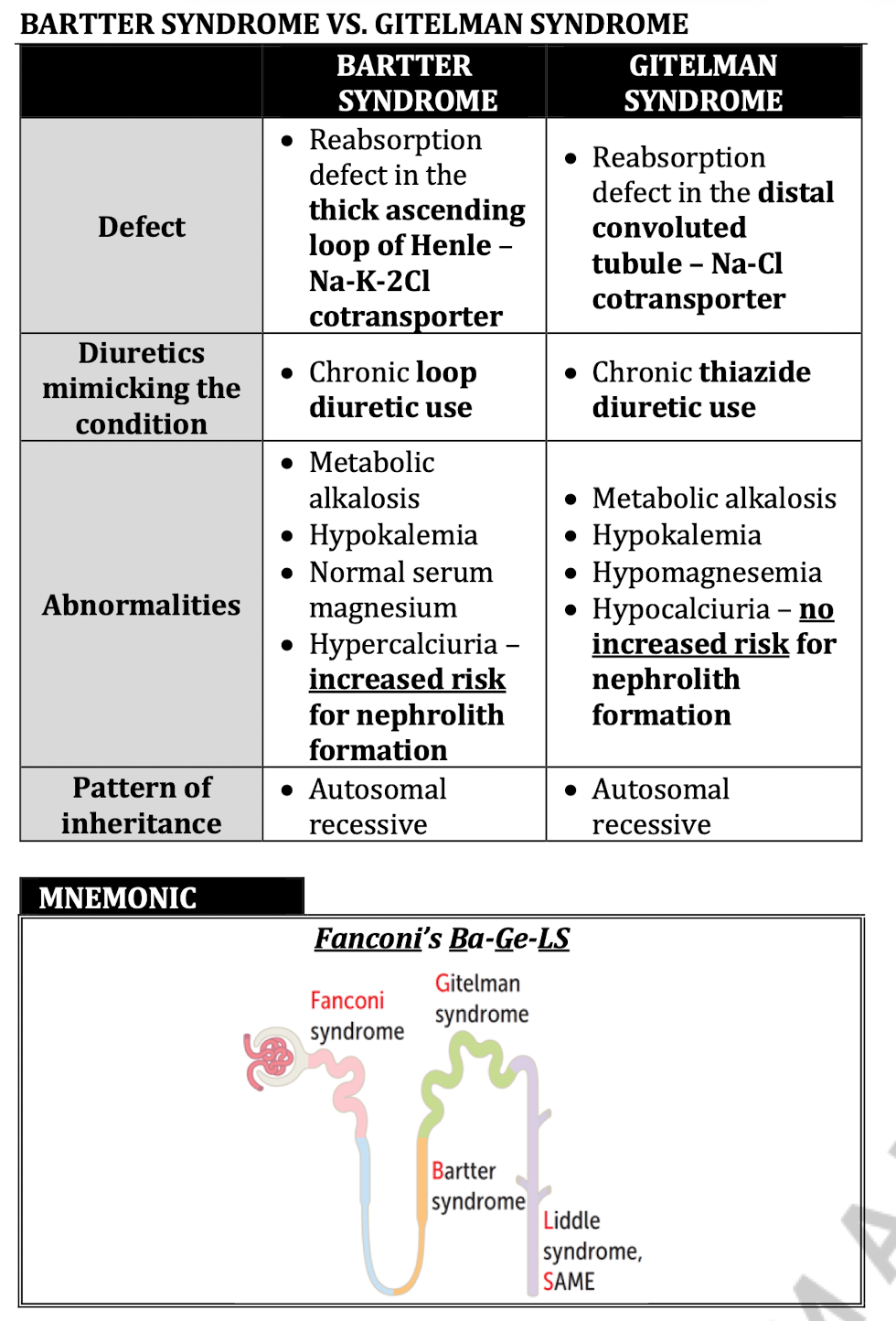
Bartter Syndrome vs Gitelman Syndrome
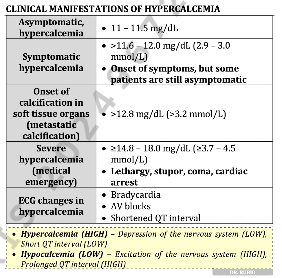
Clinical manifestations of hypercalcemia
Treatment of hypercalcemia

Clinical presentation of hypocalcemia

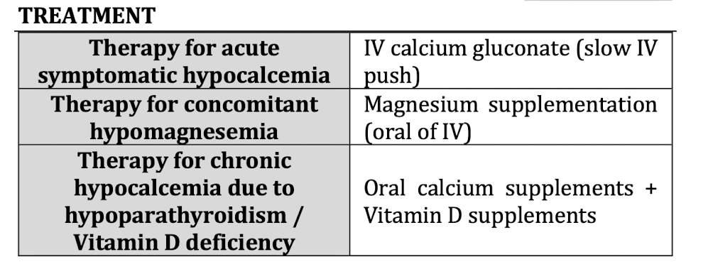
Treatment for hypocalcemia
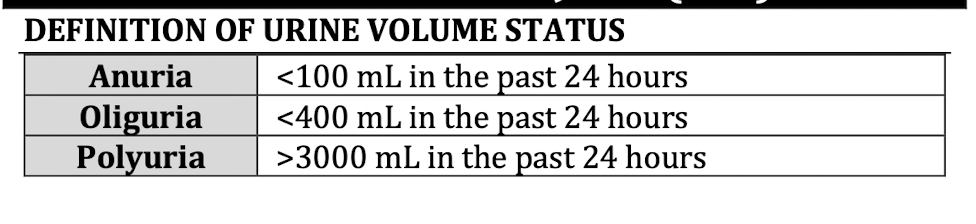
Define the ff:
Anuria
Oliguria
Polyuria
Define AKI (3)

3 Classifications of AKI
Prerenal:
Congestive Heart Failure
Liver Failure
Impaired renal autoregulation (NSAIDs ACE-I, ARBs)
Intrinsic:
Glomerular - Acute Glomerulonephritis
Tubules and Interstitium - ischemia, sepsis, infection, nephrotoxins
Vascular - vasculitis, malignant, TTP-HUS
Postrenal:
Obstructive

Most common intrinsic cause of AKI
Acute Tubular Necrosis
Urinalysis finding in Acute Tubular Necrosis
Muddy brown casts
Electrolyte profile of AKI - 5
Hyponatremia
Hyperkalemia, hypermagnesemia
Hyperphosphatemia
Hypocalcemia
Hyperuricemia
Peak GFR
120ml/min/1.73m2
Rate of decline of GFR by age
At the beginning of the third decade of life (20s), there is a normal annual decline in GFR by 1ml/min/1.73m2, GFR at age of 70 is at 70ml/min.1.73 (in a normal patient with comorbidities)
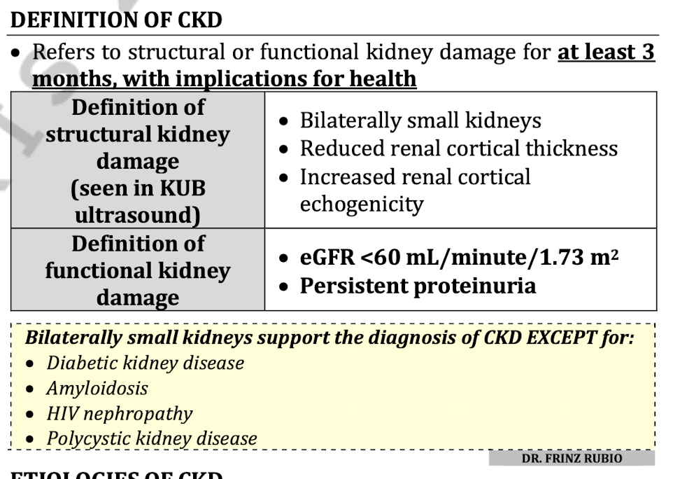
Define CKD
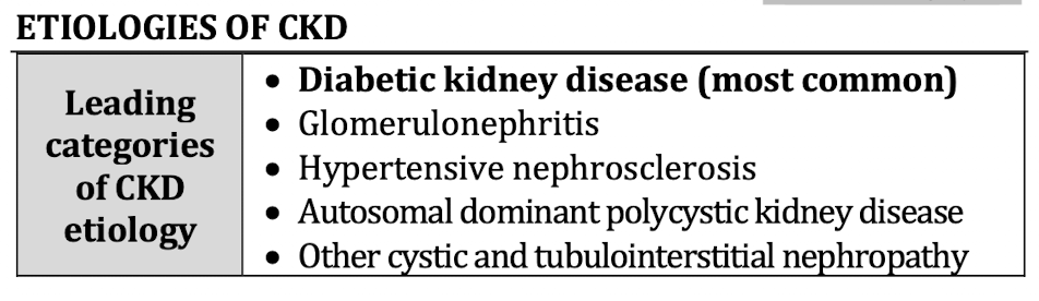
5 Etiologies of CKD
What determines CKD prognosis
GFR
Albuminuria (the higher the albuminuria >300mg/g the worse the prognosis)
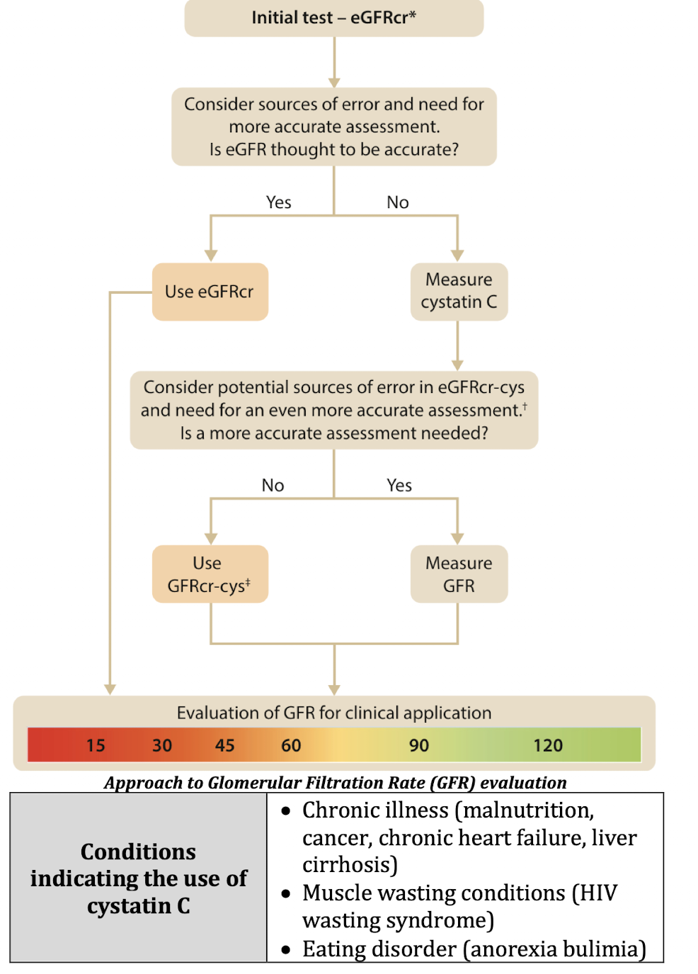
When to use Cystatin C for eGFRcr
What CKD stage do complications (laboratory) usually appear
CKD Stage 3 and 4
Stages in CKD when gadolinium use should be limited/avoided:
May cause nephrogenic systemic fibrosis (resembles skin changes seen in scleroderma)

CKD stage where anemia is universally present
Stage 4
CKD stage where peripheral neuropathy becomes clinically evident
Stage 4
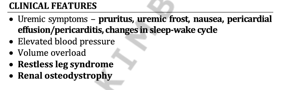
5 clinical features of CKD
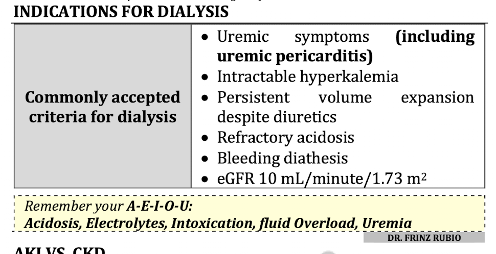
Indications for dialysis
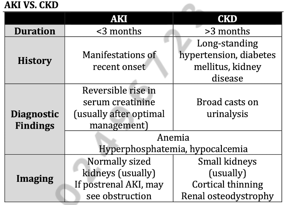
AKI vs CKD in terms of:
Duration
History
Diagnostic Finding
Imaging
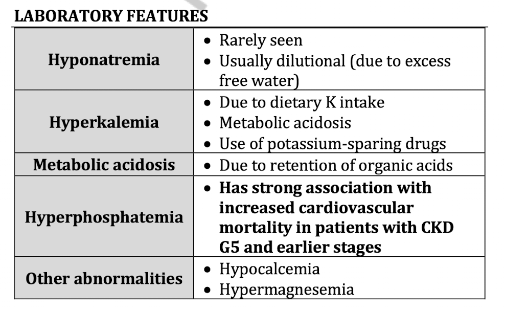
Lab Features of CKD (5)
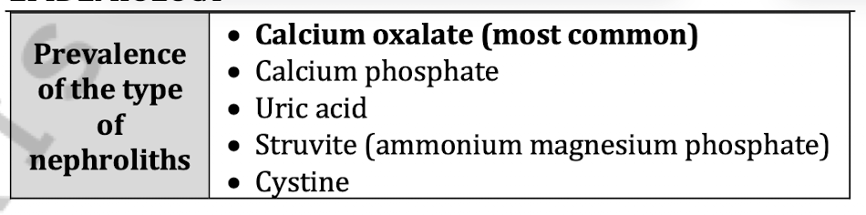
Prevalence of nephrolith types 5
Most clinically important inhibitor of calcium containing stones
Urine citrate
Risk of stone doubles in what amount of urine per day
<1L urine per day
Vit C intake among males can increase the risk for
Calcium oxalate stones
Vit C increases endogenous oxalic acid formation
Supplemental calcium intake increases risk for
calcium oxalate stone
calcium phosphate stone
Gold standard diagnostic test for nephrolithiasis
Helical CT scan without contrast
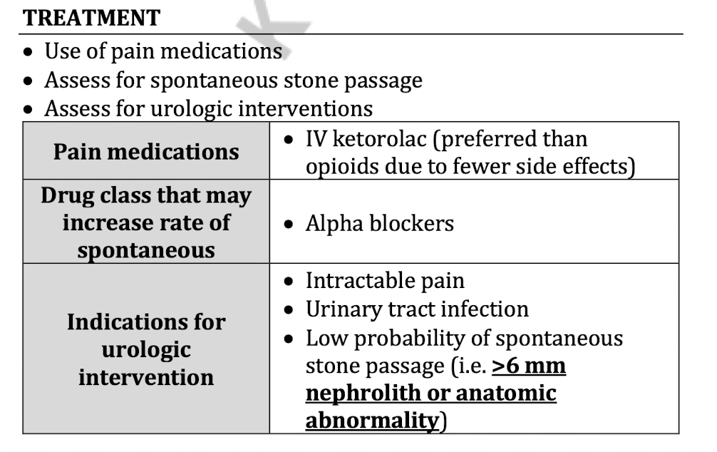
Treatment of nephrolith
medical expulsion therapy - tamsulosin 0.5mg OD PO, for distal nephrolith <6mm