bone
 Call Kai
 Learn
Learn Practice Test
 Spaced Repetition
 Match
Match Flashcards
 Knowt Play
1/73

There's no tags or description
Looks like no tags are added yet.
Name | Mastery | Learn | Test | Matching | Spaced | Call with Kai | Chat |
|---|
No analytics yet
Send a link to your students to track their progress
74 Terms
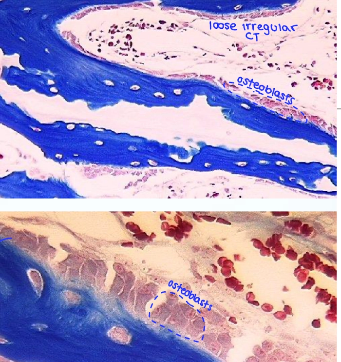
What are the 3 cells of bone?
The 3 bone cells are osteoblasts, osteocytes, and osteoclasts. Osteoblasts form bone, osteocytes maintain bone, and osteoclasts break down/resorb bone.
How can you distinguish osteoblasts, osteocytes, and osteoclasts histologically?
Osteoblasts are bone-forming cells on the inner/outer bone surfaces and are not in lacunae; osteocytes are located inside lacunae and have processes extending through canaliculi; osteoclasts are large multinucleated giant cells associated with bone-resorption/erosion lacunae.
What are osteoblasts?
Osteoblasts are bone-forming cells. They cover the inner and outer surfaces of bone, are capable of dividing, and have euchromatic DNA consistent with active cells. They are not located within lacunae.
Where are osteoblasts located?
Osteoblasts cover the inner and outer surfaces of bone and are not located within lacunae.
How can you identify an osteoblast in a histology section?
Look for active bone-forming cells lining the surface of bone. Unlike osteocytes, they are not enclosed in lacunae.
What are osteocytes?
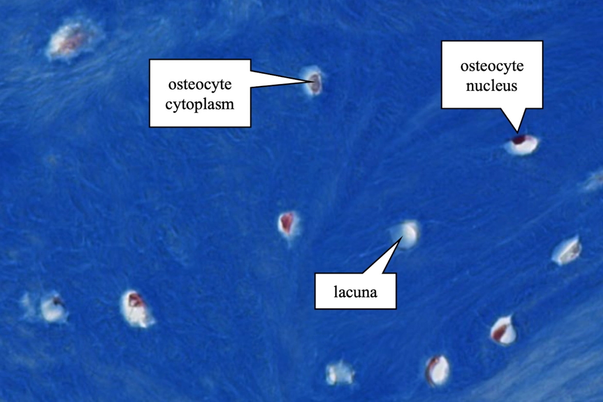
Osteocytes are bone cells derived from osteoblasts that maintain bone by secreting new matrix (osteoid). They reside in lacunae and respond to hormones.
Where are osteocytes located?
Osteocytes are located within lacunae in the bone matrix, typically at regular intervals between lamellae in an osteon.
What are canaliculi and why are they important?
Canaliculi are microscopic canals within the bone matrix through which osteocyte cytoplasmic processes extend. They connect lacunae and participate in cell-cell communication and the flow of nutrients and waste.
How can you identify an osteocyte histologically?
Identify a small cell located within a lacuna. In appropriate sections, its cytoplasmic processes extend into tiny canaliculi radiating through the surrounding matrix.
What are osteoclasts?
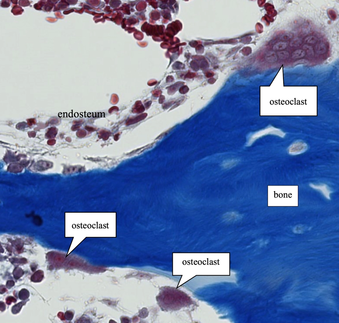
Osteoclasts are large, multinucleated giant cells that break down bone. They produce lysosomal enzymes that digest the organic collagenous matrix and organic acids that dissolve the mineralized inorganic matrix.
How can you identify an osteoclast histologically?
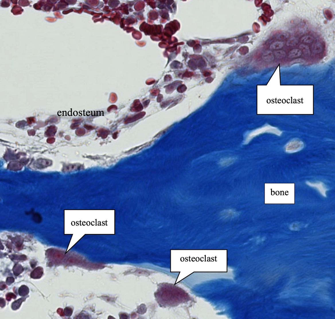
Look for a large multinucleated giant cell associated with an erosion lacuna, a space created where the cell is actively resorbing bone.
What is an erosion lacuna?
An erosion lacuna is a space created by osteoclasts as they actively resorb bone.
How do osteoclasts break down the two major components of bone matrix?
They use lysosomal enzymes to digest the organic/collagenous matrix and organic acids to dissolve the mineralized inorganic matrix.
What is the extracellular matrix of bone called?
The extracellular matrix of bone is called osteoid. It consists of organic and inorganic components; the matrix is initially secreted by osteoblasts and subsequently mineralized.
What are the organic components of bone matrix?
The organic components include type I collagen fibers plus ground substance containing glycosaminoglycans (GAGs) and glycoproteins.
What type of collagen fiber is found in bone?
Bone contains type I collagen fibers.
What is the function of type I collagen in bone?
The collagen-containing organic component contributes tensile strength, making bone more resistant to breakage under tension.
What is the ground substance of bone?
Bone ground substance contains glycosaminoglycans (GAGs) and glycoproteins.
What are the inorganic components of bone?
The inorganic component consists of hydroxyapatite crystals.
What are hydroxyapatite crystals?
Hydroxyapatite crystals are inorganic calcium phosphate crystals that precipitate and mineralize the osteoid.
What is the function of hydroxyapatite in bone?
Hydroxyapatite contributes compressive strength, making bone resistant to breakage under compression.
How do the organic and inorganic components contribute differently to bone strength?
The organic components, especially type I collagen, provide tensile strength and resistance to tension. The inorganic hydroxyapatite component provides compressive strength and resistance to compression.
What is the basic sequence of bone formation?
Bone formation occurs through endochondral ossification, beginning with osteoblast secretion of osteoid followed by mineralization of the matrix into bone.
What are the 2 types of microscopic bone architecture?
The two types are woven bone and lamellar bone.
What is woven bone?
Woven bone is immature bone that is eventually replaced by lamellar bone. It forms rapidly, so cells and collagen fibers are arranged irregularly.
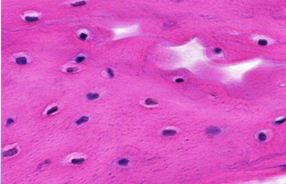
How can you identify woven bone histologically?
Woven bone lacks Haversian systems/osteons. Its rapid formation produces an irregular arrangement of cells and collagen fibers.
What is lamellar bone?
Lamellar bone is mature bone formed slowly by remodeling a woven-bone precursor. It has Haversian systems/osteons composed of organized layers of lamellae.
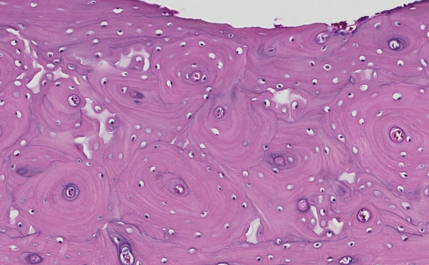
How can you identify lamellar bone histologically?
The key feature is the presence of osteons: organized concentric layers of lamellae arranged around central canals. Their circular appearance in transverse section indicates mature/lamellar bone.
What is the major histologic difference between woven and lamellar bone?
Woven bone has no osteons and has irregularly arranged cells and collagen fibers. Lamellar bone has osteons with organized concentric lamellae around central canals.
Which type of bone forms rapidly?
Woven bone forms rapidly.
Which type of bone is mature?
Lamellar bone is mature bone.
Which type of bone is eventually replaced by lamellar bone?
Woven bone is eventually replaced by lamellar bone during remodeling.
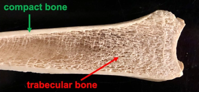
What are compact/cortical and cancellous/trabecular/spongy bone?
Compact/cortical bone is the dense outer shell of bone. Cancellous/trabecular/spongy bone consists of small internal spicules. Either gross type may microscopically contain woven bone, lamellar bone, or a mixture.
What is an osteon (Haversian system)?
An osteon is the structural unit of mature bone consisting of concentric layers of lamellae arranged around a central canal. It forms a tubular unit that runs the length of the bone.
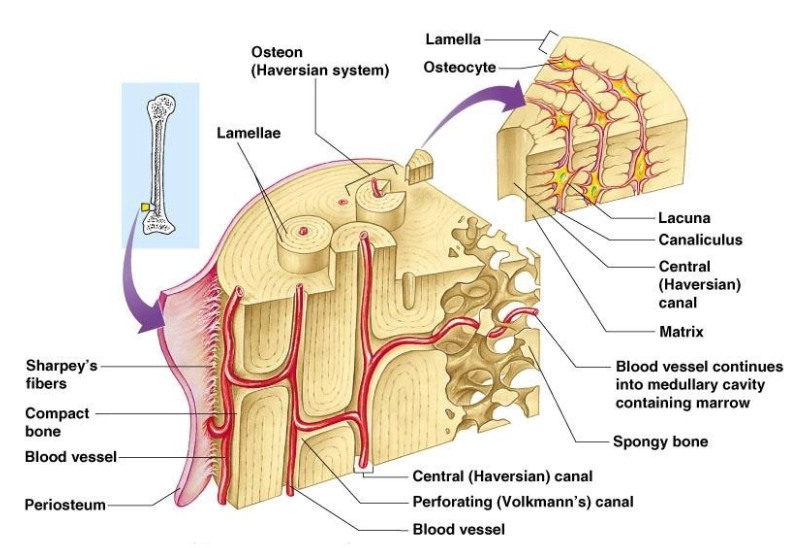
What is the central canal of an osteon?
The central canal is the structure at the center of an osteon. It contains blood vessels, lymphatic vessels, and nerve fibers.
How can you identify a central canal histologically?
In a transverse section of mature/lamellar bone, look for the central space at the center of a circular osteon, surrounded by concentric lamellae.
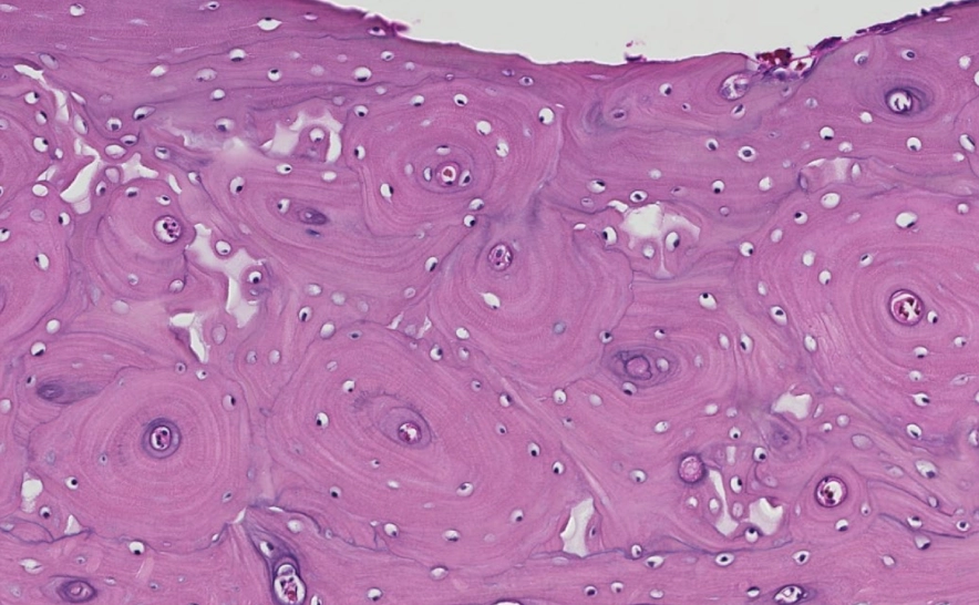
What are lamellae?
Lamellae are concentric layers of bone matrix in an osteon. Their collagen fibers alternate in direction from one layer to the next.
How can you identify lamellae in an osteon?
In transverse section, look for the concentric circular layers surrounding a central canal. These rings are the lamellae.
Where are osteocytes located within an osteon?
Osteocytes reside in lacunae located at regular intervals between the lamellae.
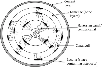
What is a lacuna?
A lacuna is a small space in the bone matrix that houses an osteocyte.
How can you identify lacunae histologically?
Look for small spaces within the bone matrix, especially between lamellae in an osteon. Osteocytes reside in these spaces.
How are lacunae different from canaliculi?
A lacuna houses an osteocyte; canaliculi are microscopic channels connecting adjacent lacunae and carrying osteocyte processes.
What are perforating (Volkmann's) canals?
Perforating canals interconnect longitudinally arranged central canals with one another and with the medullary/marrow cavity of bone.
How can you identify perforating canals histologically?
Look for canals that connect central canals with one another or with the marrow cavity rather than forming the central core of a concentric osteon.
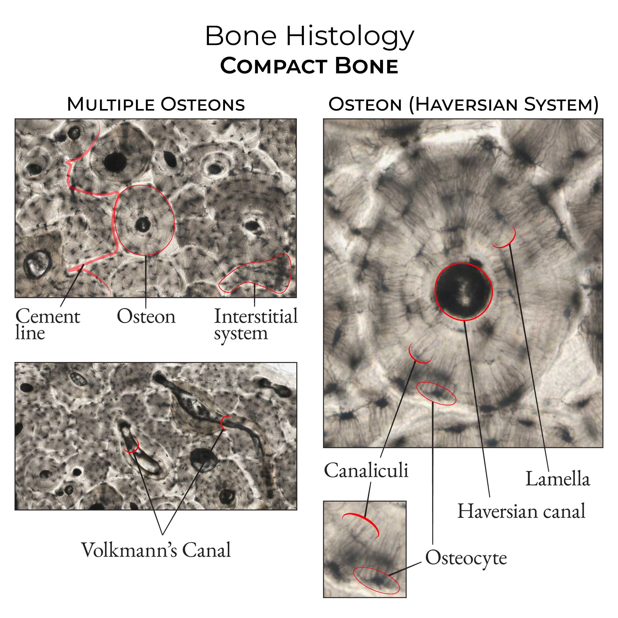
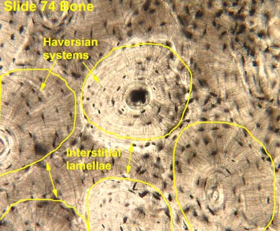
What are interstitial lamellae?
Interstitial lamellae are orphan fragments of partially degraded osteons. They do not have central canals.
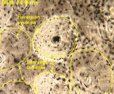
How can you identify interstitial lamellae histologically?
Look for irregular remnants/fragments of lamellar bone between intact osteons. Unlike complete osteons, they lack a central canal.
What are inner circumferential lamellae?
Inner circumferential lamellae are layers of bone arranged in a circular pattern around the marrow cavity. They are covered by endosteum, smooth the inner surface of bone, and are not part of an osteon.
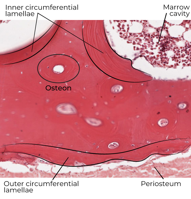
How can you identify inner circumferential lamellae histologically?
Find the circular layers immediately adjacent to the marrow cavity on the inner surface of compact bone. They are not centered around a central canal and are covered by endosteum.
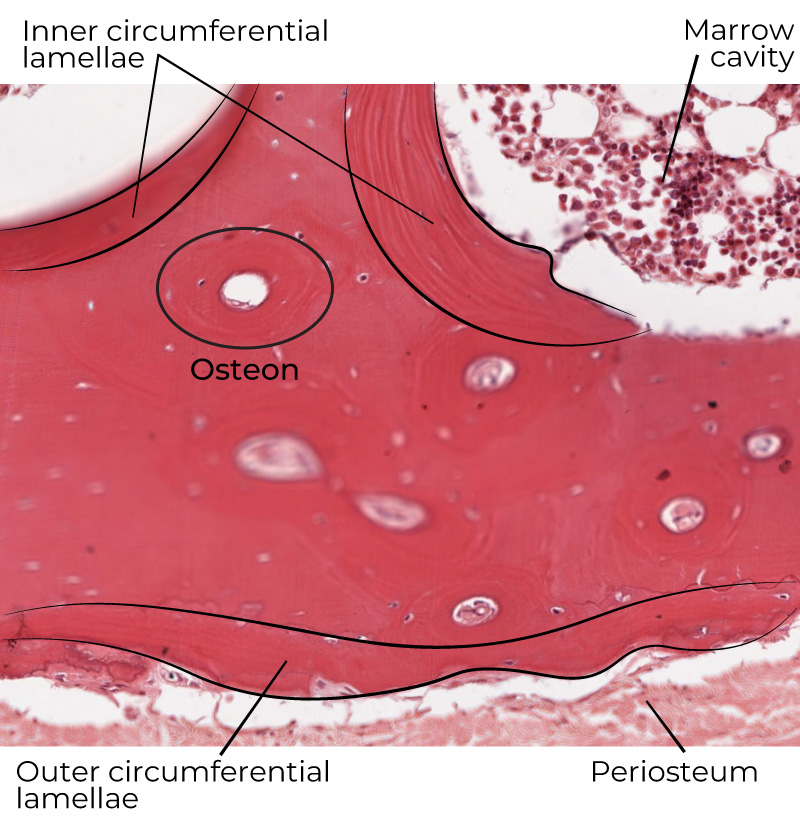
What are outer circumferential lamellae?
Outer circumferential lamellae are layers of bone arranged around the outer surface of bone. They are covered by periosteum, smooth the outer surface, and are not part of an osteon.
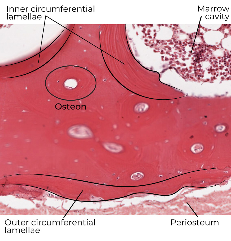
How can you identify outer circumferential lamellae histologically?
Look at the outer surface of compact bone for circumferential layers beneath the periosteum. They are not organized around a central canal.
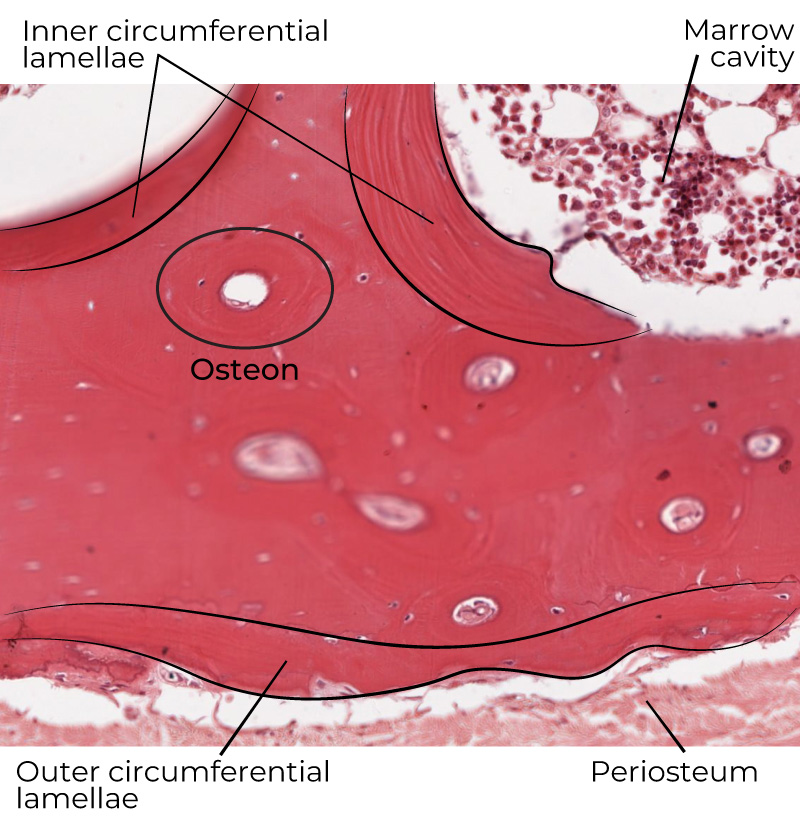
What is the difference between circumferential lamellae and osteonal lamellae?
Osteonal lamellae form concentric rings around a central canal within an osteon. Circumferential lamellae run around the inner or outer surface of the bone and are not part of an osteon.
What is endosteum?
Endosteum is the inner loose irregular connective tissue lining of bone. It covers all inner surfaces and surrounds blood vessels.
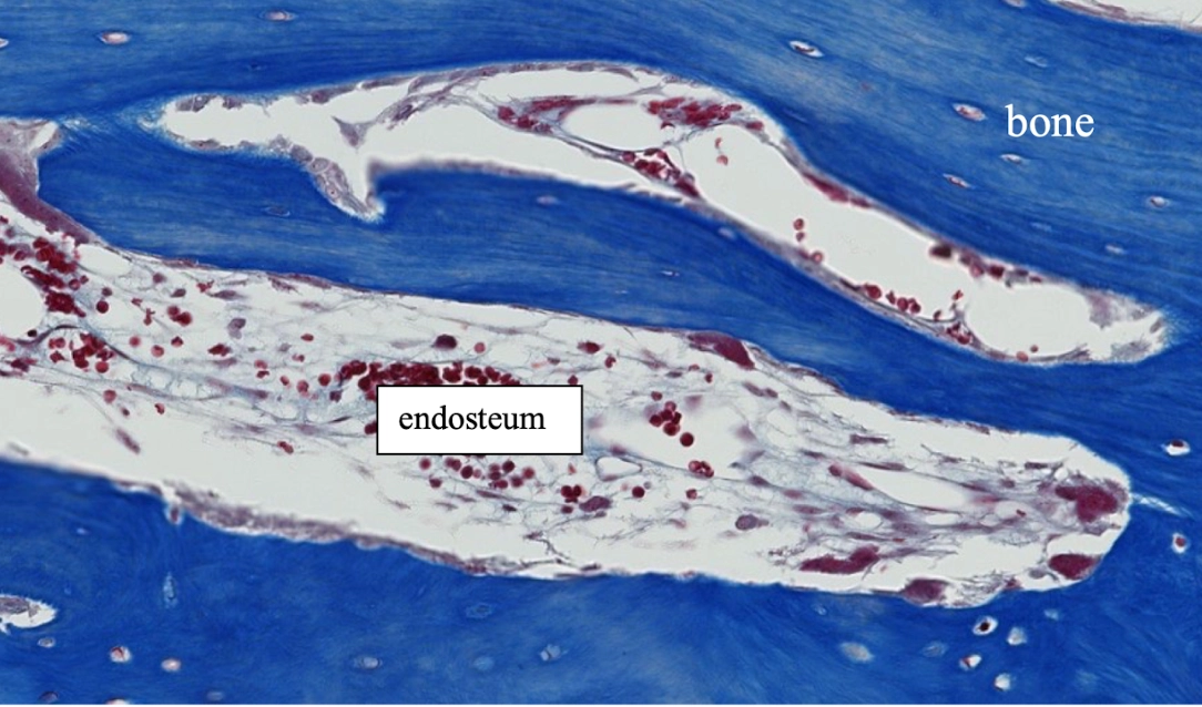
What type of connective tissue makes up the endosteum?
The endosteum is made of loose irregular connective tissue.
How can you identify endosteum histologically?
Look along the inner surfaces of bone, including surfaces associated with the marrow cavity and around blood vessels. The lining is loose irregular connective tissue.
What is periosteum?
Periosteum is the outer dense irregular connective tissue lining of bone. It is highly vascularized and innervated.
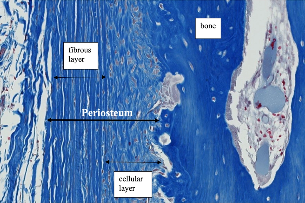
What are the two layers of periosteum?
The periosteum has an outer fibrous layer and an inner cellular (osteogenic) layer.
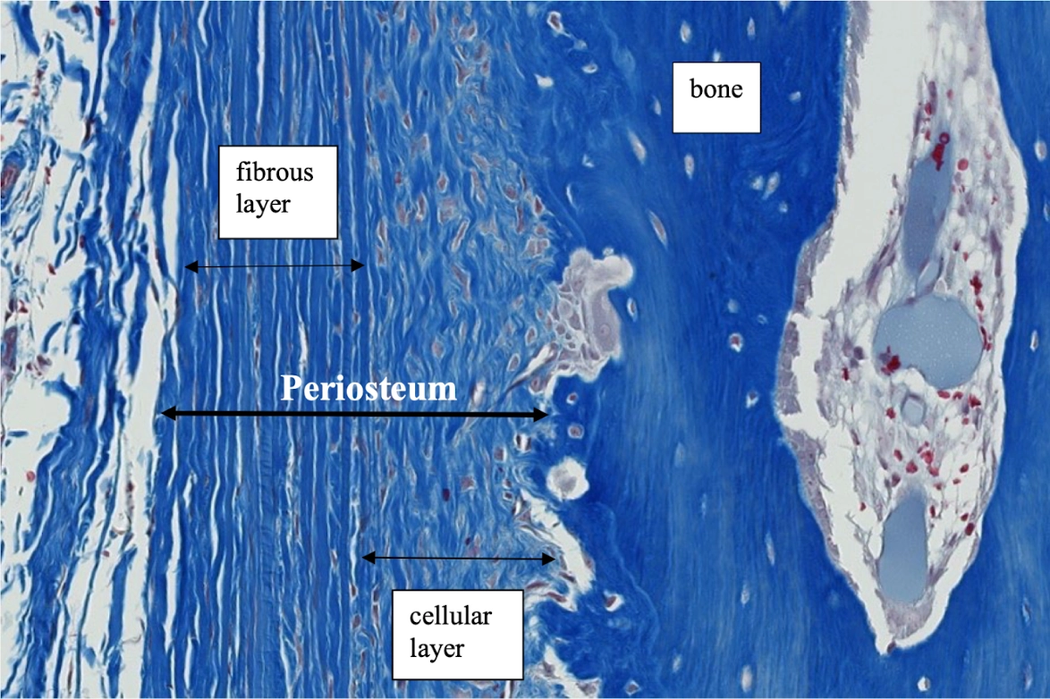
What is the fibrous layer of the periosteum?
The fibrous layer is the outer layer of dense connective tissue that acts as a capsule. Its predominant cell is the fibroblast.
How can you identify the fibrous layer of periosteum histologically?
Look at the outermost periosteal layer. It is dense connective tissue functioning as a capsule, with fibroblasts as the predominant cell type.
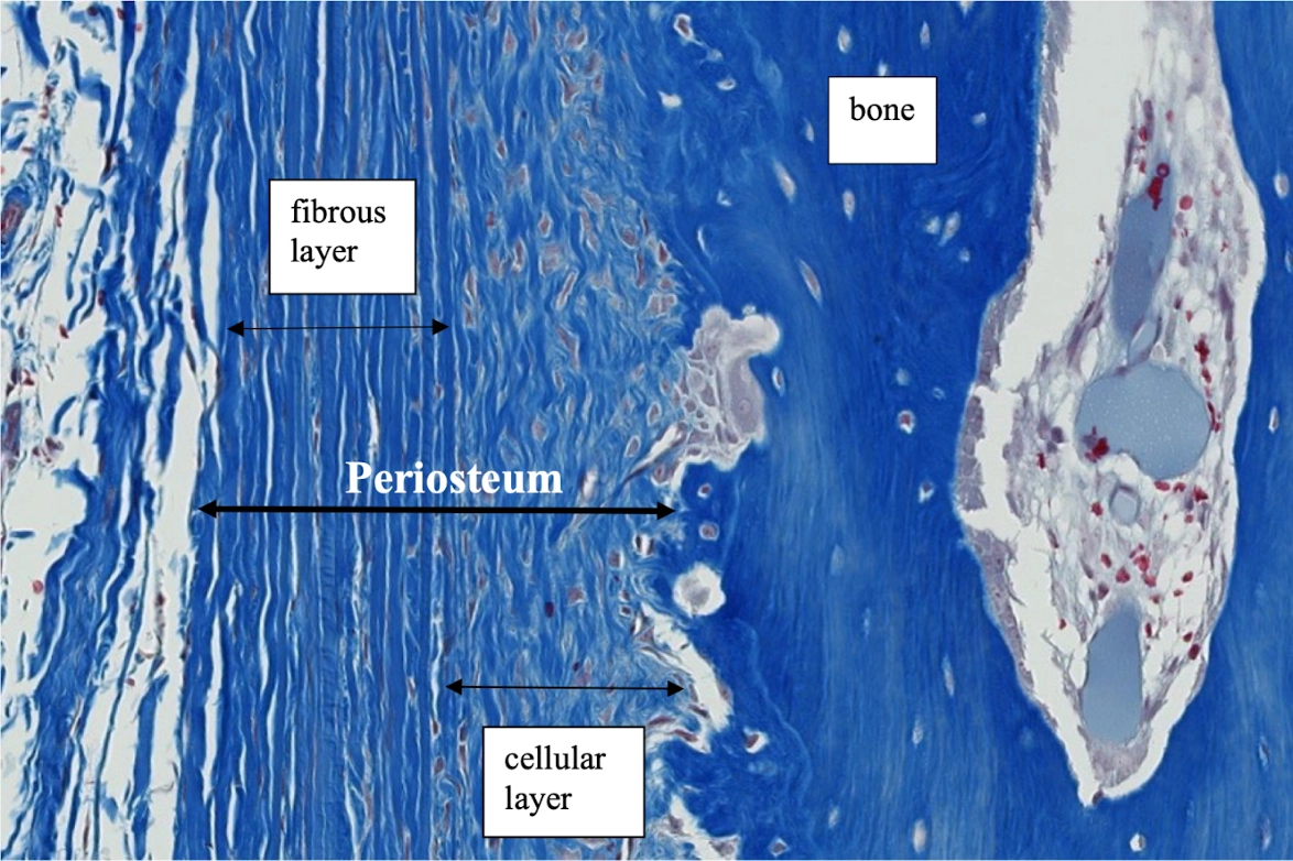
What is the cellular (osteogenic) layer of the periosteum?
The cellular/osteogenic layer lies between the fibrous layer and cortical bone. It is highly cellular and contains osteoprogenitor cells.
How can you identify the cellular layer of periosteum histologically?
It is the highly cellular inner layer of the periosteum immediately adjacent to cortical bone, between the fibrous layer and the bone.
What is the difference between periosteum and endosteum?
Periosteum is the outer dense irregular connective tissue covering of bone and is highly vascularized and innervated. Endosteum is the inner loose irregular connective tissue lining that covers internal bone surfaces and surrounds blood vessels.
What are perforating (Sharpey's) fibers?
Perforating fibers are bundles of type I collagen fibers extending from the outer fibrous layer of periosteum into the outer circumferential layer of bone.
What is the function of perforating (Sharpey's) fibers?
They anchor the periosteum to bone and also anchor some tendons and ligaments to bone, as well as the periodontal ligament of teeth.
How can you identify perforating fibers histologically?
Look for bundles of type I collagen extending from the fibrous periosteum into the outer circumferential bone layer.
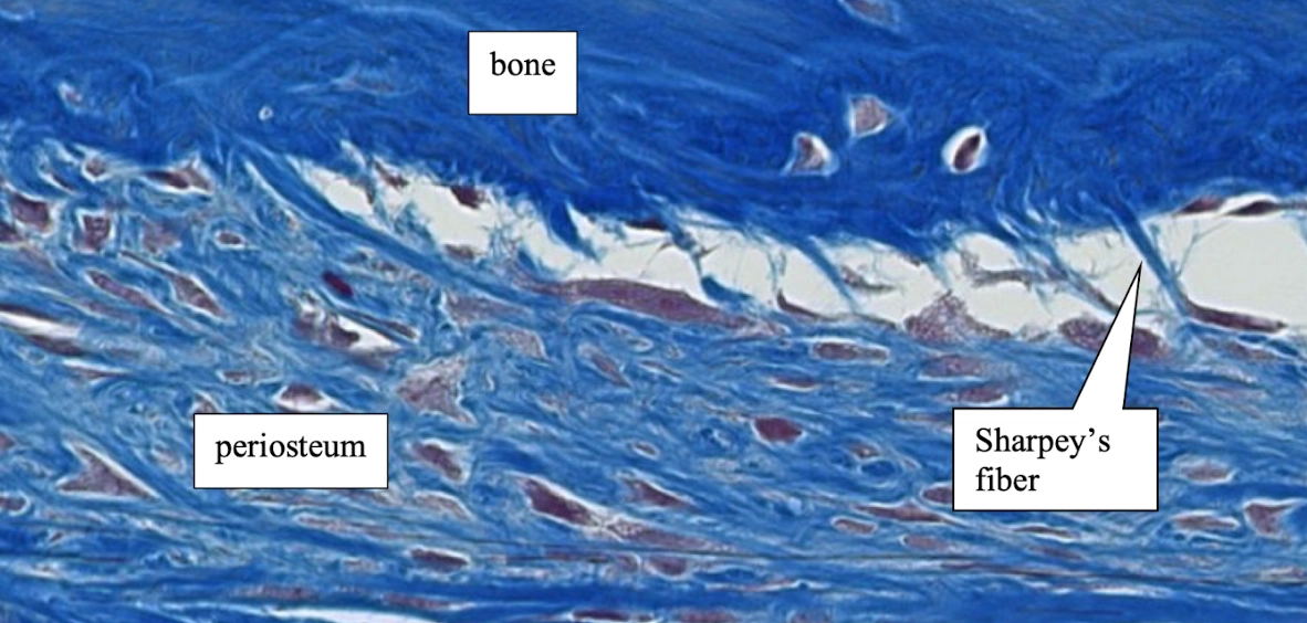
What is the relationship between periosteum and outer circumferential lamellae?
The periosteum covers the outside of the bone, while the outer circumferential lamellae form the underlying circular bone layer that smooths the outer surface. Sharpey's fibers extend from the periosteum into this layer.
What is the relationship between endosteum and inner circumferential lamellae?
The endosteum covers the inner surface of bone, while inner circumferential lamellae form circular layers around the marrow cavity and smooth the inner bone surface.
What should you look for first when identifying mature lamellar bone in a transverse section?
Look for osteons: circular structures with a central canal surrounded by concentric lamellae. Then identify lacunae/osteocytes between lamellae and canaliculi radiating between lacunae.
What should you look for first when identifying an osteon in transverse section?
Find a central canal and the concentric rings of lamellae surrounding it. Lacunae containing osteocytes lie between the lamellae.
What features should you identify within a complete osteon?
Central canal, concentric lamellae, osteocytes in lacunae, and canaliculi. The central canal contains blood and lymphatic vessels and nerve fibers.
What features distinguish an interstitial lamella from a complete osteon?
An interstitial lamella is a remnant of a partially degraded osteon and lacks a central canal. A complete osteon has concentric lamellae surrounding its own central canal.
What does a longitudinal section of an osteon emphasize?
Because osteons run longitudinally along the length of the bone, a longitudinal section emphasizes the tubular/lengthwise organization of osteons and their central canals.
What does a transverse section of an osteon emphasize?
A transverse section shows the osteon in its characteristic circular pattern: a central canal surrounded by concentric lamellae, with lacunae between the lamellae.
What does it mean that bone is highly vascular unlike cartilage?
The study guide emphasizes that bone is highly vascular, whereas cartilage is not, although both bone and cartilage have lacunae.
What is a key distinction between bone and cartilage based on the study guide?
Both have lacunae, but bone is highly vascular whereas cartilage is not.