403 Unit 1: MSK
 Note
Note Call Kai
 Learn
Learn Practice Test
 Spaced Repetition
 Match
Match Flashcards
 Knowt Play
1/625

There's no tags or description
Looks like no tags are added yet.
Name | Mastery | Learn | Test | Matching | Spaced | Call with Kai | Chat |
|---|
No analytics yet
Send a link to your students to track their progress
626 Terms
In organs, Stroma is _ and Parenchyma is _
General supportive/connective tissue
Tissue relevant for specific organ function (e.g. hepatocytes)
LO: What are the 4 basic tissue types?
Epithelial
Connective
Muscle
Nervous
The deep part of epithelial cells are anchored to the same _
Basement membrane
LO: Do epithelial cells divide?
Yes, rapidly
O2 reaches epithelial tissue via _
Diffusion (no direct blood supply)
Epithelium functions
Protection from harmful stimulus (e.g. skin protects from outside world)
Secretion (via glands)
Absorption (epithelial wall of GI tract)
Filtration (through kidney glomerulus)
LO: What factors are used to classify epithelia?
Shape
Number of layers
LO: Are epithelia vascular or avascular?
Avascular
Epithelial cell shapes
Squamous (flat), Cuboidal (cubes), Columnar (columns)
Epithelial layer types
Simple (1 layer)
Stratified (>1 layer)
Epithelial shape is classified by which layer?
The surface layer (apical)
Pseudostratified Epithelium
A single layer of epithelial cells attached to the same basement membrane that contains nuclei at different levels
Transitional Epithelium
Stretchy layers of stratified epithelium that can form a domed surface when subjected to distensive forces
LO: What are the functions of connective tissues?
Bind musculoskeletal structures (tendons/ligaments)
Provide structural support (bones and cartilage)
Protect organs (bones, cartilage, adipose)
Insulate body (adipose)
Transportation (blood)
LO: What cell types are found in connective tissues?
Fibroblasts (make fibers)
Macrophages (WBC’s which mount immune defense)
Mast cells (heparin and histamine production)
The connective tissue “matrix” is composed of _
Fibers (proteins) and Ground substance
_ connective tissues are not well vascularized
_ connective tissues are well vascularized
Cartilaginous, tendinous, and ligamentous
All other
LO: What are the 3 types of cartilage?
Hyaline, Fibrocartilage, Elastic
LO: What are the 3 types of loose connective tissue?
Areolar, Adipose, Reticular
LO: Rank the density of 1) Dense connective tissue, 2) Loose CT, 3) Liquid CT, 4) Bone CT, 5) Cartilage CT
Bone
Cartilage
Dense CT
Loose CT
Liquid CT
LO: Define tensile strength
Maximum force that a structure can withstand while being stretched (end to end)
LO: Define compressive strength
Maximum force that a structure can withstand while being compressed (end to end)
LO: Define shear strength
Maximum force that a structure can withstand while being pushed in opposite directions
Bone resistance ranking (shearing/tensile/compression):
_
_
_
Tensile
Compression
Shearing
Osseous Tissue is composed of _
Organic (collagen) and Inorganic (calcified mineral) components
Periosteum is composed of _ tissue
Dense, irregular connective
Ligaments and Tendons are composed of _ tissue
Dense connective
Red Bone Marrow function
Site of hemopoiesis (where RBC’s develop)
Yellow Bone Marrow function
Triglyceride storage in medullary cavities (hollow shaft of diaphysis)
Arteries carry blood _
Veins carry blood_
Away from the heart/Towards tissues
Towards the heart/Away from tissues
Nerves
Groups of axons bundled outside of CNS
Which component of the skeletal system is important in hemopoiesis?
A. Arteries
B. Hyaline cartilage
C. Osseous tissue
D. Red bone marrow
E. Yellow bone marrow
D. Red bone marrow
Bone cell types
Osteogenic cells
Osteoblasts
Osteocytes
Osteoclasts
Osteogenic Cells
Stem cells of bone which can mature into osteoblasts when bone remodeling is necessary
Osteogenic cells can be found in _
The periosteum, Endosteum, and Nerovascular canals
Osteoblast Cells
“Bone-Building Cells” which 1) produce organic components of bone, 2) Initiate calcification, and 3) Contribute to hormonal regulation of resorption
Osteocytes
Mature bone cells which maintain bone metabolism via nutrient/waste exchange
Osteoclasts
“Bone resorbers” which break down bony ECM at the ruffled border via protons and lysosomal enzymes
When [blood calcium] is low, _ (bone cell type) should be up-regulated
When [blood calcium] is high, _ (bone cell type) should be up-regulated
Osteoclasts
Osteoblasts
Osteogenic cells are derived from _
Mesodermal mesoschyme
Progression of a bone cell throughout its lifetime
Mesenchyme —> Osteogenic Cell —> Osteoblast —> Osteocyte
Osteoclasts are formed via _
Fusion of monocytes
In osteoporosis, bone resorption occurs at a higher pace than bone deposition. Which bone cells are outpacing the bone-building cells?
A. Osteoblasts
B. Osteoclasts
C. Osteocytes
D. Osteogenic cells
B. Osteoclasts
LO: List and describe the primary functions of the different types of bone cells
Osteogenic Cell: Differentiate into bone-building cells
Osteoblast: Build bone
Osteocyte: Maintain bony ECM
Osteoclast: Resorb bone
LO: Differentiate between the origins and development of the different types of bone cells.
Osteogenic cells, Osteoblasts, Osteocytes share direct lineage from mesenchyme
Osteoclasts form independently via monocyte fusion
LO: The organic component of osseous tissue is composed of _, which functions to _
Collagen, Provide flexibility and tensile strength to the bone (and an ECM framework)
LO: The inorganic component of osseous tissue is composed of _, which functions to _
Crystalized mineral salts (calcium, hydroxyapatite), Provide bone its hardened/calcified structure
Rough composition of bony ECM
30-40% organic components
45-55% inorganic components
15% water
Which component of bone is primarily responsible for flexibility and tensile strength?
A. Calcium mineral salts
B. Collagen
C. Hydroxyapatite
D. Osteoclasts
B. Collagen
LO: Describe the location of compact and spongy bone in the skeletal system
Generally,
Compact bone forms the external layer of bones and most of the diaphysis shaft
Spongy bone forms short/flat/irregular bones and the epiphyses of long bones
LO: Identify the alternate names for compact & spongy bone.
Compact: “Cortical” or “Dense” Bone
Spongy: “Trabecular” or “Cancellous” Bone
Compact bones are organized into _
Osteons
Osteons
Concentric lamellae of mineralized bony ECM surrounding a central canal which contains neurovasculature and lymphatics
Perforating Canals/Volkmann’s Canals
Passages that transversely cross an osteon to connect neurovasculature from the periosteum to the central canals and medullary cavity
Circumferential Lamellae
Outer layer of osteons which connect directly to the periosteum and support appositional (“thickening”) growth
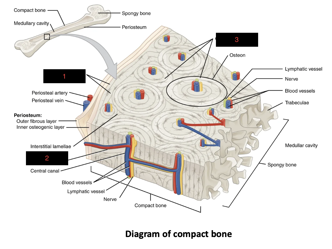
1
Circumferential Lamellae
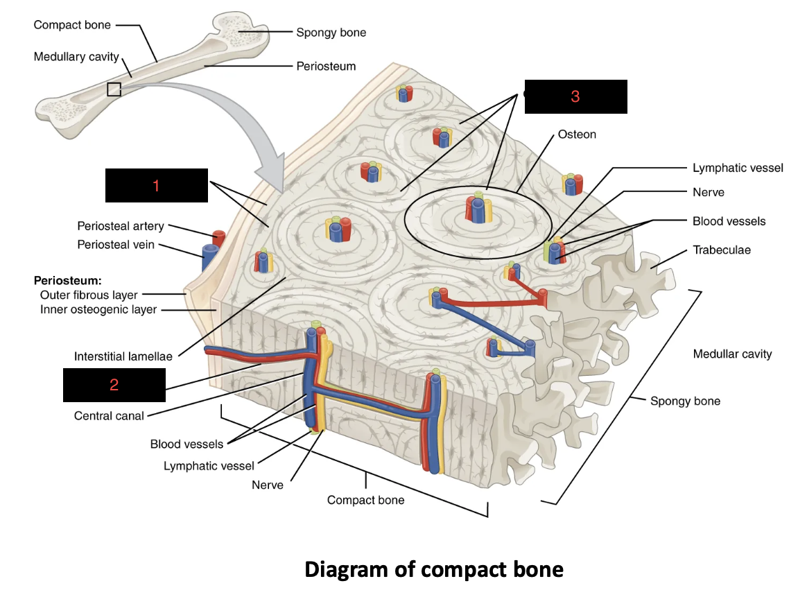
2
Perforating/Volkmann Canal

3
Concentric Lamellae
Spongy bone is organized into _, determined by _
Trabeculae (not osteons), Lines of stress
Which structures associated with compact bone directly surrounds a central canal?
A. Circumferential lamellae
B. Concentric lamellae
C. Perforating canals
D. Trabeculae
B. Concentric lamellae
LO: Explain the structural organization of compact bone, and identify the components of an osteon.
Compact bone is organized into repeating osteons with concentric lamellae, perforating canals, and circumferential lamellae
LO: Define and summarize the roles of perforating canals and circumferential lamellae.
Perforating Canals: Supply neurovasculature to medullary cavity and central canal
Circumferential Lamellae: Connect osteons to periosteum and support appositional growth
LO: Compare the organizational structure and function of spongy bone to compact.
Compact: Osteons with limited gaps for bone durability
Spongy: Trabeculae with large gaps to 1) hold marrow, 2) decrease brittleness, 3) decrease weight
Diaphysis
Body/shaft of a long bone containing mostly compact bony tissue
Epiphyses
Proximal and distal ends of long bones containing mostly spongy bone (with a compact bone cover)
Metaphysis
The region between a long bone’s epiphysis and diaphysis where growth plates reside
Growth plates are _ joints
Synchondrosis
Periosteum
A layer of dense irregular CT (outer) and cells (inner) which surround the external surface of a bone
Endosteum
The inner lining of the medullary cavity, bony canals, and spongy bones (wherever there is a hole/opening)
The articulating surface of epiphyses are covered by _
Hyaline cartilage
Medullary Cavity
The hollow space within a diaphysis where yellow bone marrow and vasculature reside
Articular cartilage is typically located on what part of a long bone?
A. Diaphysis
B. Epiphysis
C. Medullary cavity
D. Metaphysis
E. Periosteum
B. Epiphysis
LO: List and describe the primary characteristics of the three major regions of a long bone.
Diaphysis (Long shaft made of compact bone)
Epiphysis (Proximal/distal ends made of spongy bone)
Metaphysis (Contain cartilaginous growth plates in region between diaphysis and epiphysis)
LO: Explain the role of the epiphyseal (growth) plate within the metaphysis.
Ossify during development/growth of long bones
LO: Summarize the composition and location of the periosteum, endosteum, articular cartilage, and medullary cavity.
Periosteum: Dense/irregular CT (outer) and cellular layer (inner) which wrap around bone
Endosteum: Single layer of cells which lines gaps in bone
Articular Cartilage: Cover epiphyses at joints with hyaline cartilage
Medullary Cavity: Hollow space in diaphyses containing yellow bone marrow and vasculature
LO: Define ossification and list four situations in which bone formation occurs.
The process by which bone forms during:
Initial bone formation
Bone growth
Bone remodeling
Fracture repair
Two methods of ossification
Intramembranous and Endochondral
Intramembranous Ossification
When bone forms directly within mesenchyme (mesenchyme —> bone)
_ bones undergo Intramembranous Ossification
Facial bones (including mandible and fontanels), Medial clavicle
Endochondral Ossification
Mesenchyme —> Hyaline cartilage —> Bone
_ bones undergo Endochondral Ossification
Most (non-facial or medial clavicular)
LO: Differentiate between intramembranous and endochondral ossification in terms of associated connective tissue and examples of bones formed through each method.
Intramembranous: Mesenchyme —> Bone (facial bones and medial clavicle)
Endochondral: Mesenchyme —> Hyaline Cartilage —> Bone (most other bones in the body)
Interstitial Growth
Growth of long bones endogenously from ossifying cartilage at epiphyseal plates
Appositional Growth
Growth of long bones exogenously via thickening of periosteum/endosteum
LO: Compare interstitial and appositional growth in terms of location and effects on bone structure.
Interstitial: Endogenous bone growth/lengthening at epiphyseal plates
Appositional Growth: Exogenous bone thickening along periosteum/endosteum
The occipital bone of the skull forms via which type of ossification process?
A. Endochondral
B. Intramembranous
B. Intramembranous
_ bone undergoes remodeling faster than _ bone
Spongy, Compact
3 major factors affecting bone remodeling
Minerals (inorganic elements)
Vitamins (organic nutrients)
Hormones (regulation)
Minerals affecting bone remodeling
Calcium, Phosphorus, Magnesium, Fluoride, and more
Vitamins affecting bone remodeling
Vitamin A (osteoblast stimulation and collagen synthesis)
Vitamins C, K, and B12 (osseous tissue protein synthesis)
Vitamin D (absorb calcium from GI tract to build osseous tissue)
Hormones affecting bone remodeling
hGH
Thyroid Hormone
Calcitonin
Parathyroid Hormone
Sex Hormones
Bonus: Calcitriol (active Vitamin D)
hGH effects on bone (4)
Stimulate osteoblasts
Promote cell division at epiphyseal plates
Increase necessary proteins
Promote synthesis of IGF’s
Thyroid Hormone effect on bone
Stimulate osteoblasts
Calcitonin effects on bone (2)
Promote blood calcium uptake
Inhibit osteoclasts
Parathyroid Hormone effects on bone (2)
Increase osteoclast activity (increase blood [Ca2+])
Stimulate calcitriol synthesis
Calcitriol
Active form of Vitamin D which increases absorption of calcium from GI tract
Sex Hormones effect on bone
Promote osteoblast activity during puberty
If blood calcium levels are low, which hormone may be secreted in order to maintain homeostasis?
A. Calcitonin
B. Human growth hormone
C. Parathyroid hormone
D. Testosterone
C. Parathyroid hormone
LO: Explain the function of bone remodeling and identify differences in renewal rates between spongy and compact bone.
Bone remodeling is necessary to maintain stronger tissue along common lines of stress.
RateSpongy > RateCompact
LO: Explain the roles and effects of key minerals, vitamins, and hormones on bone growth and remodeling.
Minerals: Provide resources to build bone
Vitamins: Stimulate synthesis of proteins
Hormones: Regulate concentration of calcium in bones and blood