Sensory Systems: Chemical Senses
1/15
There's no tags or description
Looks like no tags are added yet.
Name | Mastery | Learn | Test | Matching | Spaced |
|---|
No study sessions yet.
16 Terms
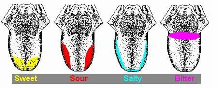
Basic tastes: salt, sour, sweet, bitter
Salt: NaCl
Sour: HCl
Sweet: Sucrose
Bitter: Quinine
Sensation of Taste
Interaction with olfactory (smell) and tactile (texture) stimuli
perception of taste is a combination of stimuli not just taste (smell, texture, temp)
Taste thresholds
Sweet: sucrose 10-2 M
Sour: HCl 9x10⁻⁴ M
Salty: NaCl 10-2 M
Bitter: quinine 8x10⁻⁶ M
High threshold: sweet and salty
Low threshold: sour and bitter (need to be easily detected for spoiled/poisonous food)
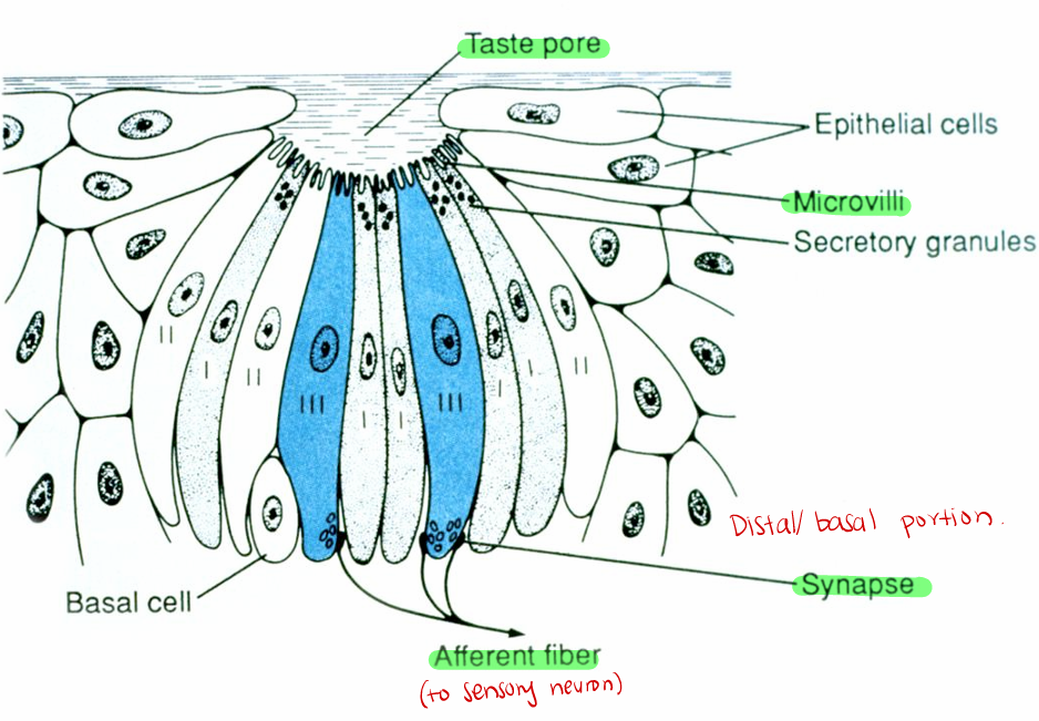
Taste Buds
Structure: receptor (specialized cell/not a neuron) and supporting cells
Receptor has synaptic vesicles at base that release neurotransmitters (communicate with afferent (sensory) nerve cells)
Cells are replaced about every 10 days (basal cells- maintenance and support)
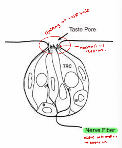
Signal Transduction in Taste Receptors
Membrane receptors on apical microvilli
Variety of primary/second messenger mechanisms
-Depends on chemical type
Salty (Na+) and Sour (H+/K+): ion channels → depolarize
Bitter and Sweet: 2nd messenger system (cyclic AMP → depolarize)
*Receptor cells do not generate action potentials. Instead, it receives a stimulus that depolarizes it, pushing the vesicle out. It is not all-or-nothing but instead is proportional to the number of receptors stimulated (generates membrane potential)
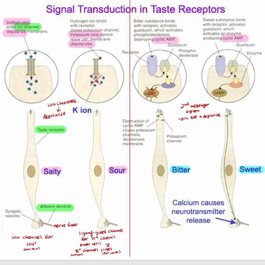
Synaptic contact generates action potential in cranial nerve ending
primary afferent is group III small myelinated nerve, with cell body in cranial nerve ganglion
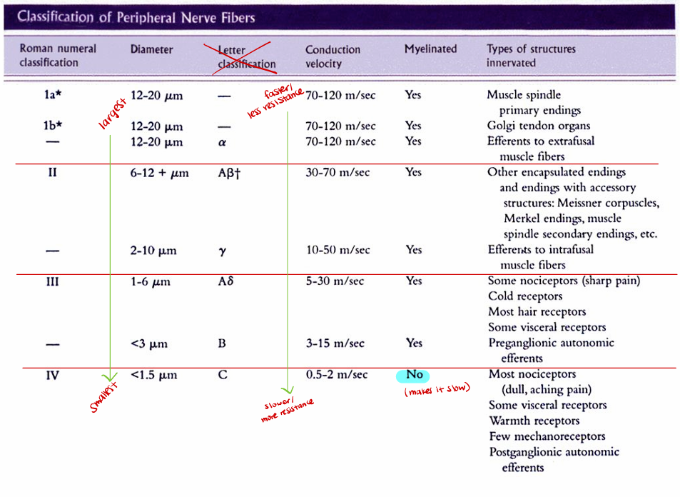
Classification of Peripheral Nerve Fibers
Largest/ Fastest/ Least resistance → Smallest/ Slowest/ More resistance
Group 1, 2 ,3 4 (unmyelinated)
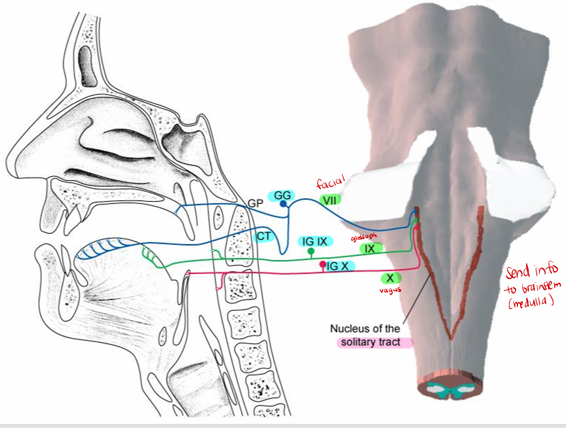
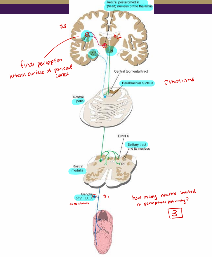
Taste Pathways (nerves → medulla)
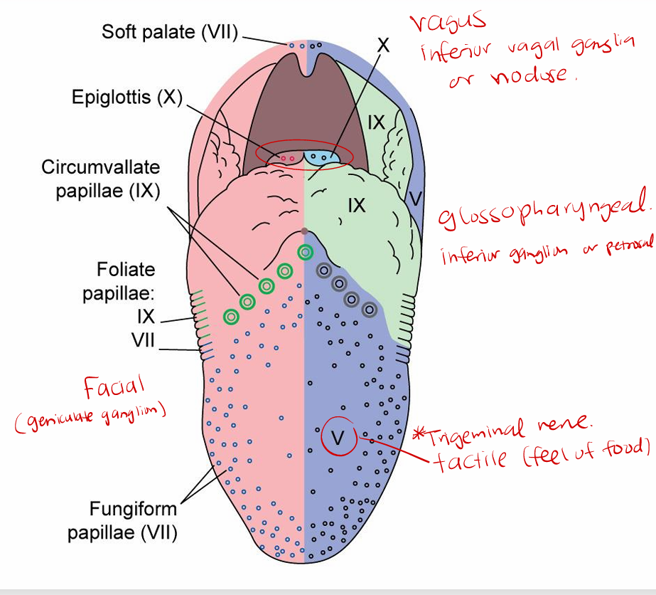
Chorda tympani nerve to facial (VII) for front 2/3 of tongue (anterior)
geniculate ganglion (outside brainstem: where cell bodies of facial nerves located)
Glossopharyngeal (IX) for back of tongue (1/3)
petrosal or inferior ganglion
vagus (X) for throat
inferior vagal (nodose) ganglion
Facial, glossopharyngeal, vagus nerves to solitary nucleus and tract of medulla
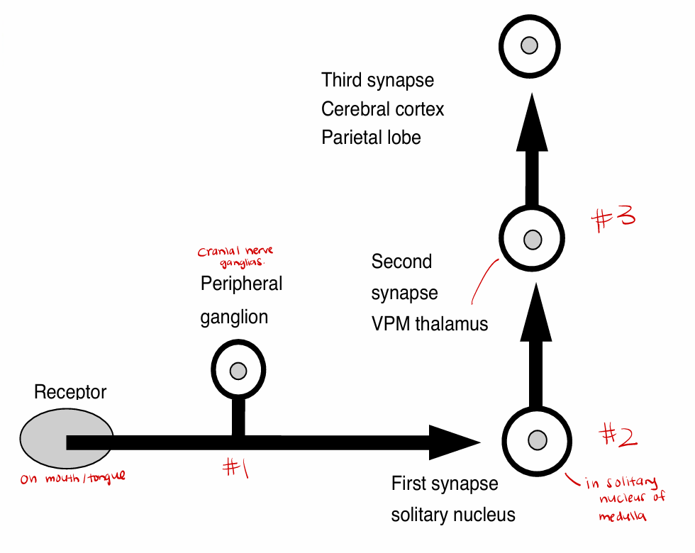
Taste Pathways (medulla→ parietal lobe)
From solitary nucleus (in brainstem/medulla):
Relayed to ventral posterior medial nucleus (VPM) of thalamus (relay station)
Perceptual path ends at lateral surface of parietal cortex (Brodmann areas 3,1,2) (final perception)
Taste reflexes to salivatory nuclei (VII, IX) (vagus (X) not connected to salivary glands)
Additional path to hypothalamus & amygdala (limbic system: emotional center)
Via parabrachial nucleus of pons
Provides input to pleasure/pain, hunger/thirst/satiety centers
Sensation of Smell
Thought to be analogous to immune system
6 general groups: floral (rose), ethereal (pears), musky (musk), camphor (eucalyptus), putrid (rotten eggs), pungent (vinegar)
Odors may directly trigger instinctual behavior, strong emotional responses/memories in humans
Direct links to “emotional” brain centers (limbic system)
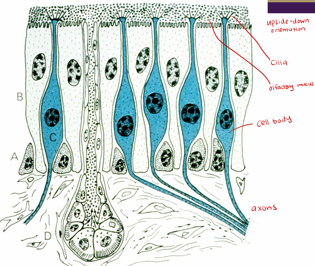
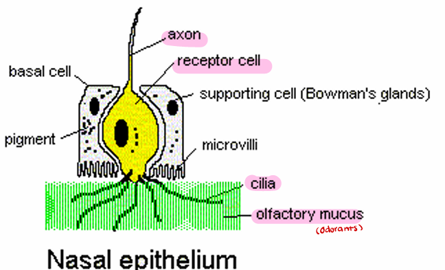
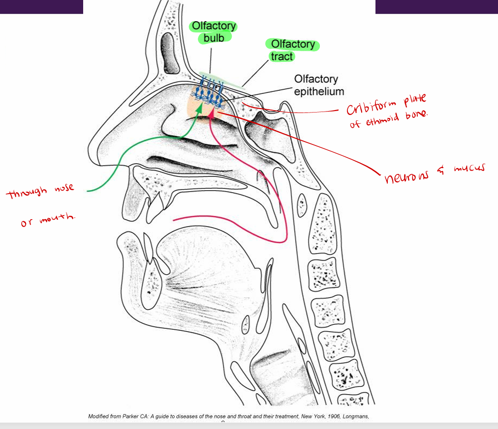
Olfactory Epithelium
Structure: receptor neurons and supporting cells
*same cell is receptor and nerve fiber cell body (no separate ganglion)
Human olfactory epithelium is about 5 cm², dog may be 100 cm²
The olfactory receptor cell is a bipolar neuron - apical dendrites project cilia into the mucus layer, axon on the basal side conducts the information to the brain
Surrounded by a basal cell for regeneration, and supporting cell for maintenance and support
Does not have separate ganglion, the receptor cell is the receptor and the nerve fiber cell body
Axons bundle up and make up CN I (olfactory nerve)
Signal Transduction in Smell Receptors
Membrane receptors on cilia
Second messenger system produces depolarization (cAMP or cGMP)
Threshold for some substances very low
In Sensation of Smell, Action potential travels via…
Action potential travels via unmyelinated fiber (CN I) (olfactory nerve)
primary afferent fiber is group IV axon
passes in bundles through cribriform plate of the ethmoid bone and synapses on olfactory bulb
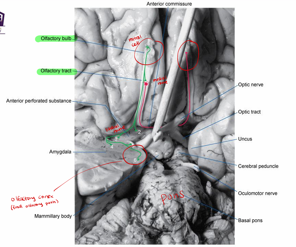
Olfactory Pathways
Fascicles of olfactory nerves (CN I) penetrate cribriform plate to enter olfactory bulb
Synapse on neurons of bulb (mitral cells)
Olfactory bulb (no longer CNI) processes and relays information (2nd order fiber) into the lateral and medial olfactory tracts
Seen on ventral surface of frontal lobe
Olfactory cell (a neuron) passes through the openings of the cribriform plate as bundles (forms CN I), enters the olfactory bulb → glomeruli of the mitral cells → olfactory tract to the olfactory cortex on parahippocampal gyrus (Brodmann area 28)
2 total neurons responsible for conducting message
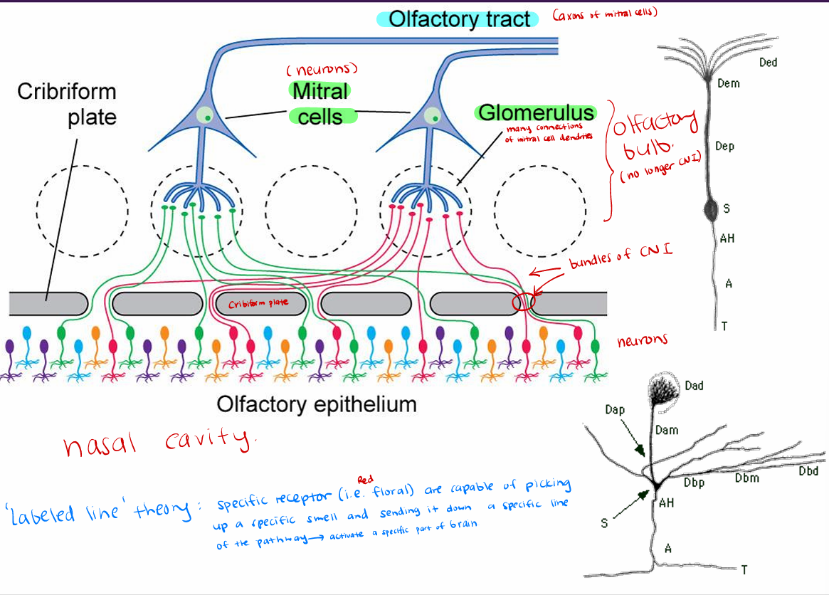
Labeled line theory
specific receptor (i.e. floral) are capable of picking up a specific smell and sending it down a specific line of the pathway → activate a specific part of brain
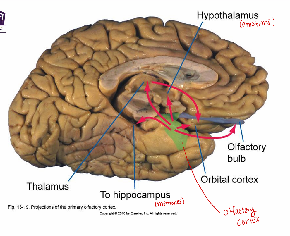
Olfactory tract to olfactory cortex
Olfactory tract to olfactory cortex on parahippocampal gyrus (Brodmann area 28), septal area, hypothalamus, limbic system
perception on parahippocampal
others for hunger/thirst/satiety, instinct memories , emotion centers
note no direct connection to sensory thalamus (no relay system)