Neurotransmitters and behaviour
 Call Kai
 Learn
Learn Practice Test
 Spaced Repetition
 Match
Match Flashcards
 Knowt Play
1/80

There's no tags or description
Looks like no tags are added yet.
Name | Mastery | Learn | Test | Matching | Spaced | Call with Kai | Chat |
|---|
No analytics yet
Send a link to your students to track their progress
81 Terms
what is a neuron
core componentof the NS (brain, spinal cord (CNS) and ganglia (PNS)
What is the soma
complex cell body
axon and dendrites extrude from it
review and processes information
dendrites
branch profusely and get thinner with each branching
receives information from other neurons
axon
leaves soma at the axon hillock
extend for great distances and has hundreds of branches
sends information to other neurons
Three types of neurons (functional)
afferent (sensory)
efferent (motor)
interneuron (association)
Afferent neuron
convey information from tissues and organs into the CNS
simplest form of neuron
stimulus can come from exteroreceptors or intereceptors
Stuctural types of sensory neuron
bipolar (retina)
somatosensory (skin and muscle)
Efferent neuron
transmit nerve impulses from brain to affectors
Where are efferent neurons located
in the lower brain stem and spinal cord
What is the structure of efferent neurons
extensive dendritic networks and large axons that connect to muscle
Interneurons
connect neurons within specific regions of the CNS
link sensory and motor neurons
allow brain and spinal cord to process and integrate information
branch extensively
why are interneurons important for reflexes
process sensory information from sensory neurons and relayed to motor neurons
what’s different about the knee jerk reflex compared to other reflexes
there is a direct connection from sensory to motor neuron
what receives synaptic signals from other neurons
soma and dendrites
how are signals transmitted to other neurons
down the axon > axon terminal > receiving dendrite/body
how do neurons communicate
neurotransmission/synaptic transmission (electrical signals that turn into chemical energy) via the synapse
what triggers the release of neurotransmitters
action potential
action potential
quick electric pulse that neurons use to communicate with each other
cell membrane of neurons
forms border of neuron
seperates the inside environment from the outside environment
controls movement of substances in and out of the neuron
external layer contain receptors (specialised proteins) that respond to specific molecules
what does extracellular fluid contain
water
ions
nutrients
signalling molecules
what is the cell membrane made out of
lipids and proteins
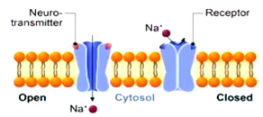
ligand-gated channel
allow ions to pass through the membrain in response to the binding of a neurotransmitter
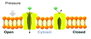
mechanically-gated channel
open in response to mechanical stress (i.e touch, hearing)
always open/leaky channel
passive ion channels that are always open to maintain homeostasis
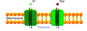
voltage-gated
activated by changes in the electrical membrane potential near the channel (i.e action potential)
located at axon and synapse
directionally propagate electical signals
iron-specific (only allow one type of ion to pass through)
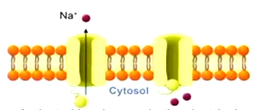
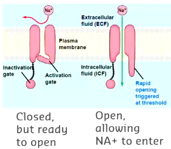
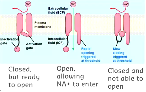
activation gate
inactivation gate
what is the function of gated channels
receive information
maintain homeostasis
detect stimuli
send message
activation gate
opens when neuron receives the right signal allowing specific ions to flow through
inactivation gate
closes shortly after channel opens stopping flow and activation of ions
what is the purpose of inactivation and activation gates
speed and efficency
ions only briefly flow into cell membrane for a period of time before it resets
what triggers the opening and closing of voltage gated channels
changing ion concentration between the intra and extracellular fluid (Na+, K+, Ca2+ and Cl-)
At resting potential, the inside of the neuron is more * relative to the outside
negatively charged
What is the charge of a neuron at resting potential
-70mv
why is intracellular fluid more negatively charged than the outside
The is a higher concentration of the protein anions and potassium inside the axon relative to the ouside
what is in the extracellular fluid
chloride and sodium
how is resting potential of the neuron maintained
ungated channels allow K+ and Cl- pass freely
Gated sodium channels keep out Na+
Graded potentials
small changes in the cell membrane
excitory: make the inside of the neuron more positive (depolarisation)
inhibitory: makes the inside of the neuron more negative (hyperpolarisation)
Depolarisation
-50mv threshold reached > voltage gated channels open > influx of sodium ions into the cell > rapid positive change in intracellular fluid (depolarisation)
saltutory conduction
when the electrical signal skips between the nodes of ranvier down the axon
faster
what stimulates the release of neurotransmitters
action potentials
what type of channels are on the post synaptic neuron
ligand gated
neurotransmitter
chemiscal must be synthesised/made in the neuron or be present in it
when released the chemical must produce a response in target
same response must occur when the chemical is experimentally placed on the target
must be a mechanim for removing the chemical following its work
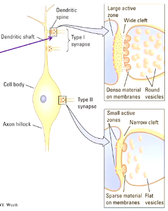
excitatory neurotransmitters
increase likelihood of action potential threshold (depolarisation)
more on dendrites
wider gap
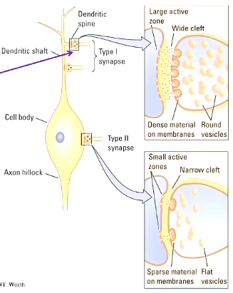
inhibitory neurotransmitter
decrease likelihood of action potential (hyperpolarise)
more on cell body
what determines whether a neurotrasnmitter is excitory or inhibitory
depends on
type of channel coupled to receptor
concentration of permeant ions inside and outside the cell (charge)
classes of neurotransmitters
small molecule neurotransmitters
peptide neurotransmitters (neuropeptides)
small molecule neurotransmitters
derived from the food we eat
eat > gut > small intestine absorb nutrients into blood > cell body of neuron
packaged ready to use in axon terminals
quickly replaced following release
communicate and quick firing rate
acetylcholine, histamine, amines (dopamine, serotonin). amino acids (glutamate, glycine)
acetylcholine
one type of small molecule neurotransmitter
functions in both PNS and CNS
neuromodulator
key NT for parasympathetic nervous system
primarly excitatory NT
acetylcholine
type of small molecule NT
fundamental to cholinergic system (network of neurons that use acetylcholine)
muscle activation (PNS)
effects as a neuromodulator upon plasticity, arousal and reward
forebrain networks: learning, decision making and attention (CNS)
serotonin
small molecule neurotransmitter
primarily an excitory neurotransmitter
found in neurons of the raphe nucei region of the pons and upper brain stem > extend to forebrain and other parts
regulate sleep and wakefulness, mood, aggression, appretite, arousal, perception of pain
implicated in anxiety, depression, cognitive symptoms of schizophrenia, memory problems, appetite regulation and psychotic experiences
peptide neurotransmitters (neuropeptides)
made on the neuron’s cell body
responsible for the synthesis of proteins in cells on ribosomes
packaged in the membrane into vesicles > transported to the axon terminal
slow production and not easily replaced
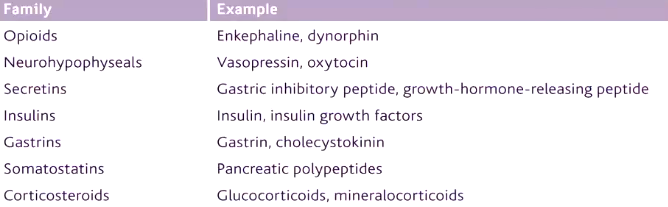
endorphins
neuropeptide
naturally occur in the body
opiod receptors and reduce pain
contribute to pleasure and wellbeing
released during exercise, stress, laughter and other rewarding experiences
motivation, emotion, attachment and stress regulation
what is the main neurotransmitter used in motor neurons to make movement possible
acetylcholine
motor neurone disease (MND)
progressive disease that attacks the motor neurons in the brain and spinal cord
amyothrophic lateral sclerosis (ALS)
type of MND
stuff muscles, muscle twitching, gradual muscle wasting
lose ability to speak, swallow and breath, progress to frontotemporal dementia
how does acetylcholine control the sympathic nervous system
activates the SNS to release noradrenaline
prepares the body for fight or flight
how does acetylcholine control the parasympathic nervous system
activates PNS
neurons release acetylcholine
prepares body for rest and digest
activating system
network of neurons that uses one primary neurotransmitter to regulate brain activity
these neurons have cell bodies clustered in specific brain regions and send axons through the brain
four activating CNS systems
cholinergic
dopaminergic
noradrenergic
serotonergic
cholinergic system
use acetylcholine to communicate with other neurons
cognitive functions: attention, memory and emotional processing
cholinergic input from the basal forebrain helps maintain sustained attention
supports attentional processing of important, including threat-related stimuli
alzheimer’s disease
neurogenerative disease characterised clinically by insideous, chronic and progressive cognitive decline
what are the two key neuronal changes that take place in the brain during alzheimer’s
loss of cholinergic cells in the forebrain (medication to increase acetylcholine in the forebrain (donepezil)
neuritic plaques in the cerebral cortex (amyloid) which are associated with neurofibrillary tangles
changes trigger inflammation and deterioration of cholinergic cells
dopaminergic system
neural pathways in the brain that transmit dopamine
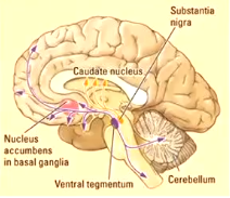
8 dopaminergic pathways
mesolimbic
mesocortical
nigrostriatal
tuberoinfundibular
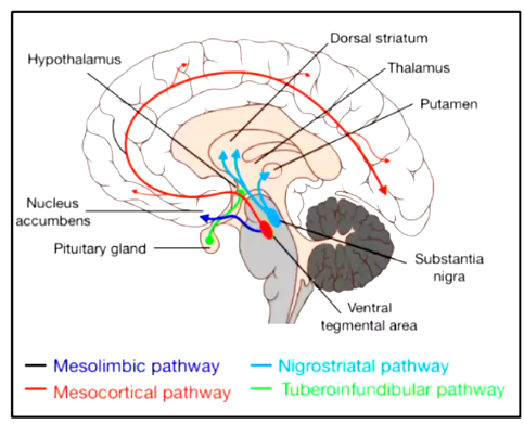
nigrostriatal pathway
projects from substantia nigra upwards to the striatum
striatim is the primary input to the basal ganglia system
parkinsons disease results in loss of dopaminergic innervation to the striatum and other basal ganglia
symptoms result from the progressive degeneration of dopamine-producing cells of the substantia nigra
mesolimbic pathway
begins in the ventral tegmental area of the midbrain and connects to the nucleus accumbens, amygdala, hippocampis and prefrontal corex
role in motivation, pleasure, reward, reinforcement and learning
addiction (too much), schizophrenia and depression (too less)
what causes addiction
increased dopamine release in the mesolimbic pathway
methamphetamine
meth blocks receptors of original neuron so seratonin cannot be reuptaken and goes to next neuron > floods it > damages receptors of the next neuron'
abnormal neuroplastic changes in brain structures
changes to white matter integrity
reduced hippocampal volume
reduction in grey matter in the cingulate cortex
limbic cortex
paralimbic cortex
regions involved in emotion, memory and decision making
overstimulation of the amygdala: aggression, distruption of inhibitory circuits, ODC, hyperactive dopaminergic signal transduction, stimulant psychosis
noradrenergic system
norepinephrine
originate in the locus coeruleus and lateral tegmental field (medulla, pons of brain stem)
emotion: depression mania stability of mood
mobilises brain and body for action
what are the behavioural affects of norepinephrine
increases arousal and alertness
promotes vigigilance
enhances formation
retrieval of memory
focuses attentions
increases restlessness and anxiety
what are the physiological effects of norepinephrine
increased heart rate and blood pressure
triggers release of glucose from energy stores
increases blood flow to skeletal muscles
reduces blood flow to the gastrointestinal system
when is norepinephrine released
norepinephrine releases lowest during sleep, rises during wakefulness and reaches its highest levels in situations of stress and danger
locus coeruleus is important for…
wakefulness
where is the serotonergic system located
originated in the dorsal and median raphe in the midbrain
dorsal raphe is on the midline of the brainstep and is one of the raphe nuclei extending to most parts of the CNS
serotonergic system responsible for producing…
serotonin
how many serotonin receptors subtypes are in the serotonergic system and what is the effect of this
15
produces diversity of signalling so one neurotransmittercan produce very different effects
what is the function of the serotonergic system
maintains activity in forebrain and plays role in wakefulness
regulate sleep/wake cycles
precursor for melatonin
regulated circadium rhythm
what is the reduced function of the serotonergic system associated with?
insomnia and depression
what is enhanced function of the seotonergic system associated with?
weight loss and suppressed diet
what psychopathological disorders is linked with serotonergic dysfunctions?
anxiety
eating disorders
schizophenia
impule constrol disorders
autism
aggressive behaviours
depression
what do antidepressents do
increase serotonin levels in the synapse
blocks reactants
what are the symptoms of high serotonin levels due to MDMA and antidepressents together
agitation
fever
sweating
muscle rigidiy
rapid heart rate
death