Microbiology Lab Quiz #1
 Call Kai
 Learn
Learn Practice Test
 Spaced Repetition
 Match
Match Flashcards
 Knowt Play
1/78

There's no tags or description
Looks like no tags are added yet.
Name | Mastery | Learn | Test | Matching | Spaced | Call with Kai | Chat |
|---|
No analytics yet
Send a link to your students to track their progress
79 Terms
Ubiquitous (describing microbes)
present or existing everywhere
Microorganisms/Microbes
Living creatures that are too small to see with the naked eye
Contamination
unintentional exposure to microbes
inoculation
intentional exposure to microbes
Culture
controlled growth of microbes in a lab
Fomite
object or surface that can harbor and transfer microbes
incubate
develop, grow
Pellicle
bacteria layer on top of incubated broth
scum/oily contact lens appearance
Sediment
bacteria layer at bottom of incubated broth
sand/salt appearance
Turbidity
cloudiness
in broth, turbidity indicates growth of microbes
Fastidious
picky eater
Morphology
shape
Descriptive texture terms (of bacteria)
moist
dry
dull
shiny
mucoid
Descriptive edge terms (of bacteria)
smooth
wavy
rough
curled
filamentous (root-like)
Motility
ability to move
bacterial movement can be tracked with TCC deeps (semi-solid media)
What is the classification for SabDex media?
Selective media
What is the purpose of SabDex media?
Grow certain microbes and prevents others
What are some unique traits of SabDex media?
Low pH
High sugar
Grows fungi
What is the classification for NA media?
Standard media
What is the purpose for NA media?
Basic nutrients to grow microbes
What are the unique traits for NA media?
None
What is the classification for TSA media?
Enriched media
What is the purpose for TSA media?
Extra nutrients to help fastidious bacteria grow
What are some unique traits of TSA media?
Good at growing bacteria from human skin
3 general arrangements of bacteria
Bacilli
Cocci
Spirrila
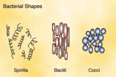
Types of bacilli
bacillus (1 rod shape)
diplobacilli (2 rods connected end to end)
streptobacilli (3+ rods connected end to end)
palisades (rods leaning on each other side by side, like books)
streptobacilli

palisade bacilli

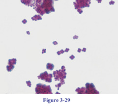
Types of cocci
coccus (single sphere)
diplococci (2 spheres connected)
streptococci (3+ spheres connected)
staphylococci (grape cluster)
tetrad (4 spheres in square formation)
streptococci

staphylococci

tetrad cocci
Types of spirilla
vibrio (comma)
spirillum
spirochete
vibrio

spirillium

spirochete

milliliter (mL)
1/1,000 L
microliter (µL)
1/1,000,000 L
P100 micropipette
10-100 µL
... 0.01-0.1 mL
P1000 micropipette
100-1,000 µL
...0.1-1 mL
Agar
Gelatin-type material derived from seaweed
Which physical form of media has 1.5% (the most) agar?
Solid media (slants and plates)
Which physical form of media has 0% (the least) agar?
Liquid media (broth)
Which physical form of media has 0.5-0.7% agar?
Semi-solid media (deep)
What is semi-solid media used for?
Test for motility or O2 requirement
Needle used to stab line of bacteria down center of media
What is solid slant media used for?
transport and storage
What is solid plate media used for?
to quantify or isolate
What is liquid media used for?
culturing large quantities of bacteria
What is a loop used for?
Inoculating liquid media and solid media
Making smears ("loop-full" of solid growth on a "loop-full" of water)
Aseptic inoculation techniques
Flame needle/loop from back to tip
Work within range of Bunsen burner
Hold tubes at sideways-ish to prevent air contamination
Flame tubes after opening and before closing
Clamshell plates
Simple staining
use a single stain to observe bacteria
ex: Direct stain, Negative stain, Capsule stain
Differential staining
use two stains to distinguish bacterial cells based on their cell wall structure
ex: Gram stain, Acid-Fast stain, Endospore stain
Mordant
something used to make dye penetrate cell wall and stick
ex: iodine, heat
What 2 types of staining did we talk about NOT heat fixing in lab?
Negative simple stains and Capsule staining
What is the charge of the outside of a bacterial cell?
-
What is the charge of the surface of a glass slide?
+
What is the charge of an acidic dye?
-
What is the charge of a basic dye?
+
Disadvantage of Direct simple stain?
Heat fixing causes some shape and size distortion
Advantage of Negative simple stain?
View true morphology and size of bacteria
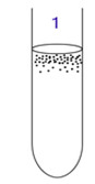
Obligate aerobe
grows only in O2 presence
Deep results: top-only growth
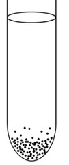
Obligate anaerobe
grows only in O2 absence
Deep results: bottom-only growth
Facultative anaerobe
grows with or without O2
Deep results: throughout growth

Gram staining
distinguishes bacteria on thickness of peptidoglycan

Gram staining process
Smear bacteria on plate
Air dry
Heat fix
1st stain
Mordant (iodine)
Decolorizer (Alcohol, medium strength)
2nd stain

Gram staining results
thick peptidoglycan positive: purple
thick peptidoglycan negative: pink

Acid fast staining
distinguishes bacteria on presence of waxy mycolic acid in cell wall

Acid-Fast staining process
Smear bacteria on plate
Air dry
Heat fix
1st stain
Mordant (heat 8 mins)
Decolorizer (Acid alcohol, strong)
2nd stain

Acid-Fast staining results
mycolic acid positive: red
mycolic acid negative: blue

Mycobacterium
a large family of bacteria that has mycolic acid in cell wall
often found in soil and water
Endospore
A thick-walled protective spore that forms inside a bacterial cell and resists harsh conditions.
Endospore staining
distinguishes bacteria from endospores (bacteria "eggs")
not all bacteria make endospores

Endospore staining process
Smear bacteria on plate
Air dry
Heat fix
1st stain
Mordant (heat 8 mins)
Decolorizer (Water, weak)
2nd stain
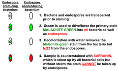
Endospore staining results
endospores: green
vegetative bacteria: red
*there will be more endospores in old cultures of endospore-creating bacteria than in young cultures

Capsule staining
Used to observe the microbial CAPSULE, a protective glycocalyx layer surrounding the cells of some bacteria and fungi
Mix of negative stain and direct stain
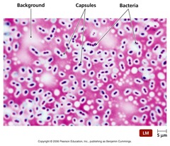
Capsule staining process
Smear bacteria on plate
Air dry
Stain
Rinse with 20% copper sulfate
Capsule staining results
capsule positive: halo around stained bacteria
capsule negative: slime glycocalyx will not show halo

Possible causes of false positive for Gram stain
-too much bacteria on plate
-too weak or too little decolorizer used
-mordant left on too long
-wrong secondary stain used
Possible causes of false negative for Gram stain
-wrong primary stain used
-mordant not on long enough
-too strong or too much decolorizer used