Chapter 11- Muscular Tissue
1/124
There's no tags or description
Looks like no tags are added yet.
Name | Mastery | Learn | Test | Matching | Spaced | Call with Kai |
|---|
No analytics yet
Send a link to your students to track their progress
125 Terms
Conductivity
the movement of an impulse from one part of the cell to another
Contractability
the ability to shorten forcibly
Extensibility
the ability to be stretched or extended (without damage)
Elasticity
tendency to recoil to a shorter length when relaxed
The sarcoplasm contains all normal organelles but also:
-myofibrils
-glycogen
-myoglobin
-an abundance of mitochondria
-sarcoplasmic reticulum
-terminal cisternae
-t tubules
myofilaments are made of contractile proteins:
actin and myosin
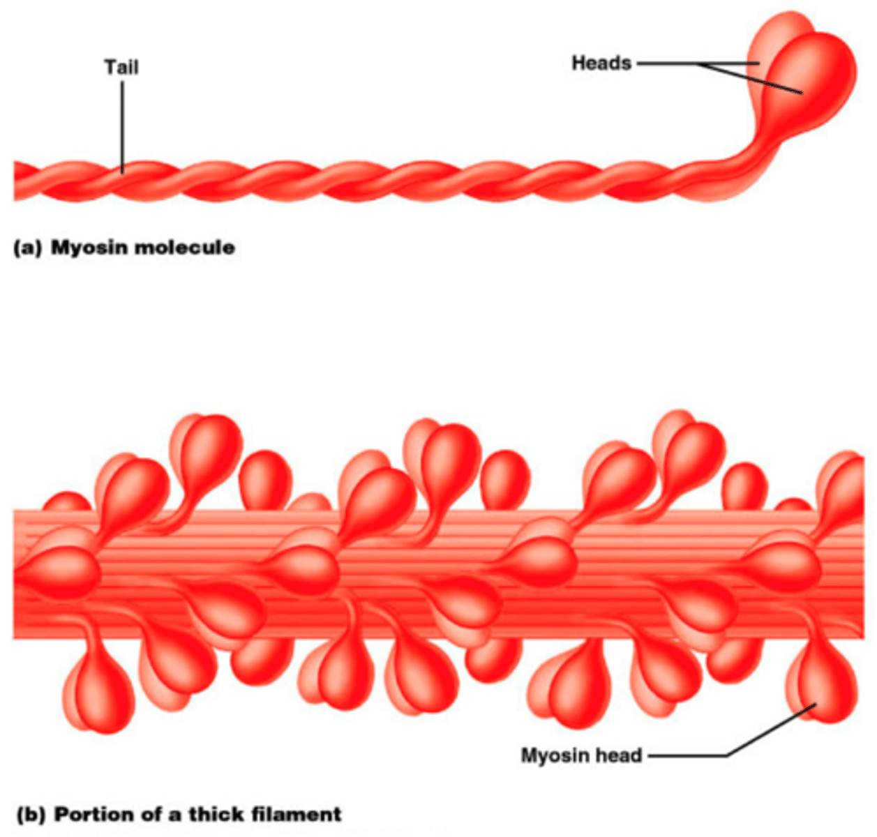
Thick myofilaments consist of:
-two heads attached to a flexible, hinge-like region attached to a tail.
-The heads contain actin-bonding sites that form cross-bridges with actin.
-has ATP bonding sites
-Tails form the cord of the filament; the heads turn outwards
Thin filaments are mostly composed of which protein?
Actin
-actin bonds to myosin
Thin filaments consist of:
-globular subunits of G-actin
-a whole string of G-actin= F-Actin (filamentous actin)
-active sites for myosin heads
-Two intertwined heads of F Actin make up the thin filaments
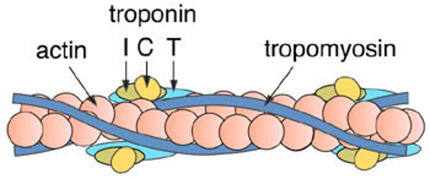
Tropomyosin
-Stabilizes actin filament
-in a relaxed muscle, blocks the actin active site; prevents actin and myosin binding
Troponin
Regulatory protein that binds to actin, tropomyosin, and calcium
Elastic myofilaments (Titin)
-Large, springy protein that helps stabilize the thick filament;
-Runs through the core of thick filaments, anchors it to Z-disc and M-Line
-Prevents overstretching and provides recoil
Dystrophin
A clinically important accessory protein associated with myofibrils
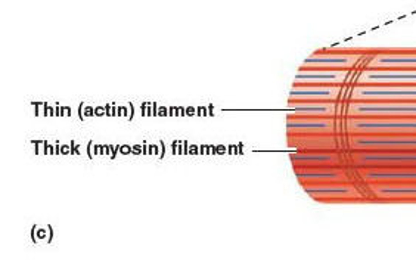
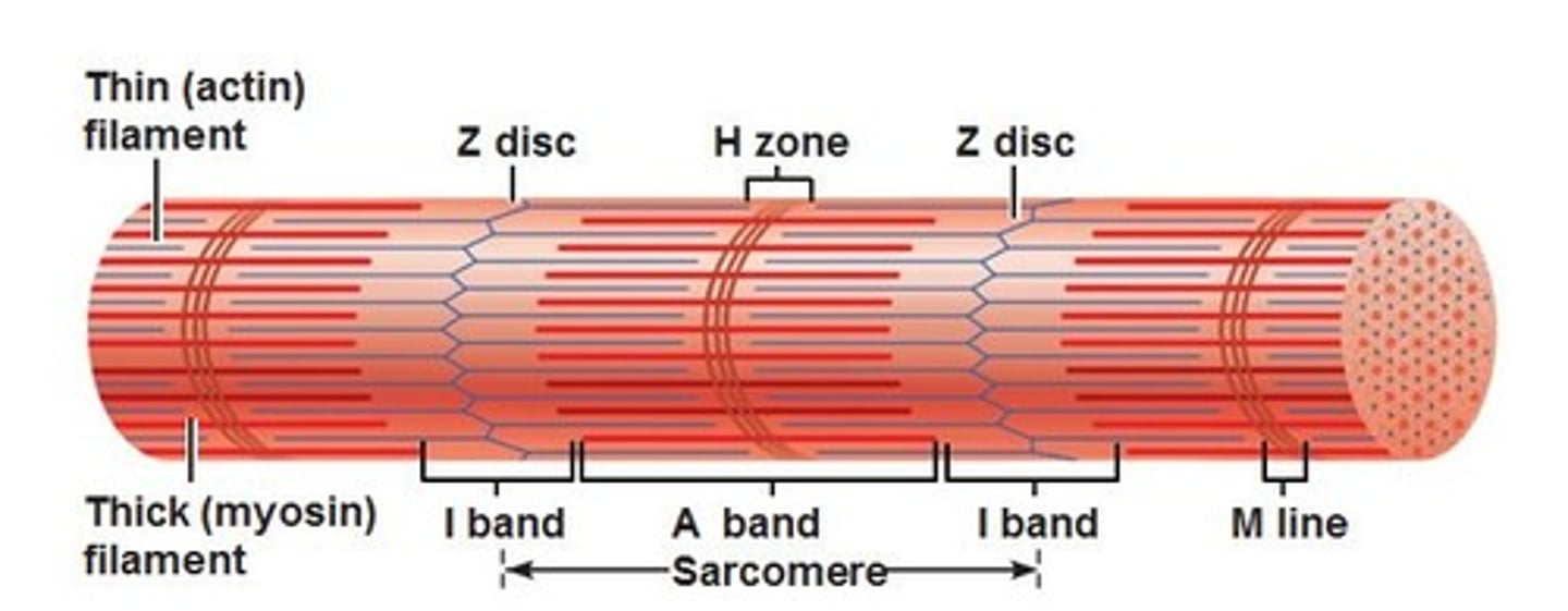
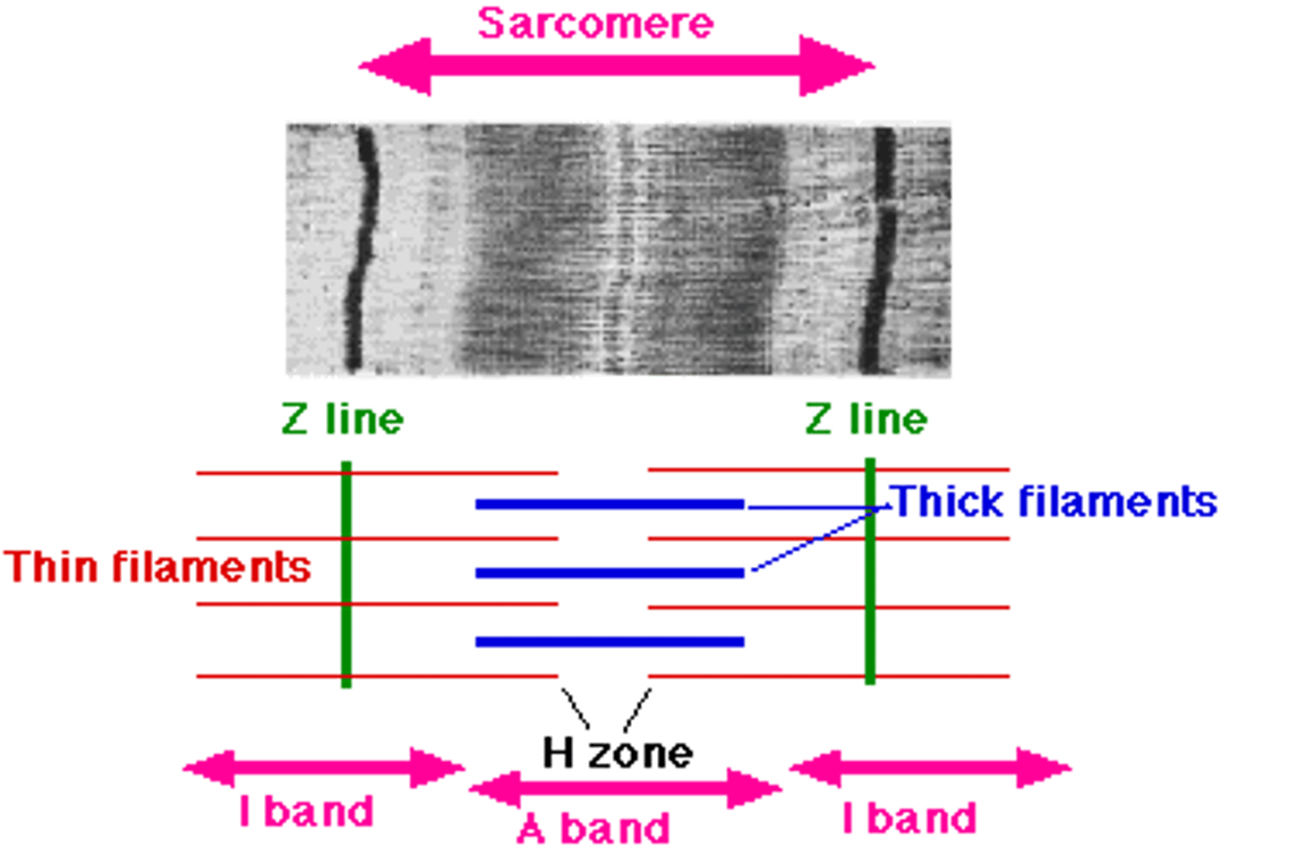
A Band
The darkest part is where thick filaments overlap an array of thin filaments

H band
middle of A band; thick filaments only
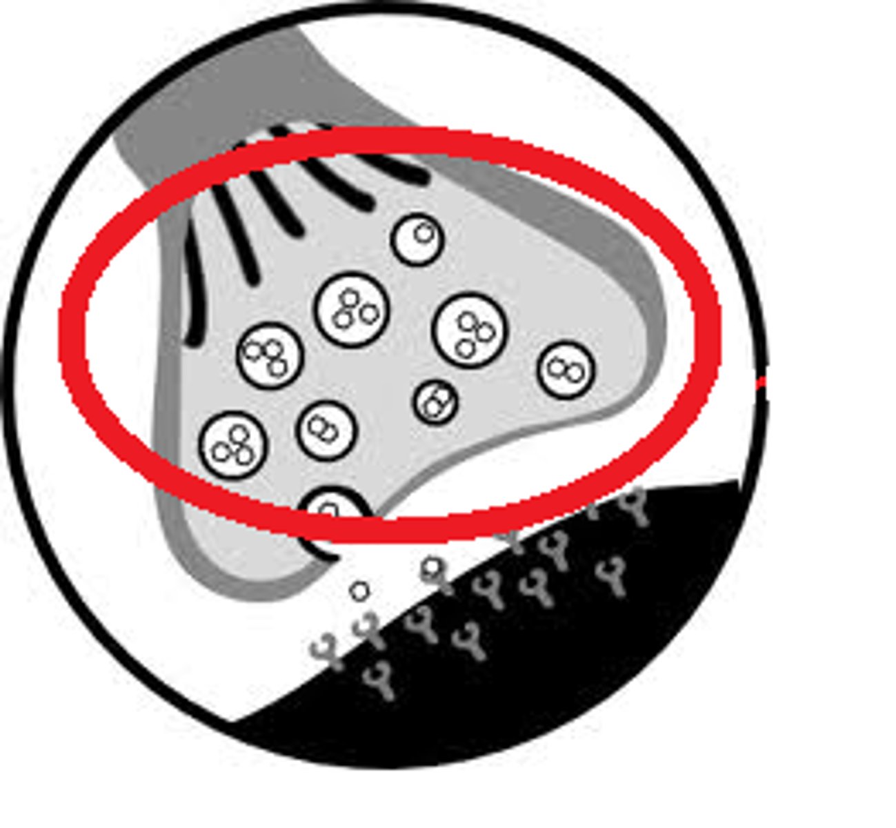
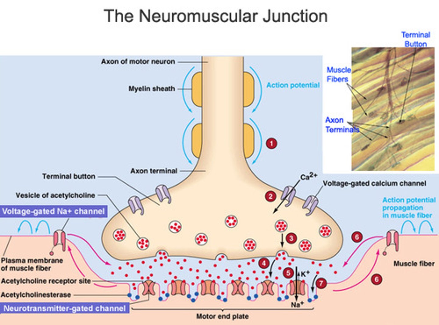
synaptic knob
Swollen end of a nerve fiber. Contains synaptic vesicles filled with acetylcholine (ACh)
What enzyme does the basal lamina contain?
Acetylcholinesterase (AChE)
-breaks down Acetylcholine (ACh)
The polarization of the membrane is due to differences of what?
Ionic Concentrations
-Intracellular Fluid (ICF) and Extracellular Fluid (EFC)
What happens during Excitation
1. Nerve AP opens voltage-gated calcium channels in synaptic knob
2. Calcium enters the knob and stimulates the release of ACh from synaptic vesicles in the cleft
3. ACh diffuses across the cleft & binds to ACh receptors in the sarcolemma.
4. Two ACh molecules bind to each receptor and its channel (changes its shape)
5. Na+ enters muscle cell; Depolarization occurs.
6. K+ exits the cell; Repolarization.
7. The quick voltage change= end plate potential (EPP)
-Voltage change in the synaptic region opens nearby voltage-gated channels; causing a chain reaction of AP. (Conduction)
Each muscle cell is supplied by?
one nerve, but each nerve supplies multiple muscle cells
Motor Unit
one nerve cell and all its associated muscle cells
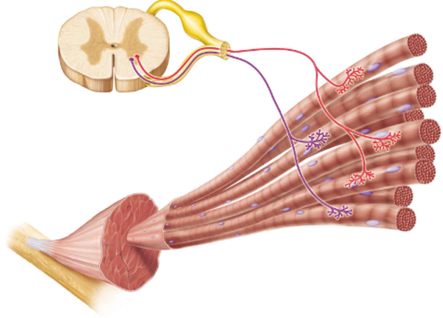
muscle fibers of one motor unit
- Dispersed throughout muscle
- Contract in unison
- Produce weak contraction over wide area
Stimulating the nerve with higher voltages produces?
stronger contractions
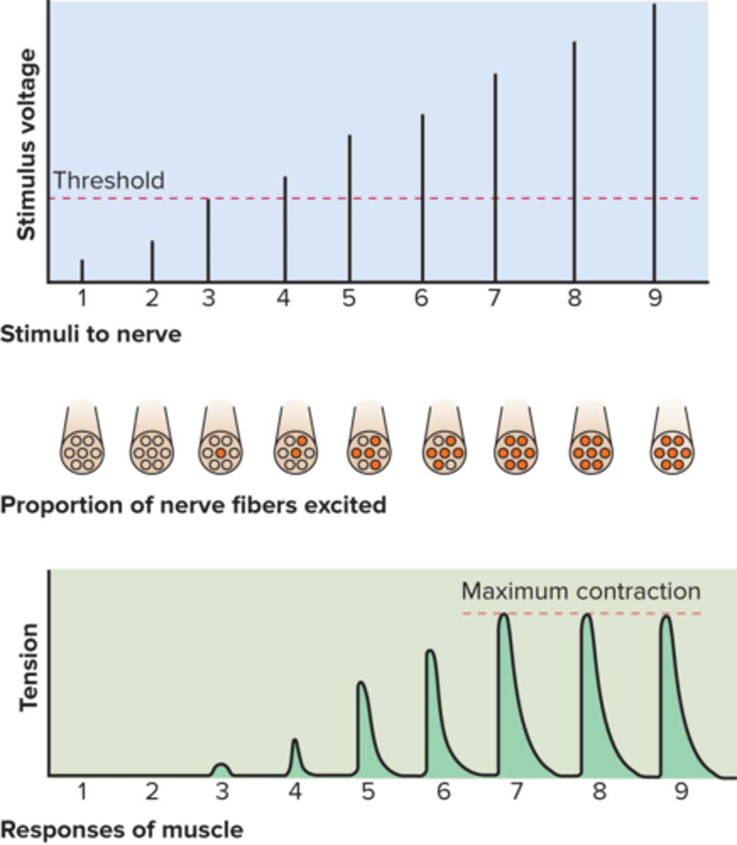
Higher voltages excite more nerve fibers which...
stimulate more motor units to contract
Immediate Energy
-used during short, intense exercise
-oxygen is briefly supplied by myoglobin but is rapidly depleted.
-Muscles meet most ATP demand by borrowing phosphate groups from other molecules and transferring them to ADP
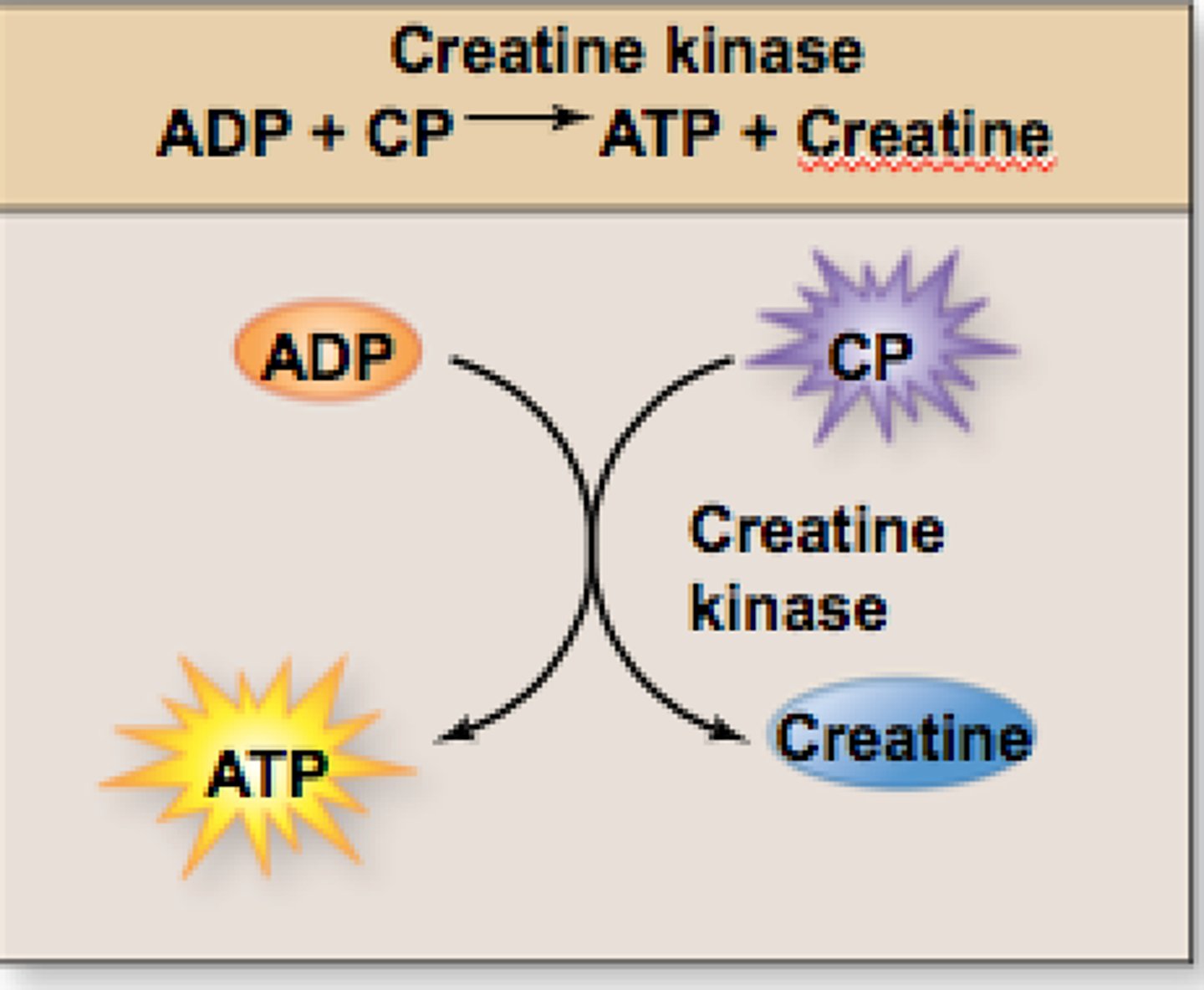
creatine kinase
obtains Pi from a phosphate-storage molecule creatine phosphate (CP) and gives it to ADP
-creates enough power for 6 seconds of sprinting.
Muscle Fatigue
the loss of contractility due to prolonged use of muscle
-leads to the physiological inability to contract a muscle
Fatigue in high-intensity exercise is thought to result from:
1. Potassium accumulation in the EFC reduces the excitability of fiber.
2. Excess ADP and P slow cross-bridge movements (actin and myosin interaction)
Fatigue in low-intensity (long duration) exercise is thought to result from:
1. Fuel depletion as glycogen and glucose levels drop.
2. Electrolyte loss through sweat can decrease muscle excitability.
3. Psychological factors of distance running
4. Central Fatigue
central fatigue
-When less motor signals are issued from brain
-Brain cells inhibited by exercising muscles' release of ammonia
Muscular strength depends on:
fascicle arrangement
-pennate are stronger than parallel; parallel are stronger than circular.
Resistance Training
builds strength only
-growth is from cellular enlargement
-muscle fibers synthesize more myofilaments and myofibrils and grow thicker
Slow twitch fibers produce more of what?
mitochondria and glycogen
Fast twitch fibers are also called
fast glycolytic (FG) or white fibers
How do slow twitch fibers resist fatigue?
aerobic ATP production
How are slow twitch fibers grouped?
Grouped in small motor units controlled by small, easily excited motor neurons.
-allow for precise movements
How are fast twitch fibers arranged?
Grouped in large motor units controlled by larger, less excitable neurons
-allowing for powerful movements.
Functional Properties of Cardiac Muscle
-contracts with regular rhythm in unison
-involuntary function, high resistance to fatigue
-contractions last long enough to expel blood
-made of cells called cardiocytes
Without a large SR or T-tubules, where does smooth muscle get its Calcium?
Ca++ needed for muscle contraction comes from ECF by way of Ca++ channels in the sarcolemma; NOT by t tubules
autonomic activity for Excitation of smooth muscle
Parasympathetic nerves secrete ACh to stimulate the GI tract smooth muscle.
Sympathetic nerves secrete norepinephrine-relaxing the muscle in bronchioles (Dilate)
Example of smooth muscle stretching:
Stomach contracts when stretched by food.
isotonic muscle contraction
muscle changes in length with no change in tension
isometric contraction
no change in length but change in tension
concentric contraction
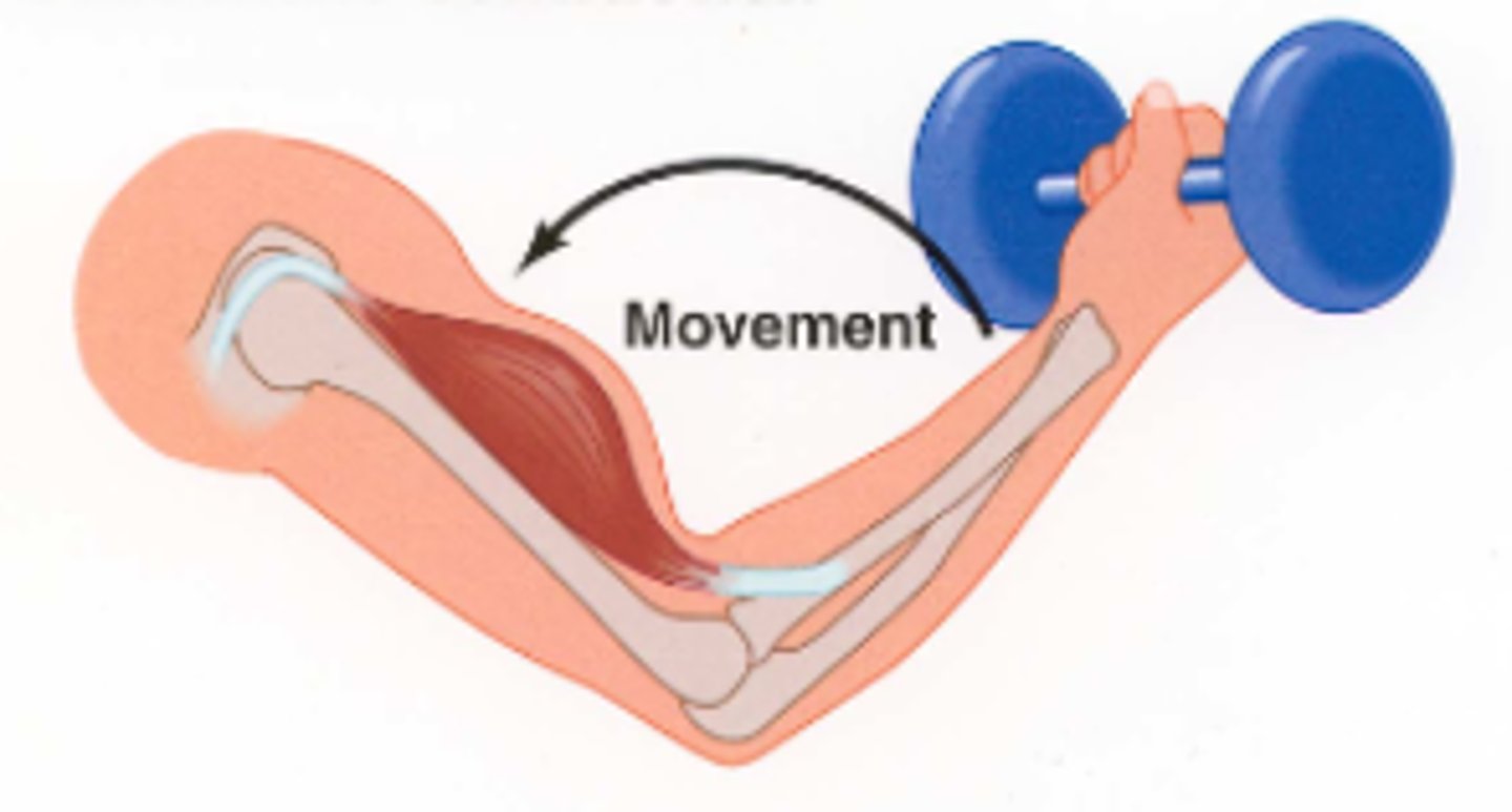
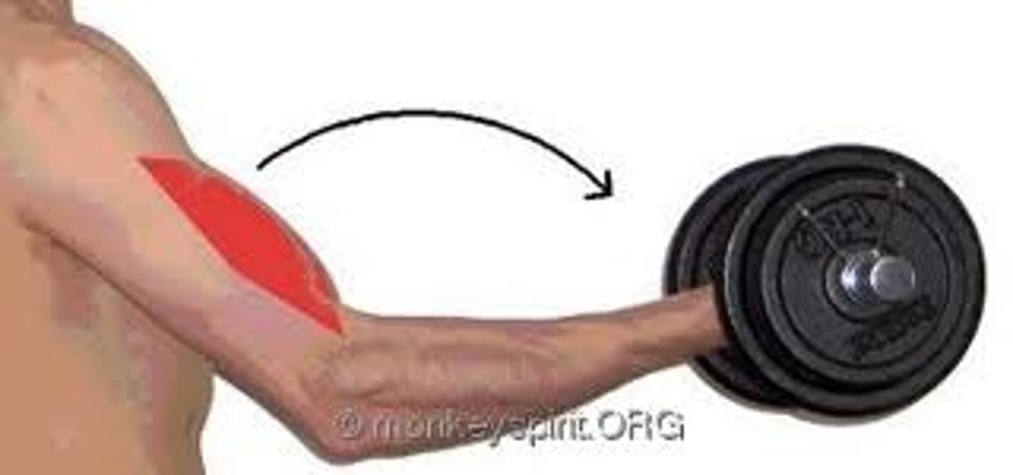
muscle shortens during the contraction but maintains tension; ex. flexing, picking up a book
eccentric contraction
muscle lengthens as it maintains tension (example: slowly lowering weight)
Excitability (responsiveness)
-ability to respond to changes in the environment
-usually a chemical signal (neurotransmitter or change in pH)
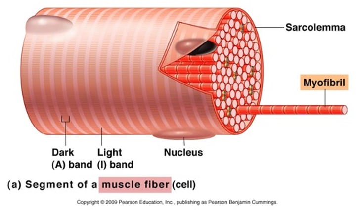
Sarcolemma
plasma membrane of a muscle fiber
Sarcoplasm
cytoplasm of a muscle fiber
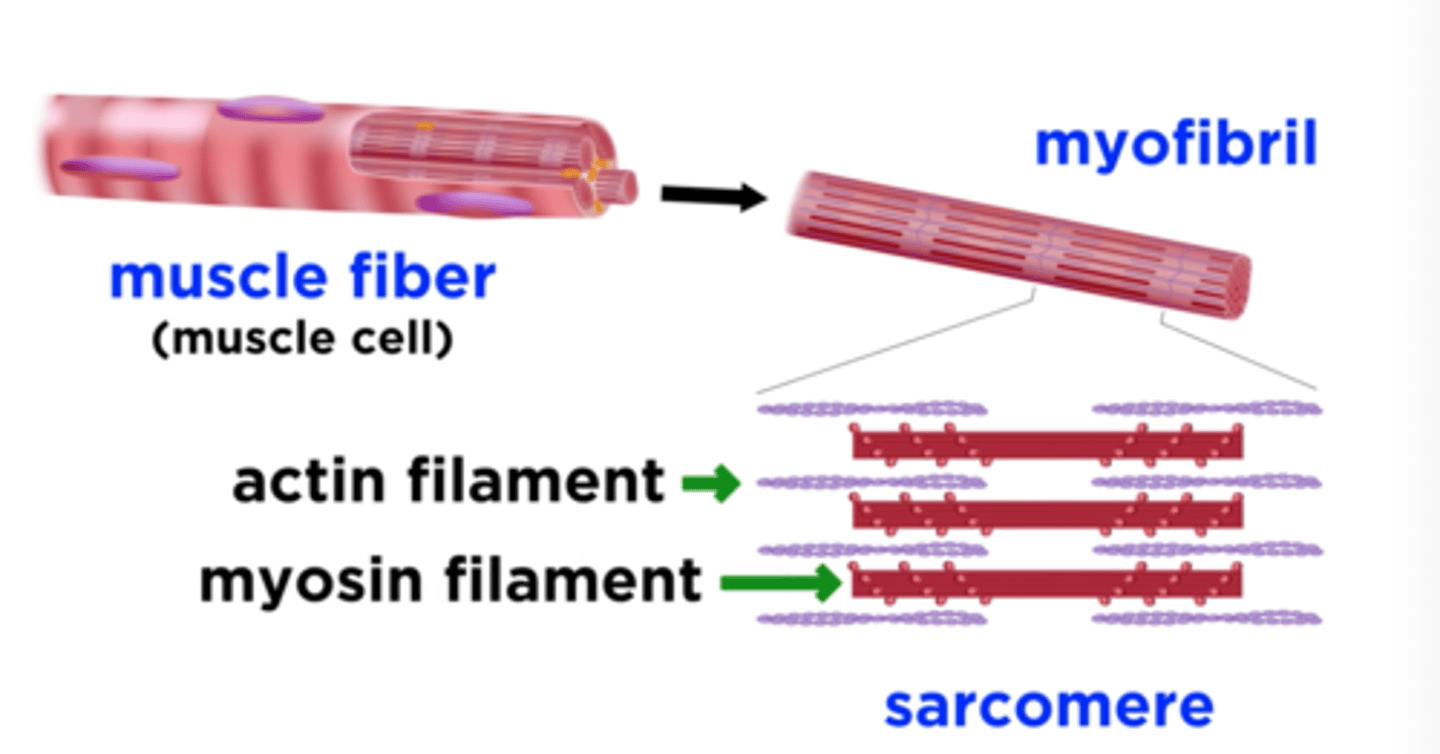
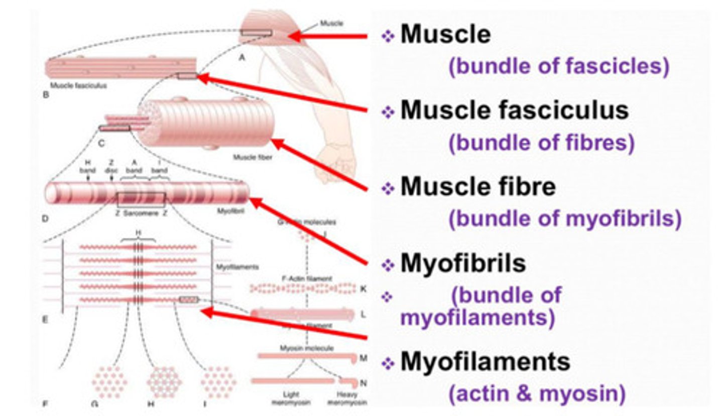
Myofibrils
Long protein cords running the length of the muscle fiber.
Serve as the functional unit of the cell.
-make up over 80% of muscle fiber volume
-contain the contractile element of the muscle
-contain myofilaments
Glycogen
A polysaccharide used for energy storage in a muscle fiber
-stored in glycosomes
-used during intensive exercise
Myoglobin
An oxygen-storing, red pigmented protein in muscle cells.
-contributes to a muscle's red color
Sarcoplasmic reticulum (SR)
The smooth ER of a muscle cell
-The SR winds around each myofibril in the muscle cell.
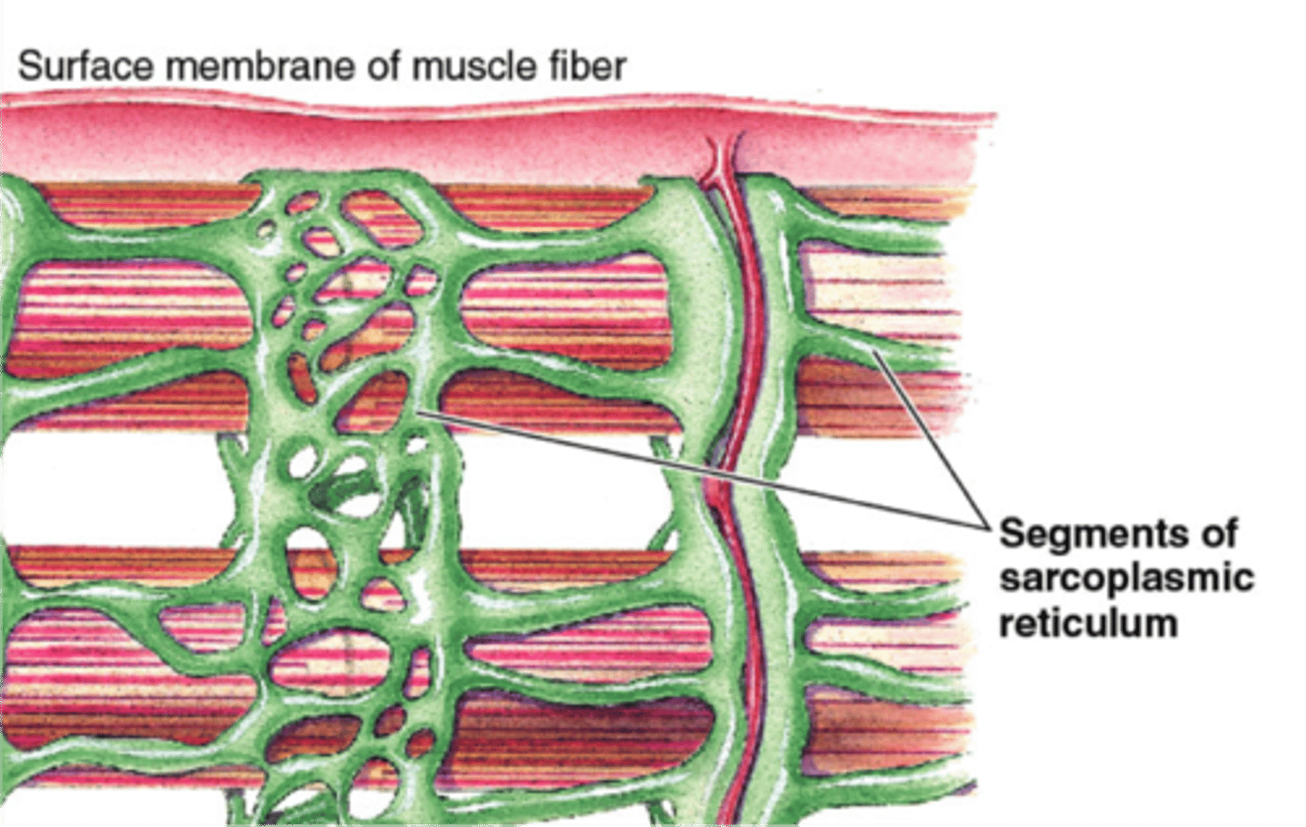
Terminal cisternae
-Dilated end-sacs of SR which cross the muscle fiber from one side to the other
-store calcium; release calcium to activate contractions
-enlarged areas of the sarcoplasmic reticulum surrounding the transverse tubules.
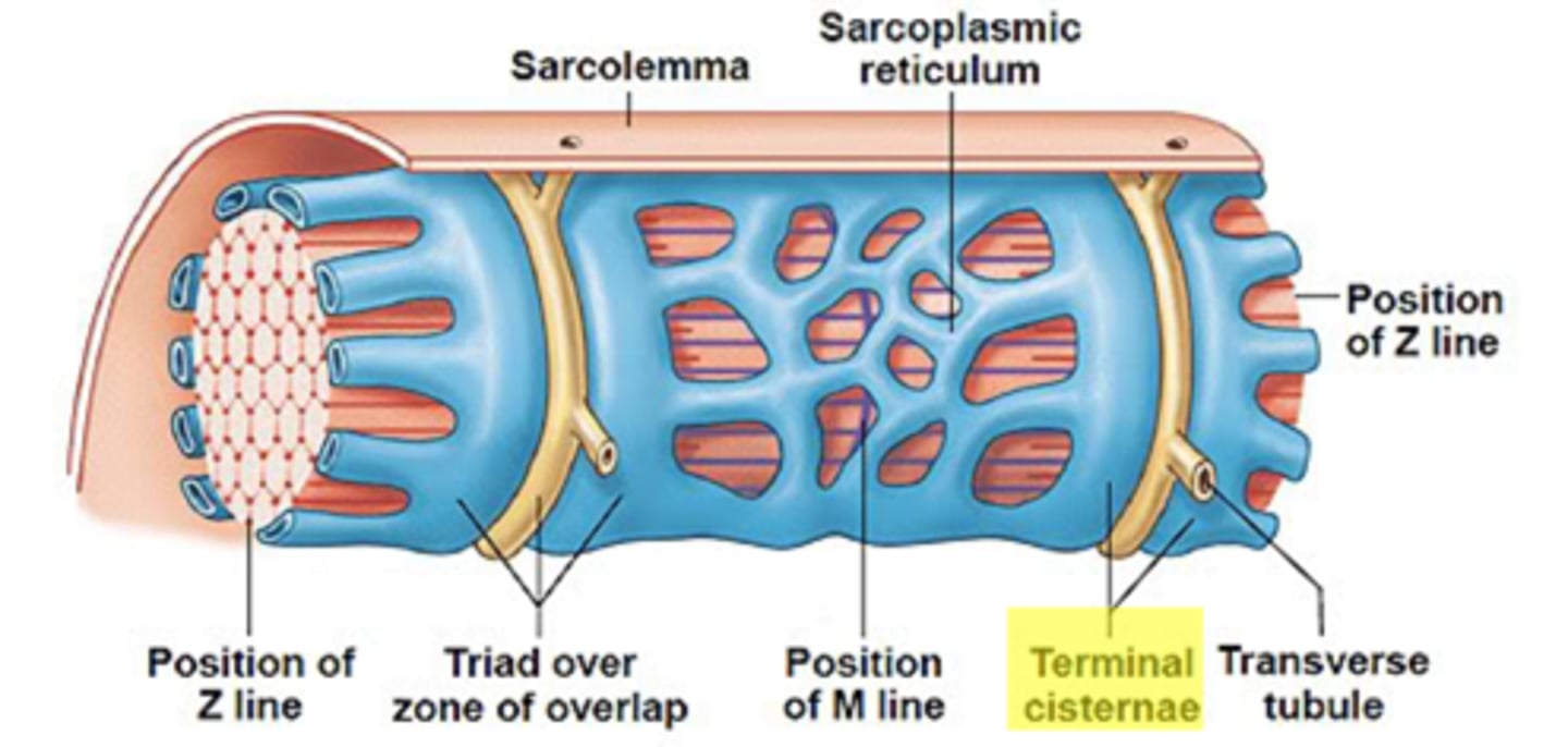
T-tubules (transverse tubules)
Tubular infoldings of the sarcolemma, which penetrate through the cell and emerge on the other side
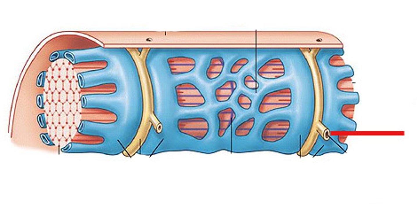
Triad
a T tubule and two associated terminal cisternae
What do contractile proteins do?
cause muscles to contract
regulatory proteins
tropomyosin and troponin
-regulate muscle contractions
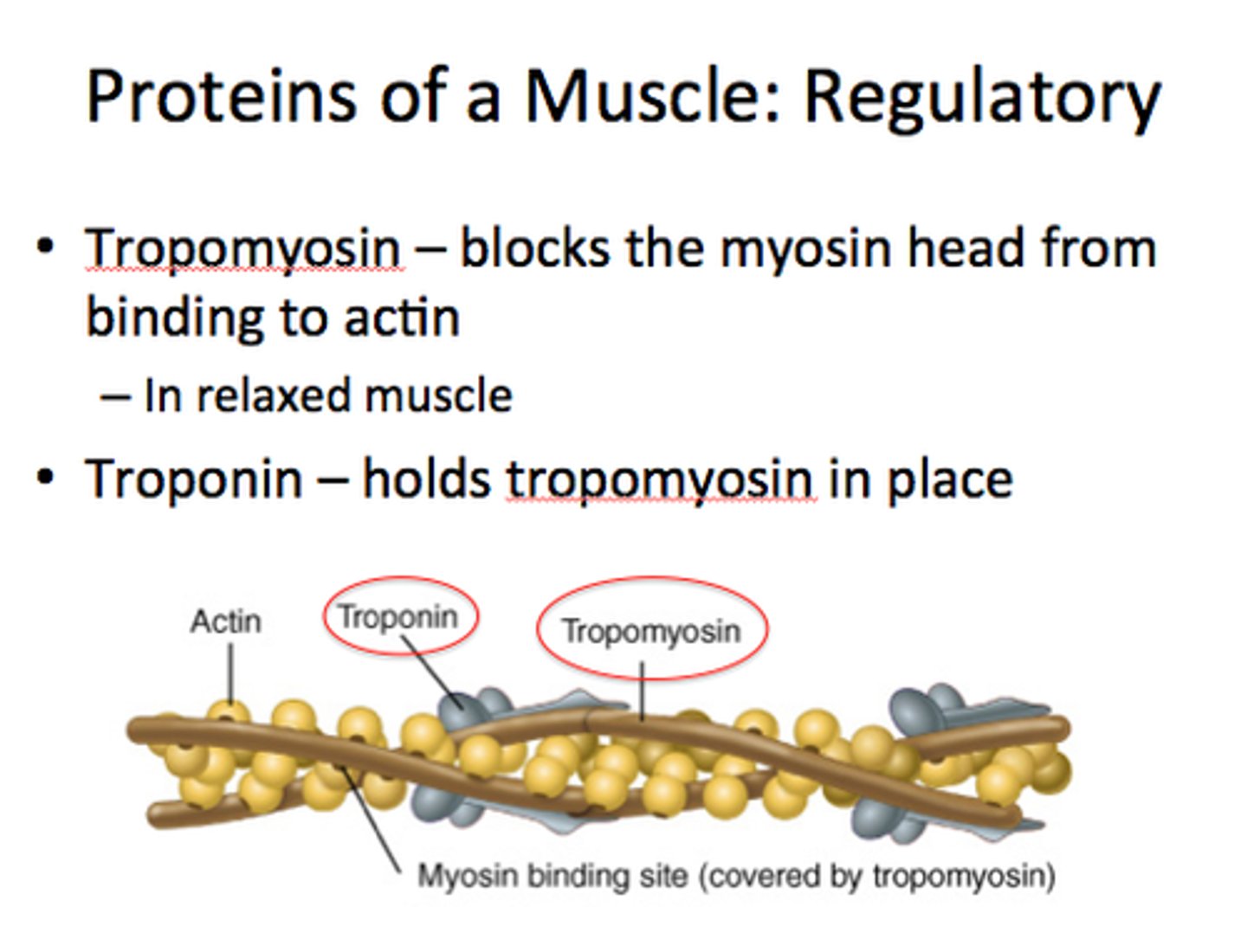
Thick myofilaments consist mainly of the protein (polypeptide):
Myosin
Sarcomere
Contractile unit of muscle
All the proteins between one Z disc and another.
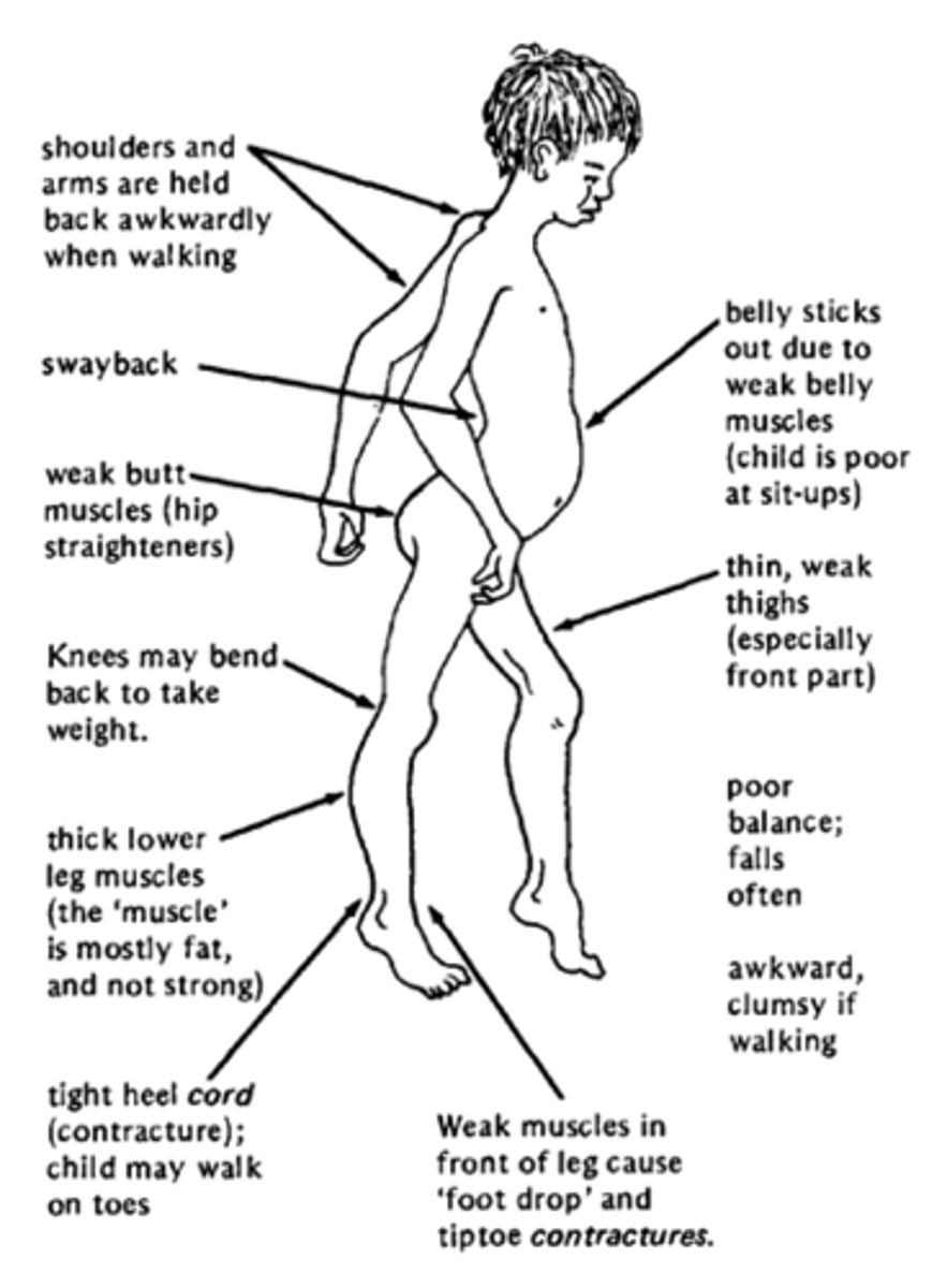
Defects in dystrophin lead to
Muscular Dystrophy
-results in the destruction of muscle cells and replacement with scar tissue
-Life expectancy is 20 years.
Striations of a muscle fiber are due to:
actin and myosin
(thick and thin filaments)
M Line
middle of sarcomere; where protein links thick filaments
- think "midline"
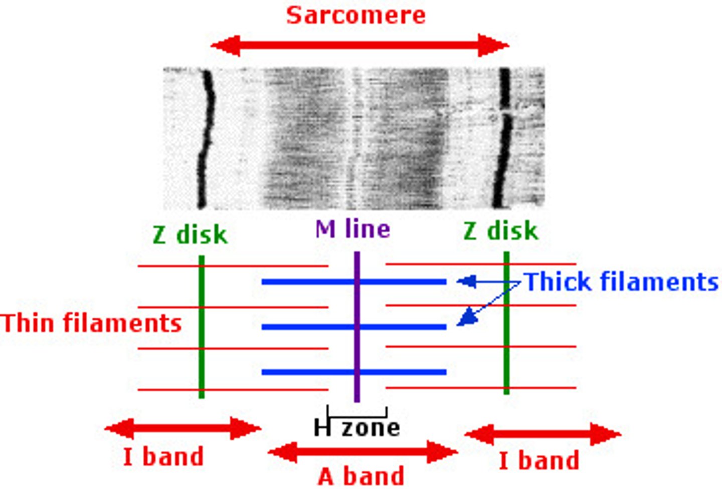
I band
thin filaments only (light)

Z disc
provides anchorage for thin filaments and elastic filaments; Bisects the I Band

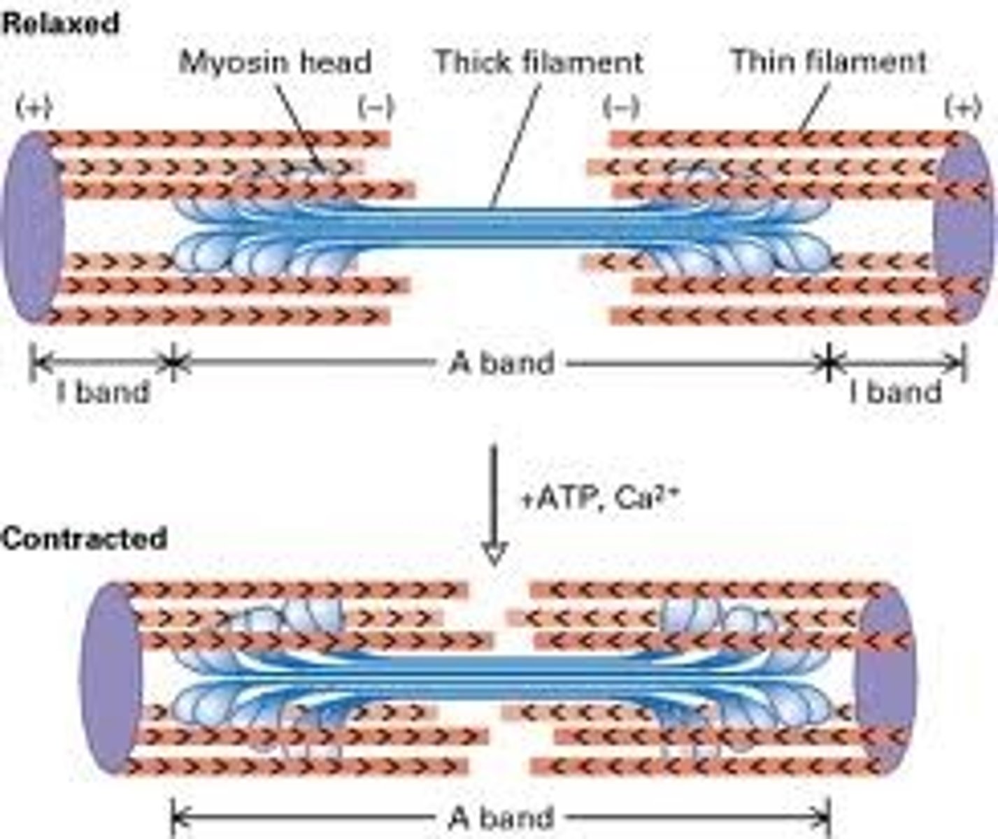
sliding filament theory
-muscle contracts when thick and thin filaments pull on one another; shortening the sarcomere
-Thick filaments (myosin) pull on thin filaments (actin) toward M Line
Muscular Hierarchy
LARGEST TO SMALLEST:
-Muscle (wrapped in epimysium)
-Fascicle (wrapped in perimysium)
-Muscle Fibers (wrapped in endomysium)
-Myofibrils
-Sarcomere
-Myofilaments
Synapse
point where a nerve fiber meets its target cell
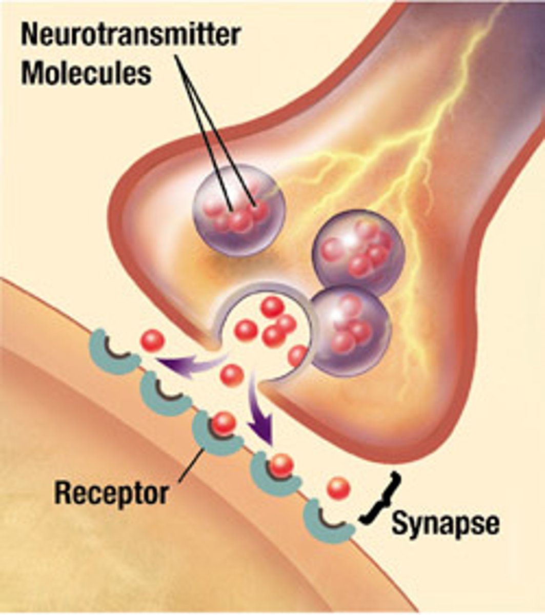
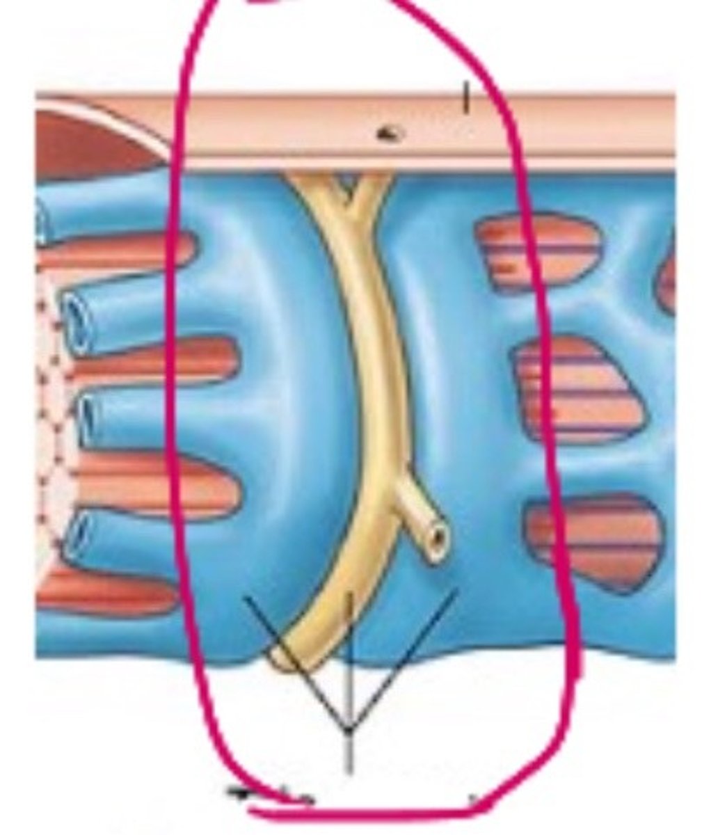
neuromuscular junction (NMJ)
when target cell is a muscle fiber
-One nerve fiber articulates with a muscle fiber at multiple positions
-stimulates the muscle fiber at several points & increases the speed of contraction.

Synaptic Cleft
the gap between the synaptic knob and sarcolemma
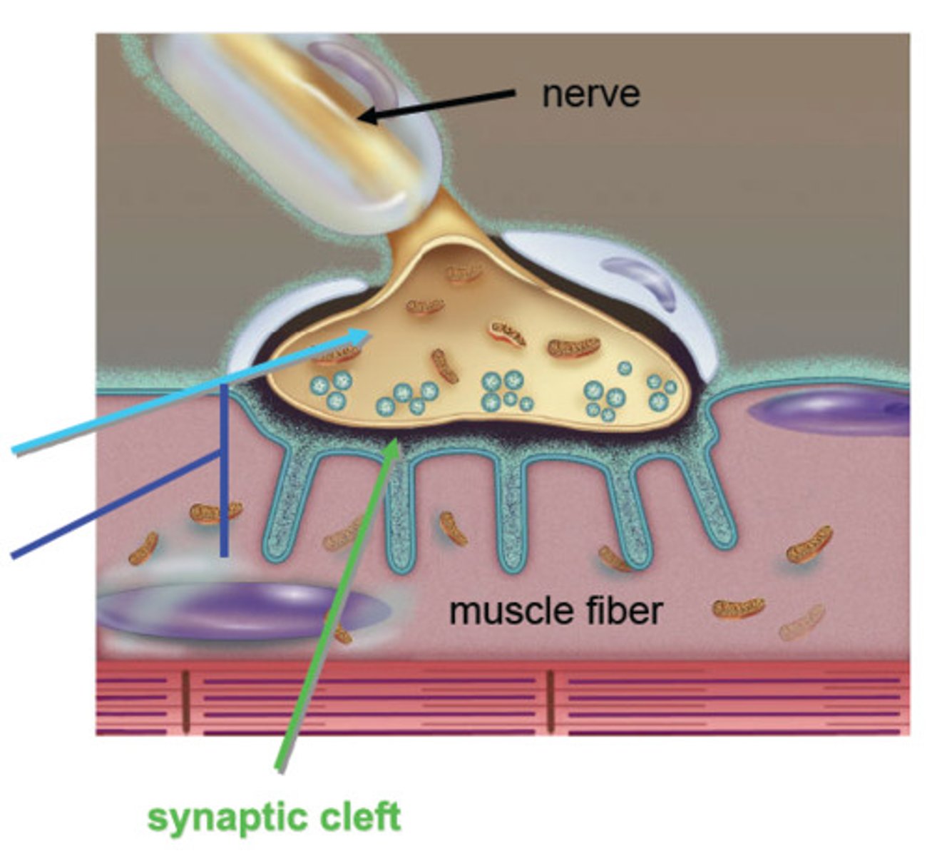
Basal Lamina
-Made of collage and glycoproteins that isolate the NMJ from surrounding tissues.
-allows for fiber relaxation

At a NMJ, what happens when a nerve send an impulse?
1. Acetylcholine from the synaptic knob into the synaptic cleft via exocytosis
2. The ACh diffuses across the synaptic cleft & binds to receptors on the sarcolemma
-Junctional folds increase surface area for ACh receptors
3. Binding of ACh begins an electrical signal on the sarcolemma.
-Causes the contraction of the muscle.
4. An electrical signal is converted to a chemical signal, back to an electrical signal.
Muscle and nerve cells are:
Electrically excitable and have electrochemical potential (like a battery)
The membranes of muscles and nerves are:
polarized (opposite charges)
Extracellular Fluid (outside of cell)
-Little K+
-Lots of Na+
-Positive Charge
Intracellular Fluid (inside the cell)
-Lots of K+
-Little Na+
-Lots of Negative Proteins
-Overall Negative Charge
The difference in charges between ICF and EFC?
-90 mV
Depolarization
process during the action potential: when stimulated, Na+ channels open and let Na+ flow down its gradient.
-The ICF charge becomes positive
Repolarization
-Potassium channels open. Letting potassium out of the cell.
-Returns to resting membrane potential
-the Na+/K+ pump resets the system
4 major steps of muscle contraction
-Excitation
-Excitation-Contraction Coupling
-Contraction
-Relaxation
What occurs during Excitation-Contraction Coupling?
1. AP spreads down T tubules.
2. Opens voltage-gated ion channels in T tubules and Ca+ channels in SR
3. Ca+ leaves the SR and enters the cytosol of muscle cells.
4. Calcium binds to Troponin in thin filaments.
5. Troponin-Tropomyosin complex changes shape and exposes active sites on Actin.
What happens during Contraction?
1. ATPase in myosin head breaks down an ATP molecule.
-(ADP and loose Phosphate remain)
2. Activates the head; "cocking" it in an extended position.
3. Head binds to actin active site forming a myosin-actin cross bridge.
4. Myosin releases ADP and P; flexes and pulls thin filament with it. Called a POWER STROKE
5. Upon binding more ATP, myosin releases actin, and process can repeat.
6. Recovery Stroke- recocks head
-each myosin head performs 5 power strokes/ second
-Each stroke utilizes one ATP
What happens during Relaxation?
1. When the nerve signal has stopped, ACh is no longer released into the synapse
2. AChE breaks down ACh and fragments are reabsorbed into synaptic knob. Stimulation of ACh stops.
3. Ca++ is actively pumped back into the SR; Ca++ binds to calsequestrin while in SR. Ca++ lost from troponin in pumped back into SR.
4. Tropomyosin blocks the active sites of actin.
5. Muscle fiber returns to resting position due to recoil of elastic components and antagonistic muscles.
small motor units
-Fine degree of motor control
-3 to 6 muscle fibers per neuron
-Ex. eye and hand muscles
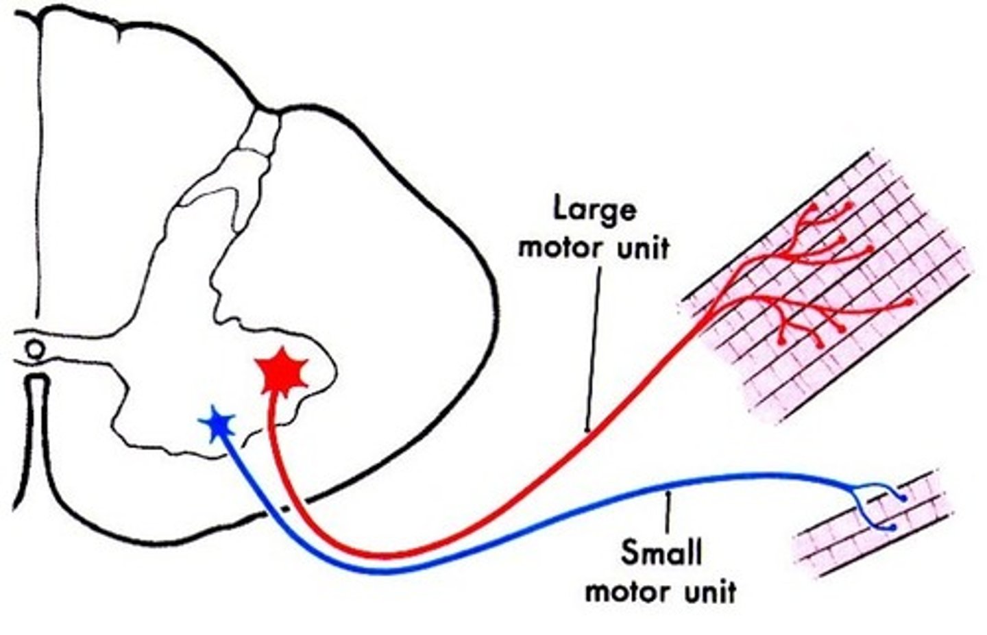
large motor units
-less control, more strength
-1000 muscle fibers per unit
Advantages of muscle fibers of one motor unit
- Motor units alternate contractions
- provides the ability to sustain long-term contractions
-prevents muscle fatigue
What do effective contractions require?
contractions of several motor units at once
Twitch
a quick cycle of contraction and relaxation when stimulus is at threshold or higher
multiple motor unit summation (recruitment)
The process of using more motor units to strengthen a contraction.
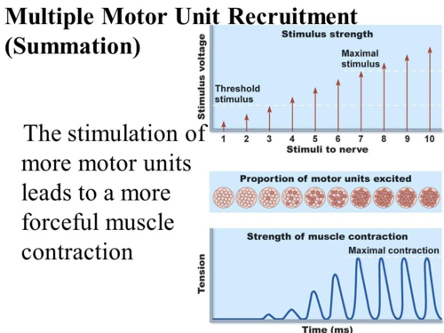
size principle of recruitment
weak stimuli (low voltage) recruit small units, while strong stimuli recruit small and large units for powerful movements
temporal summation
Higher frequency of stimulations increases contraction strength.
-low frequency stimuli produce identical twitches.
-high frequency stimuli produce temporal (wave) summation
(only partial relaxation between stimuli result in incomplete tetanus)
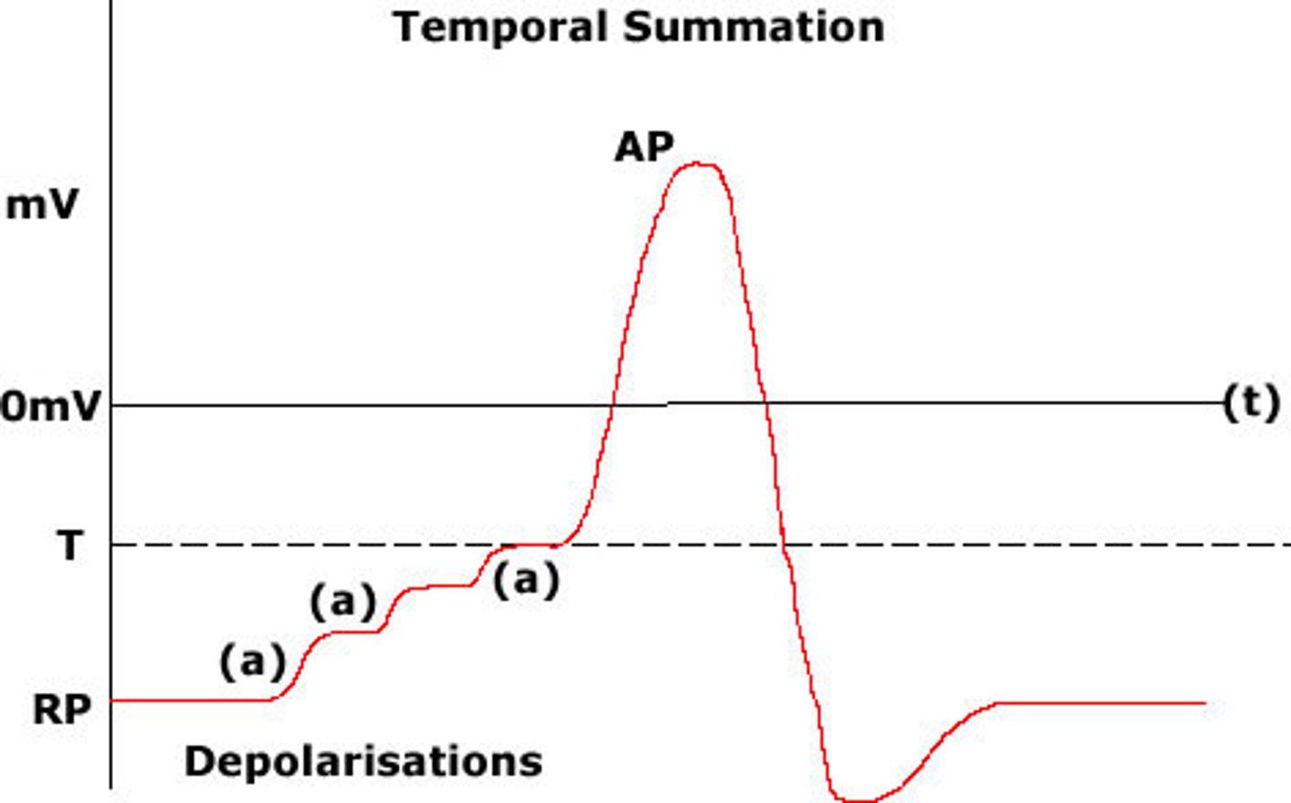
Twitch strength is also affected by:
warmer temperatures and hydration of muscles
ATP supply depends on the availability of what?
Oxygen and organic energy sources (e.g., glucose and fatty acids)
2 main pathways of ATP synthesis
anaerobic fermentation and aerobic respiration
anaerobic fermentation
-Enables cells to produce ATP in the absence of oxygen
-Yields little ATP, but lots of toxic lactic acid, a major factor in muscle fatigue
aerobic respiration
-requires a constant supply of oxygen
-produces lots of ATP, no lactic acid
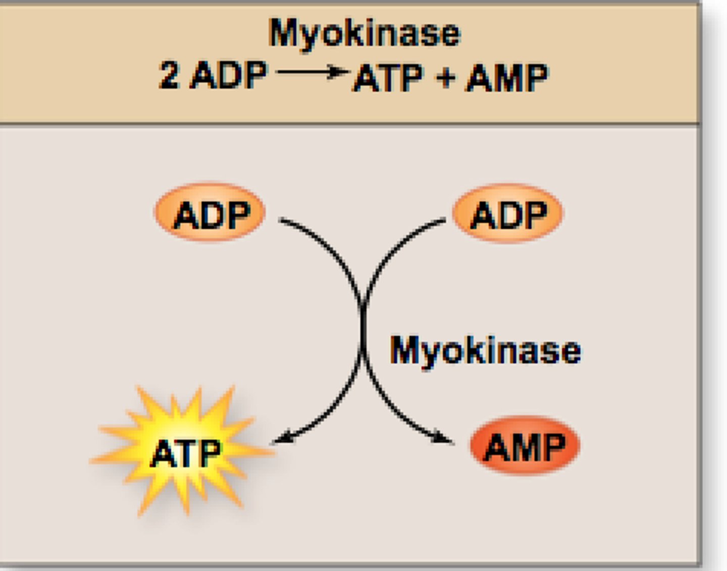
Two enzyme systems control the phosphate transfers in muscle cells
Myokinase and Creatine Kinase
Myokinase
transfers Pi from one ADP to another, converting the latter to ATP
short term energy
-As the phosphagen system is exhausted, muscles shift to anaerobic fermentation.
-muscles obtain glucose from blood and stored glycogen.
-without oxygen, glycolysis can make 2 ATP per glucose molecule consumed.
Produces enough ATP for 30 to 40 seconds of maximum activity
long term energy
After 40 seconds or so, the respiratory and cardiovascular systems deliver oxygen fast enough for aerobic respiration to meet ATP demands.
Aerobic Respiration=30-38 ATP/glucose
For 30 minutes, use = parts glucose and fatty acids; then mostly fatty acids.
Excess Post-Exercise Oxygen Consumption (EPOC)(Oxygen Debt)
We breathe heavily after exercise to :
-aerobically replenish ATP
-replace oxygen in myoglobin
-provide oxygen liver that is busy disposing lactic acid
-provide oxygen to many cells that have elevated metabolic rates after exercise.