Cell Biology Exam 1
 Call Kai
 Learn
Learn Practice Test
 Spaced Repetition
 Match
Match Flashcards
 Knowt Play
1/103

Earn XP
Description and Tags
Chapters 4 and 11
Name | Mastery | Learn | Test | Matching | Spaced | Call with Kai | Chat |
|---|
No analytics yet
Send a link to your students to track their progress
104 Terms
Amino Acid Sequence
The order of amino acids; it is unique for each protein
Primary Structure
The linear sequence of amino acids
Determination of Amino Acid Behavior
The R-group (a.k.a., side chain) of an amino acid determines both its identity and its chemical behavior
Condensation Reaction
The addition of a subunit (i.e., another amino acid) onto one end of a polymer chain, resulting in the loss of a molecule of water per subunit added
Polypeptide Backbone
The repetitive sequence N-C-C of multiple amino acids chained together
N- to C- Synthesis
The directionality of protein synthesis
Protein Folding
Hydrophobic forces (e.g., the hydrophobic side chains of many amino acids) help proteins fold into compact conformations
Urea Denaturing Experiment
Adding urea disrupts noncovalent bonds and denatures amino acids until the urea is removed; thus, we learned that proteins fold into the conformation of lowest energy
Chaperone Proteins
Assist the folding of a polypeptide chain; some can create makeshift “chambers,” isolating proteins until they correctly fold and are released
Secondary Structure
Interactions between atoms of the backbone (i.e., no side chain involvement); includes α helices, β sheets, and turns
Tertiary Structure
The full three-dimensional conformation of a protein primarily due to interactions between its R-groups
Quarternary Structure
Association of multiple polypeptide chains (i.e., it consists of more than one amino acid chain)
Number of Amino Acids per Complete Turn
3.6 amino acids per complete turn
α Helices in Lipid Bilayers
Receptors, transmembrane proteins, and transport proteins can be embedded in the cell membrane and consist of α helices; about 20 amino acids are required to span the membrane, and the R-groups that stick out of the helices are NONPOLAR
Domain
Separate regions in a tertiary structure, usually 40-350 amino acids; many proteins are composed of separate functional domains
Family
Proteins that have similar amino acid sequences and thus similar shapes
Subunit
A whole polypeptide chain; large proteins often contain more than one
Binding Site
Regions involved in the interaction between different subunits
Sickle Cell Anemia Example
A point mutation swapping Glu for Val leads to hemoglobin molecules that stick together and deform the cells; the nonpolar Val sticks to itself due to the hydrophobic effect
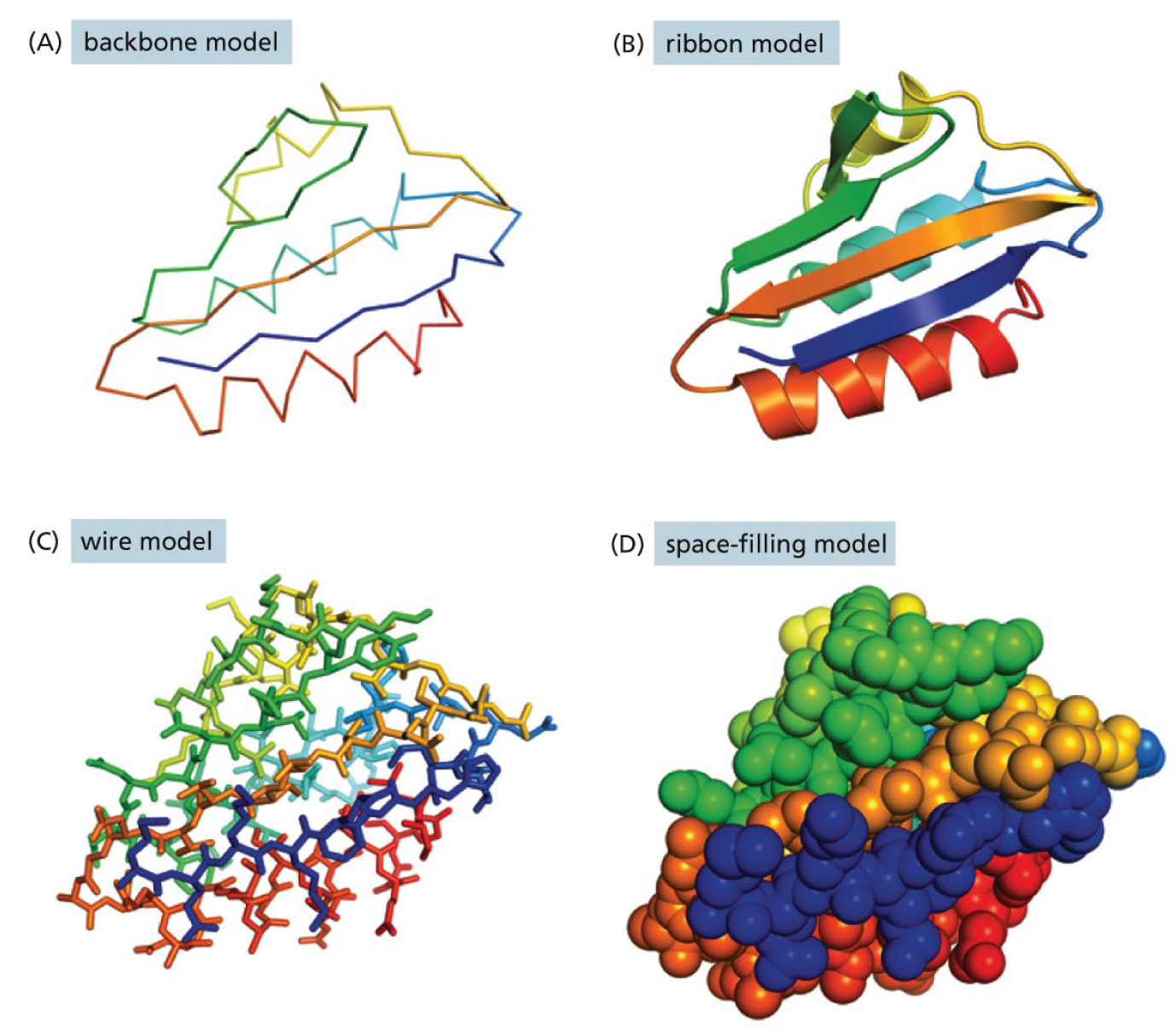
Protein Conformation Models
Backbone Model (simplest)
Ribbon Model
Wire Model (similar to Backbone Model, but the “wires” almost seem like broken benzenes in angle)
Space-Filling Model
Cross-Link Bonds
Bonds that hold together single filamentous molecules
Disulfide Bonds
These only form OUTSIDE of the cell; cytoplasm in the cell is a reducing environment, so the equilibrium of the disulfide reaction is shifted towards reduction in the cytoplasm
Protein Subunit Assemblage Types
Identical protein subunits can assemble into complex structures; dimers, helices, and rings can form depending on the shape/orientation of the binding site, and filaments, spherical shells, and hollow tubes can form from the exact same subunit
Protein-to-Molecule Binding
HIGHLY selective as the shape is important; ligands and proteins must fit perfectly, and they’re held by many weak, noncovalent bonds
Methods of Catalysis Encouragement via Enzymes
Enzymes can bind two substrates in a precise orientation to encourage a reaction between them
Enzymes can bind a substrate and rearrange its electrons in order to create reaction-favorable partial negative and positive charges
Enzymes can strain/reshape a bound substrate to force it into a more favorable transition state
Hydrolase
Enzymes that catalyze a hydrolytic cleavage reaction
Nuclease
Enzymes that break down nucleic acids by jydrolyzing bonds between the nucleotides
Protease
Enzymes that break down proteins by hydrolyzing peptide bonds between amino acids
Ligase
Enzymes that join two molecules together (e.g., DNA ligase joins two DNA strands together end to end)
Isomerase
Enzymes that catalyze the rearrangement of bonds within a single molecule
Polymerase
Enzymes that catalyze polymerization reactions such as synthesis of DNA and RNA
Kinase
Enzymes that catalyze the addition of phosphate groups to molecules (e.g., protein kinases, which bind phosphate groups to proteins)
Phosphatase
Enzymes that catalyze the hydrolytic removal of a phosphate group from a molecule
Oxido-Reductase
General name for enzymes that catalyze reactions wherein one molecule is oxidized while another is reduced (e.g., oxidases, reductases, or dehydrogenases)
ATPase
Enzymes that hydrolyze ATP
Measures of Enzyme Performance
Enzyme performance depends on how rapidly it can process its substrate; measured via V, Vmax, and Km
V
The rate of enzyme reaction
Vmax
The maximum rate a given enzyme can work at; here, all substrate binding sites are occupied
Km
The amount of substrate wherein the enzyme works at half speed; enzymes with a low Km bind tightly, and enzymes with a high Km have weak binding
Feedback Inhibition
A method by which protein function can be regulated by downstream products
Inhibitors
Reduce the activity of an enzyme; negative regulation
Activators
Increase the activity of an enzyme; positive regulation
Allosteric Regulation
When a regulatory molecule binds to an enzyme someplace other than the active site; can serve to inhibit OR to activate
*Allosteric regulation always involves conformational change
Phosphorylation
Controls protein activity by causing conformational change; one of the most important protein control mechanisms in nature as 1/3 of proteins are regulated by this process
*Can turn “on” or “off” a process
Phosphorylated Amino Acids
Serine
Threonine
Tyrosine
GTP-Binding Proteins
Proteins that are switched on/off by binding and hydrolyzing a GTP molecule; molecule is “on” upon binding GTP to the G protein, and it only turns off when GTP is hydrolyzed into GDP
ATP Hydrolysis
Allows motor proteins to produce directed movement in cells
Post-Translational (Enzyme) Modification
Greater than 100 types (e.g., acetylation, glycosylation, ubiquitination, phosphorylation, etc.); can activate/deactivate, define protein location or fate, and can modify multiple sites on certain proteins
Homogenization
Gentle mechanical procedures that rupture the plasma membranes of cells so that their contents are released
Homogenate / Extract
The “thick soup” resulting from homogenization
Homogenization Methods
Breaking apart cells with high-frequency sounds (ultrasound)
Using mild detergent to make holes in plasma membrane
Forcing cells through small holes using high pressure
Shearing cells between close-fitting rotating plunger and thick walls of a glass vessel
Centrifugation
The most widely used procedure to separate a homogenate into different parts, or fractions
*Modern centrifuges have speeds up to 100k RPM and produce forces as high as gravity x 600k
Supernatant
The smaller, less dense components of a homogenate post-centrifugation
Differential Centrifugation
Allows you to isolate different cellular components based on size and density; each step is the result of increasing speeds of centrifugation
Differential Centrifugation Results by Pellet
Pellet 1 - Whole cells, nuclei, cytoskeletons
Pellet 2 - Mitochondria, lysosomes, peroxisomes
Pellet 3 - Closed fragments of endoplasmic reticulum, other small vesicles
Pellet 4 - Ribosomes, viruses, large macromolecules
Velocity Sedimentation
Uses a sucrose gradient to top separate cellular components based on size and their sedimentation rates; as the sediments in the tube post-centrifugation separate, the tube is punctured in the bottom and allowed to drain into different tubes that move automatically below it
Equilibrium Sedimentation
Allows you to isolate different cellular components based on their buoyant density Equi; once the bands have separated based on density, they are gathered using the same draining method as velocity sedimentation
Column Chromatography
A method of fractionating proteins where a micture of proteins in solutions is applied to the top of a cylindrical column filled with a permeable solid matrix immersed in solvent; choice of matrix allows proteins to be separated according to their charge, hydrophobicity, size, or binding ability to certain groups
Ion-Exchange Chromatography
Ion-exchange columns are packed with small beads carrying either positive or negative charges; association between protein and matrix depends on pH and ionic strength of solution
Gel-Filtration Chromatography
Gel-filtration columns separate proteins according to their size; protein molecules small enough to enter the columns tiny porous beads are filtered out more slowly than larger protein molecules that cannot enter the beads
Affinity Chromatography
Affinity columns contan a matrix covalently coupled to a molecule that interacts specifically with the protein of interest; proteins can subsequently be released via pH change or concentrated salt solution, and they emerge highly purified
Electrophoresis
When an electric field is applied to a solution containing protein molecules, the proteins migrate at a speed reflective of their size and net charge
SDS Polyacrylamide-Gel Electrophoresis (SDS-PAGE)
Individual polypeptide chains form a complex with negatively charged molecules of sodium dodecyl sulfate (SDS) and thus migrate as negatively charged SDS-protein complexes through a slab of porous polyacrylamide gel; reducing agents like mercaptoethanol prevent disulfide linkages to allow movement
Isoelectric Focusing
Proteins are electrophoresed in a narrow tube of polyacrylamide gel in which a pH gradient is established by a mixture of special buffers; proteins move to a location corresponding to their isoelectric point and stay there
Two-Dimensional Gel Electrophoresis
Combines Gel Electrophoresis and isoelectric focusing in order to resolve more than 1000 proteins in a two-dimensional protein map
Antibody
Protein that bind very tightly to their target antigens; produced in vertebrates as a defense against infection
*Humans produce billions of types, each with different binding sites
Parts of Antibodies
4 Polypeptide chains with hypervariable antigen-binding sites at their ends:
2 heavy chains (dark green in image)
2 light chains (light green in image)
Variable domains
Constant domains (gray in the image)
Variable light (Vl) and Variable heavy (Vh) domains (blue in the image)
*Altering the length or amino acid sequence of the variable domains provide different antibodies
B Lymphocyte (B Cells)
Class of white blood cells responsible for antibodies; upon a B Cell’s antibody binding, it is stimulated to divide and to secrete large amounts of the same antibody in soluble form
Aggregates
Collections of antigens (can be foreign molecules, viruses, and/or bacteria) that are cross-linked by antibodies and ingested by phagocytic cells or killed by special proteins in the blood
Monoclonal Antibodies
Made by harvesting antibody-bearing B cells from an immunized animal the fusing them with a tumor cell to make a hybridoma
Increasing Antigen Detection Sensitivity
Can increase sensitivity greatly by using multiple layers of antibodies; this “sandwich” method allows smaller numbers of antigen molecules to be detected
Western Blot
An antigen of interest is separated from other molecules via electrophoresis before being incubated with labeled antibodies that bind to the antigen; allows the position of the antigen to be determined
*Separation → Transfer → Staining → Visualization
Methods of Determining Proteins’ 3-D Shape
X-Ray Crystallography
NMR Spectroscopy
Cryo-Electron Microscopy
Labeled Cell (just look at it)

Amphipathic
Molecules with body hydrophilic AND hydrophobic regions
Phospholipid Components
Phosphate-containing hydrophilic head
Two hydrophobic hydrocarbon tails, one of which is saturated (no double bonds) and one of which is unsaturated (1+ double bonds)
A kink in one of the tails which makes it harder for them to pack together and keeps membranes more fluid
Membrane Lipids
Phosphatidylcholine (more common)
Phosphatidylserine (a phospholipid)
Cholesterol (a sterol)
Galactocerebroside (a glycolipid)
Membrane Lipid Spheres
Energetically favorable conformation when in water; requires no energy to do so, keeps hydrophobic pieces away from water, and allows the membrane to self-heal
Lipid Bilayer Factors
Length of tail → shorter tails are more fluid
Number of double bonds → more double bonds are more fluid
Amount of cholesterol → cholesterol reduces fluidity
Temperature → higher temperatures increase fluidity
Scramblase
Catalyzes the transfer of phospholipids from one cytosolic layer to another in a RANDOM fashion; occurs in the ER
Flippase
Catalyzes the transfer of phospholipids from one cytosolic layer to another in a specific fashion; occurs in the Golgi Apparatus
*Can flip phospholipids, but NOT glycolipids
Golgi Apparatus
Where the asymmetric destribution of the membrane bilayer occurs; lipids are also modified here (e.g., glycosylation)
Membrane Composition in Humans
Approximately 50% protein; however there are ~50x as many phospholipids than proteins (phospholipids are much smaller)
Main Function of Membrane Proteins
Transport: getting molecules (e.g., ions, sugars, metabolites, etc.) across the membrane when they would ordinarily not be able to
*Can also serve as anchor proteins
Membrane Protein Examples
Transporters (e.g., Na+ Pump)
Ion Channels (e.g., K+ Leak Channel)
Anchors (e.g., Integrins)
Receptors (e.g., Platelet-Derived Growth Factor (PDGF) Receptor)
Enzymes (e.g., Adenylyl Cyclase)
Integral Membrane Proteins
Transmembrane
Monolayer-Associated
Lipid-Linked
Peripheral Membrane Protein
Protein-Attached
Most Common Transmembrane Protein Form
α Helices containing at least 20 amino acid residues
Hydropathy Plot
Plot that predicts trans-membrane domains; peaks are positive values that indicate hydrophobic segments of the protein
Channel Formation
Multipass transmembrane proteins can form channels
Detergent Use with Membrane Associate Proteins
Solubilizing these proteins in detergent allows their study; the proteins are brought into the solution as protein-detergent complexes
Synthetic Phospholipid Bilayer
Can be used to functionally reconstitute membrane proteins
Cell Cortex
The meshwork of proteins that lies just underneath and connects to the cell membrane; reinforces the plasma membrane
Fluid Mosaic Model
Combined mouse and human cells; initially cell membranes remained unmixed, but they steadily mixed together after time and incubation
*Membrane determined to be a fluid body!
Membrane Domain
Functionally specialized regions of a given cell’s membrane that have restricted movement
Methods of Restricting Lateral Mobility of Plasma Membrane
Tether to cell cortex inside the cell
Tether to extracellular matrix molecules outside the cell
Tether to proteins on the surface of another cell
Use diffusion barries that restric proteins to a particular membrane domain
Glycolipids
Many of the lipids on the exterior face of the lipid bilayer; these have sugars attached to their heads
Glycoproteins
Many of the membrane proteins; contain one or more short chains of sugars linked together called Oligosaccharides
Proteoglycans
Contain one or more long chains of sugars linked together called polysaccharides
Carbohydrate Layer (Glycocalyx)
The sugar layer on the outermost exterior of the cell formed by glycolipids, glycoproteins, and proteoglycans; protects the cell from physical damage and attracts water molecules