Cytology
 Call Kai
 Learn
Learn Practice Test
 Spaced Repetition
 Match
Match Flashcards
 Knowt Play
1/199

There's no tags or description
Looks like no tags are added yet.
Name | Mastery | Learn | Test | Matching | Spaced | Call with Kai | Chat |
|---|
No analytics yet
Send a link to your students to track their progress
200 Terms
There are over 200 distinguishable cell types in the body assembled into which basic tissue groups?
Epithelia
Connective tissue
Muscle
Nerve
Type of tissue that
cover body exterior surfaces and line body cavities including blood vessels
epithelia
Type of tissue that
form glands both exocrine and endocrine
have cells that tightly adherent to one another
epithelia
Type of tissue that
known for shapes: squamous, cuboidal and columnar
epithelia
Type of tissue that
functions include protection, absorption, secretion and excretion, and gas exchange
epithelia
Type of tissue that
consists of relatively few cells that are not adherent to each other
connective tissue
Type of tissue that
serve vascular system
has abundant matrix
connective tissue
Type of tissue that
functions include cohesion of structural elements
serves as a medium through which blood vessels distribute nutrients and take up metabolic waste
connective tissue
Type of tissue that
have cells involved in immune & inflammatory responses and in tissue repair following injury
connective tissue
Type of tissue that
is responsible for movement and changes in the size & shape of body organs
muscle
Type of tissue that
has elongated cells oriented parallel to each other and organized in bundles
muscle
Type of tissue that
arrange the filaments highly organized → contraction
muscle
The muscle cytoplasm or sarcoplasm is occupied mostly by _____ and ____
myofilaments actin & myosin
Cells can be various in shapes, sizes and specialized functions
However, they always share common features. List
surrounded by a membrane
have zero ( ex: in mature RBC) to many nuclei
contain organelles, which can be membrane-bound or non-membrane-bound
contain inclusions (temporary non-living structures, ex: secretory products, pigment granules, or metabolic byproducts)
The nucleus is a membrane-limited organelle that contains _____ of the cell
genetic information
T/F The nucleus has double membrane, inner and outer, in the nuclear envelope
True
Functions of the nucleus are
replication of DNA → cell division
DNA repair
RNA transcription & processing
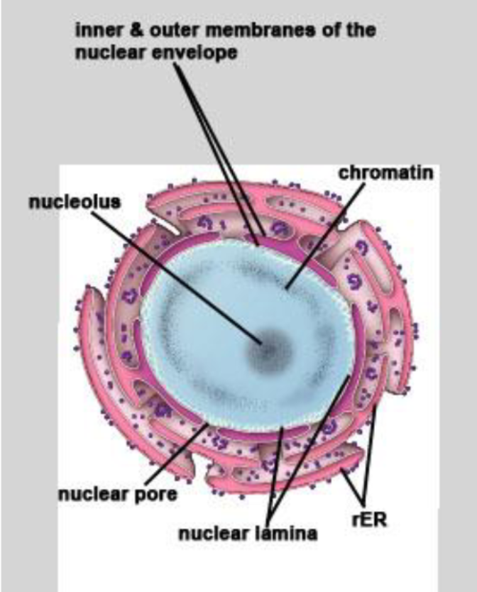
Sketch the nucleus
Three major components of nucleus are
nuclear envelope
chromatin
nucleolus
What make up the nuclear envelope?
outer nuclear membrane - continuous with ER, contains proteins involved in transport and structural support
inner nuclear membrane
nuclear pores - protein channels embedded in the membranes - regulate passage of molecules in/out the nucleus
nuclear lamina - a meshwork of lamin protein on inner membrane - give structural support and anchors chromatin
Why is the evaluation of the morphology of the nucleus important?
to determine the health of a cell through examination of normal cells characteristics
Characteristics found in normal cells are
shape: round, ellipsoid, infolded, lobulated
size: various
number of nuclei per cell: none to multi
location: central, basal, eccentric (strange)
T/F Nuclear envelope inner and outer membranes are connected with no space in between
False: they are separated by a 10-30 nm space
The space between the two unit membranes of nuclear envelope
perinuclear cisternae
T/F The nuclear pores are attached to the outer nuclear membrane only
False: the inner and outer nuclear membranes are continuous at nuclear pores
Direction of nuclear pores
bidirectional
T/F Small molecules still have to pass through the nuclear pores
F they can pass through by diffusion
Why do we call the nuclear pores highly selective?
The proteins of any size need to contain a nuclear localization amino acid sequence to be carried through an active transport (ATP or GTP hydrolysis)
Why do we call the structure of nuclear pores nucleoporins
The shape of a central cylindrical body between inner and outer octagonal rings
each ring = 8 protein particles
The basket on nuclear side formed by protein filaments that extend from each ring
Functions of nuclear lamina are
give shape and stability to the nuclear envelope
organize the interphase nucleus
structural link between chromatin and nuclear envelope
responsible for the dissolution and reformation of the nuclear envelope during cell division
position nuclear pore complexes within the nuclear envelope
Location of nuclear lamina
between the inner nuclear membrane and the peripheral heterochromatin
What make up nuclear lamina
a class of intermedia filament proteins called lamins ( lamins A, B, C)
What causes the dissolution of nuclear lamina at mitosis
lamin phosphorylation
Give me some defects of mutations in proteins of the nuclear membranes or pore complexes ( nuclear envelopathies) or lamins ( laminopathies)
Inheritable diseases
striated muscle disorders, partial lipodystrophy syndromes, peripheral
neuropathies, progeroid syndromes, developmental abnormalities & death.
Define chromatin
complex of DNA, histone and non-histone proteins found in the nucleus of eukaryotic cells
T/F Chromatins are made up of chromosomes, and carry the cell genetic material.
F Chromatins make up chromosomes
During interphase nucleus
the electron-dense portions of chromosomes are
heterochromatin
T/F Heterochromatin are constitutive (never transcribed). Only euchromatin represents genes that are being transcribed
F Heterochromatin can also be facultative ( transcription depends on cell type)
During interphase nucleus
the uncoiled or loosely packed portions of chromosomes are
euchromatin
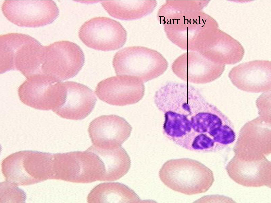
Barr body
In XX individual, one of X chromosomes is inactive and highly coiled
Under light microscope, shows up as a small drumstick of heterochromatin located adjacent to the nuclear envelope
Why is nucleolus rich in rRNA and proteins?
functions as a site of ribosomal RNA transcription and rRNA synthesis
regulates cell cycle (nucleostemin)
T/F nucleolus is surrounded by a membrane
False
T/F 1-2 nucleolus in cell determined by the synthetic activity of cell, eccentrically placed in the interphase nucleus
True
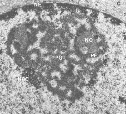
Main parts of nucleolus
NO: nucleolar organizer region of those chromosomes processing nucleolar genes
PF: pars fibrosa, dense fibrillary component
PG: pars granulosa or granular component - ribonucleo-protein particles
NAC: nucleolus-associated chromatin
How is nucleolus formed?
regions of 10 interphase chromosomes ( pairs of 5 diff chro in human) that contain the genes that encode rRNA (Nucleolar Organizing Regions) reorganize the nucleoli following cell division
What is the difference between RNA and DNA?
linear molecule
ribose sugar instead of deoxyribose
uracil base instead of thymine
product of transcription of DNA
messenger RNA (mRNA) is used for
direct the amino acid sequence of proteins
transfer RNA (tRNA) is used for
transfer the correct amino acid to the ribosome for incorporation into the protein
ribosomal RNA (rRNA) is used for
form ribosomes in association with specific protein
Ribosomes are composed of 2 subunits.
large subunit is composed of
5S
28S
5.8S rRNA
~ 49 proteins
Ribosomes are composed of 2 subunits.
small subunit is composed of
18S rRNA
~ 33 proteins
Function of ribosomal large subunit
catalyze the peptide bond formation
Function of ribosomal small subunit
bind mRNA and tRNA
T/F Under the electron microscope, the ribosome can be observed individually
F ribosomes (polyribosomes) appear as granular material, associating with outer nuclear membrane and the endoplasmic reticulum
Series of ribosome formation
in nucleolus
(1) transcription of rDNA to form pre-rRNA
(2) association of pre-rRNA with ribosomal proteins to form ribonucleoprotein
(3) cleavage of pre-rRNA into the 28S, 18S, and 5.8S rRNAs found in ribosomes
in nucleus
(4) 28S and 5.8S rRNA combines with 5S rRNA ( 5S rRNA is transcribed in the nucleus) to form the large ribosomal subunit
(5) active transport of ribosomal subunits to the cytoplasm through nuclear pores
in cytoplasm
(6) assembly of ribosomal subunits into ribosomes and polysomes in the
presence of mRNA
T/F membranes are homogenous
False
T/F membrane components are fixated
F they are in a constant dynamic flux
T/F membranes are asymmetric
True
T/F the underlying cytoskeleton partially determines the shape of the surface of the membrane
True
What is a unit membrane? What does it composed of?
The general structure of cell membranes (plasma membrane and membranes of organelles)
three distinct layers (trilaminar structure) when consisting of two electron dense lines (inner and outer leaflets) separated by an electron-lucent intermediate zone
What basic structures make up the membranes?
lipids - mainly - 50% of membrane mass - membrane form and permeability properties
proteins - membrane’s specific functions
carbohydrates - membrane’s surface recognition, facing away from the cytoplasm
The most abundant type of lipid in membranes
phospholipids
Why do we call the phospholipids amphipathic
a hydrophilic head
the hydrophobic tails
The second major lipid of membranes
cholesterol
Cholesterol function
maintains the structural integrity of membranes
The third most abundant membrane lipids
glycolipids
Where do most glycolipids locate in the membrane?
outer leaflet
peripheral proteins
associate with membranes through ionic
interactions
not across the membrane
integral proteins
buried in the lipid membrane
transmembrane proteins
Which structure perform most of the membrane’s functions
proteins
the presence of membrane proteins is indicated by
freeze fracture technique
Which membrane component contributes to the glycocalyx
carbohydrate
What are Lipid rafts
specialized membrane domains that associate with or segregate
different proteins or signaling molecules in one part of the cellular membrane
What is cytoskeleton?
A highly dynamic, complex network of protein filaments that organizes the cytoplasm and determines cell shape
functions of cytoskeleton
cell motility
organelle and vesicle transport
muscle contraction,
cell division
endocytosis/phagocytosis
providing mechanical strength
Thickest cytoskeleton component
microtubules
Thinnest cytoskeleton component
microfilaments
Strongest cytoskeleton component
intermediate filaments
Why do we call microfilaments actin filaments?
composed of actin which is the major cytoskeletal protein of most cells (~5-10% of total cell protein)
Polarization of actin protofilemnt
G-actin (globular protein) that binds head to tail
Polarization of F-action filament
two actin protofilaments twist around each other (right-handed helix)
Depolarization of actin filaments
ATP hydrolysis in G-actin
T/F a new branch of actin filaments can grow out of the current one
True
Functions of actin-binding proteins
1/ regulate the assembly and disassembly of actin; examples: cofilin,
profiling, gelsolin
2/ influence filament dynamics; for example, actin capping proteins such as
cap Z and tropomodulin regulate filament growth
3/ organize actin filaments into bundles (fimbrin, α-actinin) or networks
(filamin, spectrin, fodrin, dystrophin)
4/ actin-motor proteins utilize ATP to move along the actin filament towards
the plus end; examples myosin I and II
Functions of actin
cell attachment (focal adhesion)
cell-cell adhesion
cell surface modifications (example, microvilli and stereocilia)
movement/contraction
T/F intermediate filament can be polarized
False
T/F intermedia filaments are composed of a variety of proteins whose expression is cell-type specific
True
Functions of intermedia filaments
structural support & mechanical strength
Classes of intermediate filaments
Class 1: Keratins – epithelial cells
Class 2: Vimentin & vimentin-like – fibroblasts, muscle, glial cells, nerve stem cells
Class 3: Neurofilament – neurons
Class 4: Lamins – nucleus (in all nucleated cells)
Class 5: Beaded filaments – eye lens fiber cells
T/F microtubules can branch out from the one and another
False
T/F inside of microtubules are hollow
True
What make up microtubules
from 13 parallel protofilaments, each protofilament is composed of
dimers of α and β tubulin subunits
Microtubules are polarized with a plus end and a minus end.
Which end is fast growing end?
plus end
Microtubules are polarized with a plus end and a minus end.
Which end is slow growing end?
minus end
Which microtubular end is embedded in the MTOC (microtubule-organising center or centrosome)
minus end
What make up centrosome
fibrous centrosome matrix containing 50 or more copies of the γ-tubulin ring (γ-TuRC)
a pair of centrioles arranged at right angles to each other. Each centriole is composed of nine triplets of MT
Functions of microtubules
maintenance of cell shape
transport of organelles
separation of chromosomes during mitosis/meiosis
ciliary/flagellar movement
T/F many functions of microtubules are due to the property of dynamic instability; the rapid conversion between a growing and shrinkage state.
True
Functions of microtubules-associated proteins (MAPs)
bind to and organize MTs into bundles
stabilize MTs against disassembly
mediate interactions of MTs with other cellular components
serve as molecular motors that are responsible for the movement
of organelles, vesicles, etc.; they utilize ATP to move “cargo” in a single direction along the filament
kinesin movement toward ___ end
plus
dynein movement toward ___ end
plus