Vascular Exam 1 - Hemodynamics and Cerebrovascular (Notes)
 Call Kai
 Learn
Learn Practice Test
 Spaced Repetition
 Match
Match Flashcards
 Knowt Play
1/180

There's no tags or description
Looks like no tags are added yet.
Name | Mastery | Learn | Test | Matching | Spaced | Call with Kai | Chat |
|---|
No analytics yet
Send a link to your students to track their progress
181 Terms
We typically differentiate types of vessels based on their _____ of the vasular system, meaning are they venous or arterial, and their _____.
side, size
Larger, flow away from heart; oxygen rich
Arteries
Smaller arteries, they slow down flow velocity; increased resistance to flow due to their decreased radius
Are called resistance vessels because they can contract or dilate to control flow and peripheral resistance
Arterioles
Arterioles have increased _____ to flow due to their decreased radius.
resistance
Smallest vessels
Nutrient exchange (delivery), gas exchange, waste removal, vessel “hand shake”
Connect the arterial side to the venous side
Capillaries
Smaller veins, collect blood from capillary bed and send it to the veins
Venules
Larger, flow towards the heart (right atrium); high capacitance/storage for blood
Most of the blood of the circulatory system is housed in these (approximately 70%)
Veins
Innermost vessel wall layer; the layer that actually touches blood
Made of endothelial cells supported by elastic lamina
Functions:
Permeability
Antithrombogenic
Vasoreactivity
*Valves in veins; this layer forms valves
Tunica intima
What layer of the vessel wall forms valves?
Tunica intima
Projections into the vessel lumen
Ensure one-way flow back up to the heart
Only found in veins
Valves
Middle muscular vessel wall layer
Made of smooth muscle cells and collagen
Thicker in arteries than in veins; this is the thickest layer in arteries
Functions:
Vasoconstriction and dilation (arteries)
Tunica media
What is the thickest layer in arteries that allows for vasoconstriction?
Tunica media
Arteries _____ or _____ in response to the cardiac cycle (exercise = _____, cold weather = _____)
contract, dilate, dilate, contract
Outer vessel wall layer
Made of connective tissue, collagen, vasa vasorum
This layer contains the vasa vasorum, which supplies blood to the vessel wall itself
So this layer is sometimes referred to as “the blood vessel of the blood vessel”
Tunica adventitia
This, located in the tunica adventitia, supplies blood to the vessel wall itself
Vasa vasorum
Where does the aorta start?
LVOT
The aorta starts at the _____ where it exits the _____ _____. It becomes the _____ aorta then the _____ _____ giving rise to three branches: the _____ (_____) artery, the _____ _____ _____ artery, and the _____ _____ artery. Then it becomes the _____ _____ aorta. Once the aorta passes below the _____, it is classified as the _____ aorta. The _____ aorta has several branches, the first being the _____ _____, then the _____, the _____ arteries, the _____, and then it bifurcates into the _____ arteries.
LVOT, left ventricle, ascending, aortic arch, brachiocephalic, innominate, left common carotid, left subclavian, descending thoracic, diaphragm, abdominal, abdominal, celiac axis, SMA, renal, IMA, iliac
Technically, the first branches of the aorta are the _____ _____ at the aortic _____.
coronary arteries, root
The brachiocephalic artery branches into the _____ _____ _____ artery and the _____ _____ artery
right common carotid, right subclavian
The left/right common carotid artery bifurcates into the left/right _____ and _____ carotid arteries, which then terminate in the _____ _____ _____ at the base of the brain.
internal, external, Circle of Willis
The right vertebral artery branches from the _____ _____ _____
right subclavian artery
The left subclavian artery gives rise to the _____ _____ _____
left vertebral artery
Anatomy – Upper Extremity Arteries
The right subclavian artery is a continuation of the _____ _____ after the _____ _____ _____ artery branches off of it
Once the subclavian artery passes below the _____, it becomes the _____ artery
The _____ artery continues to the bicep area and becomes the _____ artery
At the level of the elbow or the _____ fossa, the _____ artery bifurcates into the _____ and _____ arteries
_____ = thumb side
_____ = pinky side
brachiocephalic artery, right common carotid, clavicle, axillary, axillary, brachial, antecubital, brachial, radial, ulnar, Radius, Ulna
Anatomy – Lower Extremity Arteries
The common iliac arteries bifurcate into the _____ and _____ iliac arteries
The _____ iliac artery is also called the _____ artery
The _____ iliac artery becomes the _____ _____ artery once it passes below the _____ _____
The _____ _____ artery bifurcates into the _____ (_____) and the _____ (_____) arteries
The _____ _____ artery continues down the medial thigh until it passes behind the leg to the _____ or the _____ fossa. Here it gives rise to the _____, which consists of the _____ _____ artery (ATA), _____ artery, and _____ _____ artery (PTA).
The ATA, peroneal, and PTA continue down the leg to the _____. The PTA and peroneal are more _____ and the ATA is more _____.
The ATA eventually comes down on top of the ankle and becomes the _____ _____ (DP) artery
internal, external, internal, hypogastric, external, common femoral, inguinal ligament, common femoral, deep, profunda, superficial, femoral, superficial femoral, knee, popliteal, trifurcation, anterior tibial, peroneal, posterior tibial, foot, medial, lateral, dorsalis pedis
Veins (IVC and SVC) terminate at the…?
right atrium (RA)
When scanning arteries and veins in lab, we will always use the terms proximal, mid, or distal to describe what part of the vessel we are looking at. _____ is closest to the heart and _____ is furthest from the heart.
Proximal, distal
Anatomy – IVC
The IVC terminates at the _____ _____ of the heart
The IVC is formed by the confluence of the _____ _____ veins, which continue superiorly, accepting additional _____ until the IVC gets to the heart
The _____ veins are the first tributary in relation to the heart (most _____) followed by the _____ veins
The left gonadal vein empties into the _____ _____ vein
The right gonadal vein empties directly into the _____
The common iliac veins are formed by the confluence of the _____ and _____ iliac veins
right atrium, common iliac, tributaries, hepatic, proximal, renal, left renal, IVC, internal, external
Anatomy – SVC/Upper Extremity
Opposite the IVC in the right atrium is the _____ _____ _____ (_____)
The SVC is the final destination for all _____ extremity and _____ venous blood before dumping into the _____ _____
The SVC is formed by the confluence of the _____ (_____) veins
The _____ veins are formed by the confluence of the _____ and _____ _____ veins
In the venous system, there is a _____ and _____ brachiocephalic (innominate) vein while in the arterial system, there is only a _____ brachiocephalic (innominate) vein
The subclavian vein is formed by the confluence of the _____ vein and the _____ vein (a _____ vein)
The _____ vein is at the bicep. The brachial vein is the confluence of the _____ and _____ veins.
The _____ vein is another _____ vein that confluences with the brachial vein to form the _____ vein
_____ veins of the arms and legs are paired with an artery
A lot of times, there are _____ veins to _____ artery
superior vena cava, SVC, upper, cerebrovascular, right atrium, brachiocephalic, innominate, brachiocephalic, subclavian, internal jugular, right, left, right, axillary, cephalic, superficial, brachial, radial, ulnar, basilic, superficial, axillary, Deep, two, one
Anatomy – Lower Extremity Veins
The common iliac vein is formed by the confluence of the _____ and _____ iliac veins
The external iliac vein is formed by the confluence of the _____ and _____ _____ veins
The external iliac vein becomes the common iliac vein once it passes under the _____ _____, just like on the arterial side
The _____ _____ vein is the confluence of the deep (profunda) femoral vein and the superficial femoral vein
The superficial femoral vein is located down the _____ aspect of the thigh
The _____ vein is located at the level of the knee
There is a confluence of veins at the _____ fossa of the _____ _____ vein (ATV) and the _____ _____ _____
The tibial peroneal trunk consists of the _____ _____ vein, the _____ _____ vein, and the _____ vein
internal, external, deep, superficial femoral, inguinal ligament, common femoral, medial, popliteal, popliteal, anterior tibial, tibial peroneal trunk, anterior tibial, posterior tibial, peroneal
The highest pressure in the vascular system is in the…? (100-140 mmHg)
Left ventricle / ascending aorta
Rapid rise in pressure of the left ventricle
Cardiac contraction
Once the left ventricle pressure exceeds the _____ pressure, the _____ _____ (_____) opens and blood is pumped out to the body
aorta, aortic valve, AoV
The pressure wave that travels rapidly throughout the vascular system decreases in pressure as it travels _____.
distally
Cardiac output (CO) =
stroke volume (SV) x heart rate (HR)
Amount of blood ejected in one beat
Stroke volume
How fast the heart is beating
Heart rate
Although the left ventricle generates the force, the _____ _____ between the venous and arterial sides of circulation creates movement!
pressure difference
The lowest pressure in the body is found in the…? (3 to 8 mmHg)
right atrium (RA)
Blood tends to flow from areas of _____ pressure to areas of _____ pressure
high, low
The movement of any fluid requires a _____ and a _____/_____ _____.
pathway, pressure/energy difference
The amount of flow depends on the _____/_____ _____ and _____ (peripheral).
pressure/energy difference, resistance
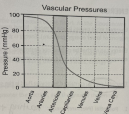
There is a significant drop in _____ at the arteriole/capillary level due to increased _____.
Why? Because there needs to be a decreased _____ and slowing of fluid at the level of the capillaries in order for nutrient exchange to happen effectively. Also, capillaries have very thin walls and would not be able to sustain high _____ or high _____.
pressure, resistance, pressure, pressure, velocity
Blood will move from one point to another in response to differences in “_____” energy
Total energy = _____ energy + _____ energy
_____ = stored or resting energy
_____ = work or energy of motion
total, potential, kinetic, potential, kinetic
_____ energy in the vascular system is primarily represented by intravascular pressure, which distends vessels. This pressure is supplied by the contraction of the heart.
Potential
_____ energy in the vascular system is presented by the velocity of moving blood.
Kinetic
Realistically there is always some energy “loss” in the vascular system as a result of _____ and _____.
viscosity, inertia
Resistance to flow, blood thickness; increases with increased hematocrit (concentration of RBCs)
Viscosity
Law of motion; occurs when blood is forced to change direction or velocity
Inertia
The slower the rate of flow, the higher the pressure, and the faster the rate of flow, the lower the pressure.
P = 4v2
Q = V x A
Bernoulli’s Principle
Velocity and pressure in the vascular system (Bernoulli) are _____ related.
inversely
We describe the type of _____ in a vessel based on how the layers of blood are traveling relative to each other.
flow
A type of flow where all of the layers flow in parallel lines.
Has two types:
Parabolic
Steady/Plug
Laminar

Type of laminar flow; layers along the vessel wall are slower due to friction, middle layers are faster
Parabolic
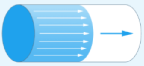
Type of laminar flow; all layers moving at the same velocity
Steady/Plug
Non-laminar flow; chaotic
_____ = areas of swirling flow (helical flow)
Turbulent, Eddies
Velocity variations with the cardiac cycle, which cause the vessel diameter to expand or contract affecting flow direction; this type of flow reflects flow above and below the pulse wave baseline
Typical of arterial flow
Pulsatile
Velocity variations with respiration
Typical of venous flow
Phasic

What causes the difference between plug and parabolic flow?
Friction
The smaller the vessel, the _____ the friction
GREATER
Turbulent flow occurs when _____ _____ is exceeded
_____ _____ is an abstract, dimensionless number that is used to determine when flow will become turbulent
_____ or greater = flow is turbulent
Reynold’s number, Reynold’s number, 2000

Turbulent flow is often seen _____ to a significant stenosis or at a vessel _____
distal, bifurcation
_____ _____ (_____): How much blood is moving
Milliliters per minute OR liters per minute
_____ (_____): How fast the blood is moving (speed); rate of change in position over time
Centimeters or meters per second
_____ (_____): Cross sectional dimension of a vessel
mm or cm squared
Flow Volume, Q, Velocity, V, Area, A
Defines the relationship between flow, pressure gradient, vessel radius, vessel length, viscosity
Poiseuille’s Law
According to Poiseuille’s Law, the change in pressure and vessel radius are _____ related to flow.
directly
According to Poiseuille’s Law, the viscosity and vessel length are _____ related to flow.
inversely
What factor has the GREATEST effect on flow?
Vessel radius
Poiseuille’s Law:
Q = ΔPπr4 / 8Lη
Vasoconstriction – vessel contracts
Radius _____
_____ resistance to flow
Vasodilation – vessels relax or dilate
Radius _____
_____ resistance to flow
decreases, MORE, increases, LESS
Resistance Equation:
R = 8Lη / πr4
Resistance and flow are _____ related.
inversely
The highest point of a pulse wave tracing (flow profile)
Peak systolic velocity (PSV)
Where the pulse wave tracing (flow profile) stops before a new waveform begins
End diastolic velocity (EDV)
The time it takes to get to peak systole from one waveform to the next
You would measure from the diastole of the first waveform to the peak systole of the next waveform
Acceleration (AC) time
A calculated measurement that assesses blood flow resistance within an artery
It essentially means more or less variation between peak systole and end diastole
Resistive Index (RI)
A higher resistance waveform is going to have a _____ resistive index (RI)
A lower resistance waveform is going to have a _____ resistive index (RI)
higher, lower
Resistive Index (RI) formula:
RI = (PSV - EDV) / PSV
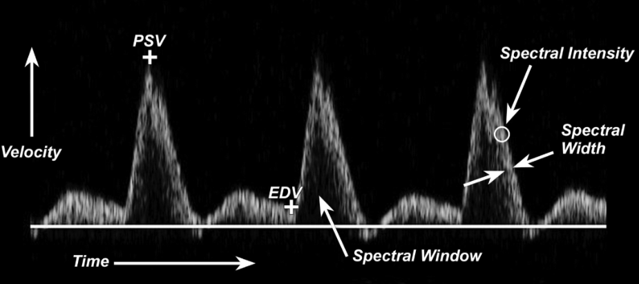
We can tell if flow is turbulent or laminar based on the _____ _____
The more open (black) the spectral window, the _____ turbulent the flow
The more filled in the spectral window, the _____ turbulent the flow
We can tell how much blood we are sampling (how much blood is going through our pulse wave sample gate) based on how _____ the white part of the tracing is (spectral width)
A thicker tracing = a _____ gate
A thinner tracing = a _____ gate
spectral window, less, more, thick, wider, smaller
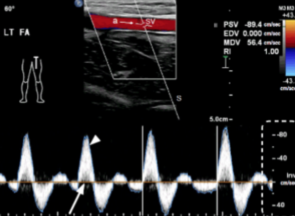
Does the image show pulsatile or phasic flow?
Pulsatile (Variable velocity and direction based on the cardiac cycle)

Does the image show pulsatile or phasic flow?
Phasic (Continuous/steady flow that is dependent on respiratory variations)
A surgical procedure to remove fatty plaque buildup from the inside of a blocked or narrowed artery
Endarterectomy
The CCA has a _____ resistance when it’s closer (proximal) to the heart, BUT we will characterize it as a _____ resistance vessel.
higher, low
High or low resistance?
ICA =
Vertebral Arteries =
ECA =
low, low, high
The _____ of the CCA is where the CCA is slightly bigger before it bifurcates into the _____ and _____
bulb, ICA, ECA
True or false: You should pulse wave the bulb of the CCA
False (unless pathology is visualized in the bulb)
Differentiating ICA/ECA
The ICA is usually _____, _____ resistance, usually always has color fill since it feeds the _____ (constantly _____), and is usually more _____
The ECA is usually _____, _____ resistance, typically has a _____ moment where it doesn’t fill with color (PULSES/FLASHES) because it doesn’t have constant _____, and is typically more _____
The ECA has _____ branches (outside the brain)
The ICA branches once it is inside the _____, so we wouldn’t see those
***Vascular anatomy is notorious for not being like the textbook, meaning the ICA and ECA can switch on which one is more medial and which one is more lateral
larger, low, brain, perfusing, lateral, smaller, high, small, perfusion, medial, extracranial, brain
Carotid Interpretation % Stenosis Grading Chart
Normal values with NO stenosis (ICA PSV, Plaque Estimate, ICA/CCA Ratio, ICA EDV)
<180 cm/s, None, <2.0, <40 cm/s
Carotid Interpretation % Stenosis Grading Chart
Values with <50% stenosis (ICA PSV, Plaque Estimate, ICA/CCA Ratio, ICA EDV)
<180 cm/s, <50%, <2.0, <40 cm/s
Carotid Interpretation % Stenosis Grading Chart
Values with 50-69% stenosis (ICA PSV, Plaque Estimate, ICA/CCA Ratio, ICA EDV)
180-230 cm/s, >50%, 2.0-4.0, <100 cm/s
Carotid Interpretation % Stenosis Grading Chart
Values with ≥ 70% stenosis but < near occlusion (ICA PSV, Plaque Estimate, ICA/CCA Ratio, ICA EDV)
>230 cm/s, >50%, >4.0, >100 cm/s
Carotid Interpretation % Stenosis Grading Chart
Values with near occlusion (ICA PSV, Plaque Estimate, ICA/CCA Ratio, ICA EDV)
High/low/undetectable, visible, variable, variable
Carotid Interpretation % Stenosis Grading Chart
Values with total occlusion (ICA PSV, Plaque Estimate, ICA/CCA Ratio, ICA EDV)
Undetectable, no visible lumen, N/A, N/A
Use a _____ than _____ box for color!
taller, wide
Where is the Circle of Willis located?
The base of the brain
What are the first branches of the aorta?
Coronary arteries
What are the two branches of the brachiocephalic (innominate) artery?
Right common carotid artery, right subclavian artery
The vertebral arteries are a branch off of the _____ arteries on both the right and left side!
subclavian
Common carotid arteries (CCA) become the _____ and _____ carotid arteries
ICA – primary blood supply to the _____
_____% of CCA blood
Typically _____ vessel (width)
NO _____ branches!
The first branch of the ICA is the _____ artery
Typically located _____
_____ resistance
ECA – primary blood supply to the _____, _____, and _____
_____ extracranial branches
Typically _____ vessel (width)
The first branch of the ECA is the _____ _____ artery
Typically located _____
_____ resistance
internal, external, brain, 80, bigger, extracranial, ophthalmic, laterally, Low, face, neck, scalp, 8, smaller, superior thyroid, medially, High
The ophthalmic artery supplies the _____ artery of the _____
The ophthalmic artery branches into the _____, _____, and _____ arteries
retinal, eye, Supraorbital, Frontal, Nasal

The ICA is divided into 3 segments:
_____ segment – extends from CCA bifurcation to the carotid canal of the petrous portion of the temporal bone
_____ segment – Courses through the petrous portion of the temporal bone and is inaccessible to ultrasound (US)
_____ (_____ _____) segment – consists of three areas: parasellar, genu (bend), supraclinoid
Cervical, Petrous, Cavernous, carotid siphon

Label the following ICA segments (1 → 7):
Cervical, Petrous, Cavernous, Parasellar, Genu, Supraclinoid, Ophthalmic artery