Cells, Tissues, and Cardiovascular Anatomy Flashcards
 Call Kai
 Learn
Learn Practice Test
 Spaced Repetition
 Match
Match Flashcards
 Knowt Play
1/95

Earn XP
Description and Tags
Vocabulary terms covering basic human anatomy, anatomical directions and planes, cardiac structure, circulation pathways, heart function metrics, ECG interpretation, medical devices, and cardiovascular pathologies.
Name | Mastery | Learn | Test | Matching | Spaced | Call with Kai | Chat |
|---|
No analytics yet
Send a link to your students to track their progress
96 Terms
Cell
The basic structural and functional unit of the human body.
Tissue
A group of similar cells that work together to perform a specific function.
Connective Tissue
Tissue that supports, connects, protects, or binds other tissues and organs; examples include bone, blood, cartilage, and adipose tissue.
Epithelial Tissue
Tissue that covers body surfaces, lines organs and cavities, and forms glands.
Muscular Tissue
Specialized tissue that contracts to produce movement, including skeletal, cardiac, and smooth muscle.
Organ
A structure made of two or more tissue types that performs a specific function.
Inferior
Toward the feet or below another structure.
Superior
Toward the head or above another structure.
Anterior
Toward the front of the body.
Posterior
Toward the back of the body.
Dorsal
Toward the back side of the body.
Ventral
Toward the front or belly side of the body.
Medial
Toward the midline of the body.
Lateral
Away from the midline of the body.
Proximal
Closer to the point of attachment or origin of a body part.
Distal
Farther from the point of attachment or origin of a body part.
Transverse Plane
A horizontal plane that divides the body into superior and inferior portions.
Coronal Plane
A vertical plane that divides the body into anterior and posterior portions.
Sagittal Plane
A vertical plane that divides the body into left and right portions.
Pulmonary Loop
The pathway of blood between the heart and lungs for gas exchange.
Systemic Loop
The pathway of oxygenated blood from the heart to the body and the return of deoxygenated blood back to the heart.
Right Atrium
Receives deoxygenated blood from the superior and inferior vena cava and the coronary sinus.
Right Ventricle
Pumps deoxygenated blood to the lungs through the pulmonary artery.
Left Atrium
Receives oxygenated blood from the pulmonary veins.
Left Ventricle
Pumps oxygenated blood to the entire body through the aorta.
Atrial Septum
The wall separating the right and left atria.
Ventricular Septum
The wall separating the right and left ventricles.
Atrioventricular (AV) Valves
Valves located between the atria and ventricles that prevent blood from flowing backward into the atria.
Tricuspid Valve
The right atrioventricular valve, located between the right atrium and right ventricle.
Mitral Valve
The left atrioventricular valve, located between the left atrium and left ventricle.
Semilunar Valves
Valves between the ventricles and major arteries that prevent blood from flowing back into the ventricles.
Pulmonic Valve
The valve between the right ventricle and pulmonary artery.
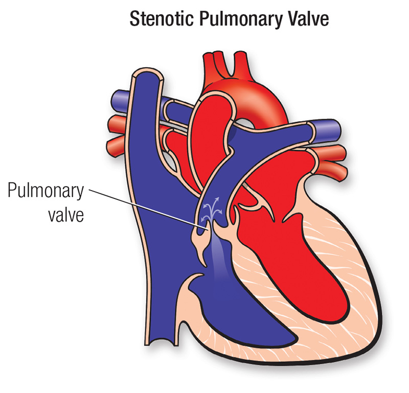
Aortic Valve
The valve between the left ventricle and aorta.
Pericardium
A protective sac surrounding the heart.
Epicardium
The outer layer of the heart wall.
Myocardium
The thick muscular middle layer of the heart that contracts to pump blood.
Endocardium
The thin inner lining of the heart chambers and valves.
Blood Vessels
Tubes that transport blood throughout the body.
Arteries
Blood vessels that carry blood away from the heart; most carry oxygenated blood.
Arterioles
Small branches of arteries that regulate blood flow into capillaries.
Capillaries
Tiny blood vessels where oxygen, nutrients, carbon dioxide, and waste products are exchanged between blood and tissues.
Veins
Blood vessels that carry blood toward the heart; most carry deoxygenated blood.
Cardiac Conduction System
A group of specialized cells that generates and conducts electrical impulses to control the heartbeat.
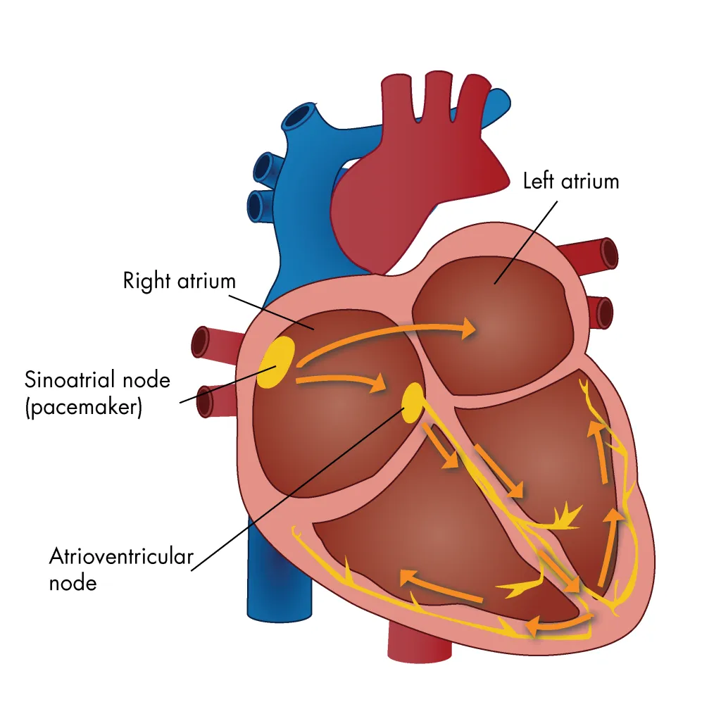
Sinoatrial (SA) Node
The heart's natural pacemaker which initiates the electrical impulse that causes the atria to contract.
Atrioventricular (AV) Node
Receives the electrical impulse from the atria and briefly delays it before sending it to the ventricles.
Stroke Volume (SV)
The amount of blood pumped by one ventricle during one heartbeat.
Preload
The amount of stretch in the ventricular muscle fibers before contraction, closely related to ventricular filling.
Afterload
The resistance the ventricle must overcome to eject blood.
Contractility
The strength of contraction of the heart muscle.
Heart Rate (HR)
The number of heartbeats per minute.
Cardiac Output (CO)
The amount of blood pumped by the heart in one minute.
Cardiac Output Formula
CO=Stroke Volume×Heart Rate
Normal Cardiac Output
Approximately 4–8/min in adults at rest, depending on the individual.
ECG / EKG
A test that records the electrical activity of the heart.
ECG Strip
A visual recording of the heart's electrical activity over time.
P Wave
Represents atrial depolarization, which leads to atrial contraction.
QRS Complex
Represents ventricular depolarization, which leads to ventricular contraction.
T Wave
Represents ventricular repolarization, when the ventricles electrically recover.
Normal Sinus Rhythm (NSR)
A normal heart rhythm originating from the SA node, generally with a heart rate of 60–100 beats per minute.
12-Lead ECG
A diagnostic test that views the heart's electrical activity from 12 different perspectives.
V1–V6
The six precordial or chest leads that examine the heart's electrical activity from different locations across the chest.
RA
Right arm electrode.
LA
Left arm electrode.
RL
Right leg electrode, usually used as the ground electrode.
LL
Left leg electrode.
Pacemaker
A device that uses electrical impulses to help maintain an appropriate heart rate and rhythm.
Permanent Pacemaker
A surgically implanted device that provides long-term electrical stimulation to the heart when the natural conduction system is inadequate.
Temporary Pacemaker
A short-term pacing system used when an abnormal heart rhythm is expected to be temporary or while waiting for a permanent device.
Implanted Cardioverter-Defibrillator (ICD)
An implanted device that detects dangerous ventricular rhythms and can deliver electrical therapy to restore a normal rhythm.
Left Ventricular Assist Device (LVAD)
A mechanical pump that helps the left ventricle pump blood to the body, often used in severe heart failure.
Ischemia
Inadequate blood flow and oxygen delivery to a tissue.
Embolus
A substance, such as a blood clot, that travels through the bloodstream and may block a blood vessel.
Thrombus
A blood clot that forms and remains attached within a blood vessel or the heart.
Aneurysm
A weakened area of a blood vessel wall that bulges or enlarges.
Atherosclerosis
A disease in which fatty deposits and other substances build up within artery walls.
Trauma
Physical injury caused by an external force.
Infarction
Tissue death caused by an interruption of blood supply and oxygen.
ASCVD
Atherosclerotic Cardiovascular Disease, caused by the buildup of atherosclerotic plaque in arteries.
Plaque
A buildup of fats, cholesterol, calcium, and other substances within an artery wall.
Coronary Artery Disease (CAD)
Narrowing or blockage of the coronary arteries, reducing blood flow to the heart muscle.
Peripheral Artery Disease (PAD)
Narrowing or blockage of arteries supplying the limbs, especially the legs.
Stenotic
Abnormally narrowed.
Rupture
A break or tear in a structure, such as a blood vessel or aneurysm.
Superimposed Thrombus
A blood clot that forms on top of an existing abnormality, such as an atherosclerotic plaque.
Ventricular Tachycardia (V-Tach)
A rapid heart rhythm originating in the ventricles that can reduce the heart's ability to pump blood.
Bradycardia
An abnormally slow heart rate, generally below 60 beats per minute in adults.
Atrial Fibrillation (A-Fib)
An irregular and often rapid rhythm caused by disorganized electrical activity in the atria.
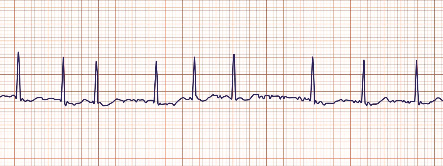
Ventricular Fibrillation (V-Fib)
A chaotic electrical rhythm in which the ventricles quiver instead of contracting effectively, requiring immediate emergency treatment.
Pulseless Electrical Activity (PEA)
Electrical activity is present on the ECG, but the heart is not producing an effective pulse or circulation.
Asystole
The absence of detectable electrical activity in the heart, commonly called a flatline.
Heart Failure
A condition in which the heart cannot pump enough blood to meet the body's needs.
Pulmonary Congestion
Excess blood and fluid buildup in the lungs, often caused by increased pressure from left-sided heart failure.
Edema
Abnormal accumulation of fluid in body tissues.
Hemophilia
An inherited bleeding disorder caused by a deficiency or dysfunction of certain clotting factors.
Coagulation Factors
Proteins in the blood that work together in a series of reactions to form a blood clot.
Sickle Cell Disease
An inherited disorder in which abnormal hemoglobin causes red blood cells to become rigid and sickle-shaped.