Pathology of the skin
1/113
There's no tags or description
Looks like no tags are added yet.
Name | Mastery | Learn | Test | Matching | Spaced |
|---|
No study sessions yet.
114 Terms
State two limitations of a biopsy.
Cannot determine cause of allergies
Cannot determine precise endocrinopathy
State and define the condition
Congenital hypotrichosis is the developmental alopecic disorder with hair loss, that can occur at birth or develop around 1 month of age, affecting head, trunk, ventrum and accompanied by abnormal dentition.
State and define the condition
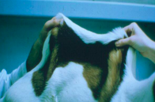
Collagen dysplasia is a complex group of connective-tissue disorders that results in the reduced tensile strength of affected tissues.
State the condition and the affected species
Epidermolysis bullosa- A group of diseases characterized by blistering of the skin due to defects in the proteins that anchor the epidermis to the dermis (e.g., collagen VII or keratin). The pathogenesis is a genetic defect in collagen, basement membrane, or keratin.
Affected species: Canine (Collie), Ovine (Suffolk & South Down)
What are the three forms of Epidermolysis bullosa (EB)?
Simplex (intra-epidermal split)
Junctional (split at the dermo-epidermal junction)
Dystrophic (split below the dermo-epidermal junction)
State the condition and the affected species.
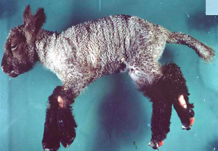
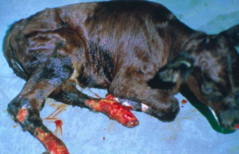
Epitheliogenesis imperfecta- A congenital disease characterized by incomplete formation of the epidermis in localized areas (usually legs and feet) and hoof/claw defects in affected areas.
Affected species: Equine, Bovine, Swine
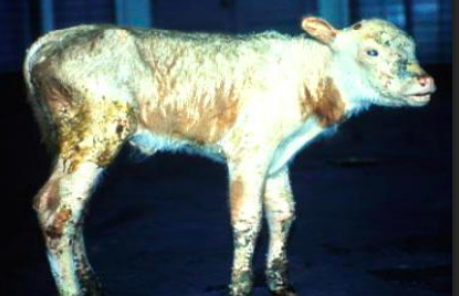
What is Ichthyosis (Fish-Scale Disease), its gross appearance and species affected?
A disease of epidermal differentiation. It is a congenital, inherited disease resulting in a build-up of keratin (hyperkeratosis).
Gross lesions include thick, horny scales, often described as "fish-scale" skin.
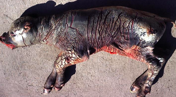
Chianina calf

State the condition, lesions, species affected and clinical signs
Lethal Acrodermatitis: An inherited disease characterized by marked parakeratosis of all footpads.
Dogs usually die of bronchopneumonia at around 15 months of age.
Bull Terriers
What factors determine the extent of solar damage?
Thickness of the keratin layer
Amount of protective hair
Degree of pigmentation.
Compare acute vs. chronic solar damage
Acute solar dermatitis (sunburn) is uncommon in animals without prior photosensitization.
Chronic solar exposure causes solar elastosis and fibrosis (thickened, wrinkled skin) and various tumours (SCCs, Hemangiomas/Hemangiosarcomas, Melanomas).
What is photosensitization and its basic pathogenesis?
It is caused by long-wavelength UV radiation activating photodynamic chemicals deposited in the skin, leading to free radical damage.
Describe Type I Photosensitization (Primary).
Caused by ingesting pre-formed photodynamic substances (e.g., helianthrone or furocoumarin from St. John's Wort) or drugs converted to photoreactive metabolites (e.g., phenothiazine or tetracycline).
Describe Type II Photosensitization.
Caused by abnormal porphyrin metabolism (accumulation of photodynamic agents). Examples: bovine congenital porphyria and bovine erythropoietic porphyria (also seen in pigs and cats).
Describe Type III Photosensitization (Hepatogenous).
Caused by liver damage (e.g., due to sporidesmin in sheep/cattle). The liver cannot excrete Phylloerythrin (a chlorophyll breakdown product), which then deposits in the skin and causes damage when exposed to UV light.
What are the chronic lesions associated with chronic sun exposure?
Solar elastosis and fibrosis (thickened, wrinkled skin). Neoplasms including SCCs (cattle, horses, sheep, cats), Hemangiomas/Hemangiosarcomas (dogs), and Melanomas (goats).
What are the key features of Acral Lick Dermatitis (ALD)?
Involves acral areas (extremities). Caused by excessive licking (considered a canine variant of obsessive-compulsive disorder).
Results in thickened skin and recurrent bacterial infections. Treatment focuses on behavioural modification.
What are the features of Psychogenic Alopecia in cats?
A behavioral disease in young to middle-aged nervous female cats. Characterized by chronic grooming leading to alopecia without changes in the epidermis or dermis. Typically affects the ventral abdomen and medial thighs.
What is Callus and what is its secondary complication?
Seen mostly in large breed dogs over bony prominences. Caused by hard bedding. Fibrosis leads to follicular plugging, which predisposes the area to bacterial furunculosis and callous pyoderma.
What is Skin Fold Dermatitis (Intertrigo) and its mechanism?
Occurs whenever two skin surfaces are in apposition (e.g., lips, tail, vulva, udder/thigh). The combination of friction and moisture causes a secondary bacterial infection.
What are the three main categories of skin defenses?
1. Physical barrier (hair shafts, keratin).
2. Chemical barrier (fatty acids, immunoglobulin).
3. Microbial barrier (resident bacteria prevent colonization).
What is a superficial bacterial skin infection?
Infection confined to the epidermis (or follicular epidermis). Neutrophilic pustules are visible histologically. Crusts form from dead neutrophils. Most do not produce systemic disease and respond readily to therapy.
What is Superficial Pustular Dermatitis? (bacterial infection)
Also called impetigo. It is most common in young animals.
What is Superficial Canine Pyotraumatic Dermatitis?
Also called hot-spot dermatitis. It is initiated by self-trauma (e.g., due to allergies). It is a focal area that is usually intensely painful. Hot weather and a dense coat predispose dogs to this condition.
What is Superficial Dermatophilosis and its pathogenesis?
Caused by Dermatophilus congolensis. Also called mud fever or lumpy wool.
Chronic dampness damages the epidermis, allowing the bacteria to colonize.
Hallmark: Alternating layers of neutrophils and keratin colonized by 'train-tracks' of bacteria.
What is Superficial Exudative Epidermatitis of pigs?
Also known as Greasy Pig Disease. Abrasions are infected by Staphylococcus hyicus in piggeries with poor hygiene. Causes marked exudative, malodorous lesions that spread to cover the entire body. It can be fatal in neonates but is typically mild in older piglets.
What is Superficial Fleece Rot?
Long fleece traps moisture, allowing colonization by Pseudomonas aeruginosa.
What is Superficial Equine Pastern Dermatitis?
Chronic damp housing allows bacterial infection of the pastern and heel by multiple bacterial species. Improved housing prevents recurrence
What defines a deep bacterial skin infection?
Bacteria are present within the dermis. They are less common and more difficult to treat than superficial infections. Can cause regional lymphadenitis and often result in residual scarring.
What is the pathogenesis of Staphylococcal Folliculitis and Furunculosis?
Trauma (around pressure points) causes blocked follicles. Blocked follicles become infected and rupture, releasing bacteria into the dermis. German Shepherd dogs are predisposed (likely due to an IgA deficiency).
What is a Cat Bite Abscess?
Oral bacteria are deposited into the dermis by puncture wounds from a cat bite.
What are the features of Mycobacterial infection?
Associated with Granulomatous dermatitis. Feline leprosy is probably due to mouse bites. Often recurs following excision, and has a guarded prognosis.
What is Botryomycosis?
Most often a staphylococcal infection due to secondary wound infection or poor hygiene (e.g., equine castration wounds). Hallmark is 'sulphur granules' (bacteria surrounded by antibody) visible grossly.
What is the pathogenesis and lesion of Pox Viruses?
Pox viruses cause proliferative, crusting, and ulcerative lesions. They are important causes of disease in birds (Avian pox) and can cause umbilicated lesions (e.g., Lumpy Skin Pox in cows/humans).
What are the features of Parapox Viruses (Contagious Ecthyma)?
Also called scabby mouth. Affects younger sheep and goats. Virus invades via abrasions (rough herbage) around the mouth and eyes (sometimes udder). Causes proliferation of the epidermis with marked crusting. Zoonotic infection is called 'orf'.
What is Papular Stomatitis?
A Parapox virus causing stomatitis in calves and mammary papillitis in adults. Zoonotic disease known as 'milker's nodule'.
What are the features of Papillomavirus?
Causes bovine fibropapillomas (common on penis/vulva) and canine oral papillomatosis. Histologically, shows proliferative fronds of epidermis supported by dermal folds. Equine sarcoids are caused by bovine papillomavirus.
How are fungal skin diseases classified?
Cutaneous (fungi restricted to the surface/follicular keratin)
Subcutaneous (fungi in the dermis)
Systemic (multiple organ involvement)
What are the features of Dermatophytosis (Ringworm)?
Microsporum canis is the most common cause. Fungi cause lysis of the hair shaft. It is seen in young animals crowded in a humid environment. Lesions show focal crusting and alopecia. It is a zoonotic disease.
What is Canine Malassezia Dermatitis?
Caused by an overgrowth of Malassezia (normal ear inhabitant) often predisposed by seborrhea. Common complaints include increased odor. Affects the ventrum, preauricular areas, and feet. 'Peanut' yeasts are visible on ear swabs.
List common Subcutaneous fungal infections.
Fungal mycetomas (dermal granulomas with fungal hyphae) in Persian cats.
Cryptococcus neoformans in cats (rarely confined to the skin).
What is the significance of Parasitic Skin Disease?
Very common in practice. Treatment is a large source of income. It is a common cause of allergic dermatitis in companion animals.
What are the features of Demodicosis (Demodex canis)?
Normal inhabitants of hair follicles.
Disease develops when the immune system cannot suppress mite numbers.
More often in young animals.
If generalized in older animals, it may be secondary to an underlying immunosuppressive disease.
Initial lesions are alopecic crusting with mild/no pruritus.
What are the hallmarks and diagnosis of severe Demodicosis?
Severe cases develop furunculosis and secondary pyoderma. There is a genetic component (affected animals should not be bred). Diagnosis is by skin scraping.
What are the features of Sarcoptic Mange (Sarcoptes scabiei)?
Causes intensely pruritic skin disease. It is a true contagious disease (no underlying disease required) with zoonotic potential. Mites live in surface keratin. Self-trauma lesions initially involve the ears, head, and neck. Responds well to treatment.
What are the features of Fleas?
Common cause of allergic dermatitis in cats and dogs (dorso-lumbar area, base of tail). Heavy burdens rarely cause anaemia in young animals.
What are the features of Chorioptic Mites?
Live on the surface causing crusting and thickening. Affects the lower limbs, scrotum, perineum, and udder.
What are the features of Flies as a cause of disease?
Can cause allergies in cats (mosquitos) and horses (stable flies). Causes Myiasis (fly-strike in sheep).
What is Pythiosis (Swamp Cancer)?
Caused by Pyhtium insidiosum. It results in a Pyogranulomatous dermatitis in horses.
What is Diamond Skin Disease?
Caused by Erysipelothrix rhusiopathiae. Vasculitis causes cutaneous infarcts. Also causes fibrinous and proliferative arthritis and vegetative valvular endocarditis.
What are the features of Scrapie in relation to skin?
Causes alopecia secondary to the infection (due to rubbing/itching).
What are the two main types of immune-mediated skin disease?
1. Allergic (Hypersensitivity) disease (Prevalent in companion animals).
2. Autoimmune disease (Extremely rare, less than 2% of cases).
What is Allergic Skin Disease (Hypersensitivity)?
An abnormally severe reaction against an antigen (allergen) caused by excessive production of IgE.
What are the three unique manifestations of Feline Allergic Dermatitis
Miliary dermatitis.
Feline eosinophilic granuloma.
Feline eosinophilic plaque.
What are the features of Miliary Dermatitis (Cats)
Multiple small, hard nodules especially over the dorsum.
Histologically: intraepidermal eosinophilic and neutrophilic pustules covered by a serocellular crust.
What are the four main groups of allergens in order of frequency (dogs/cats)
Flea.
Atopy (environmental).
Food.
Contact allergies.
What are the features of Atopy (Environmental Allergy)
Caused by environmental allergens (similar to asthma in humans). Often has a seasonal pattern. Atopic animals are classically foot-chewers and face rubbers.
Non-seasonal and tends to develop in younger animals. Least responsive to corticosteroids. Can also have gastrointestinal signs.
Rare (1.4% of cases). Caused by the production of antibodies against self-antigens. Histology is required for diagnosis, with sampling of developing lesions being important.
What is Pemphigus Foliaceus and its features?
Autoimmune disease causing pustules in the superficial epidermis. Affects the face, ears, and feet.
Breed predilection in Oriental breeds (especially Chows).
Histologically: intact pustule contains non-degenerate neutrophils and acantholytic cells.
What is Pemphigus Vulgaris?
Autoimmune disease causing vesicles deep within the epidermis. Affects the oral cavity and mucocutaneous junctions.
What is Discoid Lupus Erythematosus (DLE) and its distribution?
Caused by lymphocytes activated by UV exposure. Affects the nasal planum. Predisposed breeds: Collies and German Shepherds
What is Systemic Lupus Erythematosus (SLE) and its mechanism?
Caused by systemic deposition of antigen-antibody complexes (Type III Hypersensitivity) due to antinuclear antigen (ANA). Leads to immune complex deposition in the glomeruli, skin, and joints.
What is Dermatomyositis?
An immune-mediated disease affecting Collies and Shelties. Lesions typically occur on the face and lips.
What is Uveodermatologic Syndrome (VKHS)?
An autoimmune response to melanin with a predominance of histiocytic cells. Leads to depigmentation and uveitis (which causes blindness if untreated). Affects Akitas and Chows mostly.
What is Erythema Multiforme?
A reaction with numerous causes, but most often triggered by drugs
What is Sebaceous Adenitis?
A disease most common in Standard Poodles. Lesions include crusting and alopecia, especially on the dorsum.
How are endocrinopathies in companion animals typically divided?
1. Hypothyroidism: Variable, non-diagnostic skin lesions; blood tests are more diagnostic than skin histology.
2. Non-thyroid-dependent endocrinopathies: All produce similar histological lesions; diagnosis depends on history, signalment, and ancillary tests.
What are the features and skin lesions associated with Hypothyroidism (dogs)?
It is the most common but most over-diagnosed canine endocrinopathy. Skin lesions are variable, likely secondary to altered skin defenses. May cause reduced hair growth after clipping, increased dermal mucin, and seborrhea.
What are the similar histological lesions shared by Non-Thyroid Endocrinopathies?
Follicles in telogen phase with loss of the hair bulb, and follicular atrophy and plugging.
What is Hyperadrenocorticism (Cushing's Disease) and its systemic signs?
An endocrinopathy that spares the head and extremities. Systemic signs include PU/PD (Polyuria/Polydipsia), muscle weakness leading to a pot-belly, polyphagia, and hepatomegaly.
What is Calcinosis Cutis?
Dystrophic calcification of the dermis that is strongly suggestive of Cushing's disease (Hyperadrenocorticism). It presents as crusting plaques within thickened skin.
Which dogs are affected by Cyclical Flank Alopecia?
Bulldogs and Boxers.
What is the most common cause of Sex Hormone Responsive Dermatosis?
Hyperestrogenism due to adrenal neoplasia in ferrets. It is also seen due to ovarian or testicular tumours in dogs.
What are the features of Growth-Hormone Responsive Dermatosis?
Lack of Growth Hormone (GH) in an adult animal can result in alopecia. Lack of GH can be due to adrenal or adenohypophyseal dysfunction. It is very difficult to diagnose (typically a diagnosis of exclusion).
How does starvation (nutritional disorder) affect hair/skin?
Hair is protein, so insufficient dietary protein prevents hair production.
Copper deficiency causes light hair colour and loss of wool crimp (steely wool in sheep).
Zinc deficiency results in parakeratosis (thickened, scaly skin).
What is Laminitis and what causes its pathological effects?
Inflammation of the hoof laminae. If severe, necrosis and edema prevent adhesion of the hoof epidermis to the dermis. This causes uncontrolled proliferation of the epidermis (malformation of the hoof) and allows underlying bones to rotate.
What is Primary Seborrhea and its pathogenesis?
An idiopathic disease (mostly dogs, especially Cocker Spaniels) with a genetic basis. It involves increased turnover of epidermal cells (excess keratin). This excess keratin allows bacteria and yeasts to proliferate, causing increased odor.
What is Secondary Seborrhea?
When increased epidermal turnover is caused by an underlying condition such as an allergy or endocrinopathy.
What is "Puppy Strangles" (Juvenile Sterile Granulomatous Dermatitis)?
An idiopathic disease that presents as nodular swelling in the dermis involving the face, ears, and mucocutaneous junction. Mandibular lymphadenopathy is common. It is thought to be immune-mediated because it responds to corticosteroids.
What is Plasma Cell Pododermatitis?
An idiopathic disease only in cats causing swelling of one or multiple paws. Cytology reveals lots of plasma cells. The primary lesions are not painful. Some cases resolve spontaneously, while others respond to corticosteroids.-
Why is the diagnosis and treatment of skin tumours important in practice?
They are the most important tumours in companion animal practice and a good source of income. Cytology is often useful for diagnosis, while histology is required to formulate a treatment plan and check margins (inking margins confirms complete surgical excision).
What is a Nevus (Non-neoplastic tumour)?
Focal adnexal hyperplasia. It can trap follicles, resulting in rupture and secondary inflammation. Excision is curative.
What is Sebaceous Gland Hyperplasia?
Presents as multiple wart-like masses, especially on Cocker Spaniels, Beagles, and Poodles. Most common on eyelids and limbs.
What is a Follicular Cyst?
Malformed follicles that fill up with keratin. Can cause inflammation if they burst. Animals often develop multiple cysts.
What is Calcinosis Circumscripta?
A non-neoplastic tumour presenting as nodules overlying extremities or in the mouth of young large breeds, especially German Shepherds.
What is a Histiocytoma?
The most common canine skin tumour. A proliferation of macrophages. Tumours spontaneously regress, a process accelerated by aspiration for cytology. Seen most commonly on the head and extremities of young dogs.
What are the features of a Lipoma?
The second most common canine tumour (also common in cats). Cytology enables diagnosis. Histology is required only if infiltrative lipoma is suspected.
What are the features of a Hemangioma?
Often solar-induced in dogs. Usually dark purple and clinically mistaken for melanomas. Consists of well-differentiated blood vessels.
What are the three main groups of skin neoplasms?
1. Epithelial neoplasms (from adnexa or epidermis).
Round cell neoplasms (from inflammatory cells: MCT, lymphosarcoma, plasmacytoma).
Mesenchymal neoplasms (from supporting cells: hemangiopericytoma, sarcoid, fibrosarcoma).
What are the features of Adnexal Tumours (Epithelial)?
Very common in cats and dogs. Can be from sebaceous or apocrine glands. Malignant neoplasms derived from adnexal structures are very rare.
What are Basal Cell Tumours (Trichoblastomas)?
Develop from follicular epithelial cells at the base of the follicle. Most common around the head. Cystic and pigmented subtypes are more common in cats. Basal cell carcinomas are very rare.
What are the features of Follicular Tumours (Epithelial)?
Most are benign. All contain keratin and can ulcerate if they rupture. Intracutaneous cornifying epithelioma develops on younger dogs.
What is the significance and pathogenesis of Squamous Cell Carcinoma (SCC)?
The most common malignant epithelial tumour. Induced by sunlight. Develops in non-pigmented areas (e.g., horses: vulva, prepuce; cats: ears, eyelids, nose). Often invasive, making complete excision difficult, but rarely metastasizes.