Correlative Imaging - Sonography
 Call Kai
 Learn
Learn Practice Test
 Spaced Repetition
 Match
Match Flashcards
 Knowt Play
1/27

There's no tags or description
Looks like no tags are added yet.
Name | Mastery | Learn | Test | Matching | Spaced | Call with Kai | Chat |
|---|
No analytics yet
Send a link to your students to track their progress
28 Terms
How are sonographic images created?
As sound waves enter the body, they interact with the tissue and body fluids. Depending upon the type of tissue, the sound waves either reflect, refract, or transmit.
Piezoelectric Effect
Energy conversion from pressure to electric and electric to pressure
Transducer
Changes one form of energy into another
Sound Wave
Expansion and compression of the piezoelectric crystal
Frequency
Measure of how many complete cycles of vibrations occur in one second
Measured in Hz
Range of transducer frequencies commonly used in diagnostic medical sonography (DMS)
1,000,000-20,000,000 Hz (1-20 MHz)
Key events in the history of sonography
1940's/1950's - WWII SONAR ultrasound
1970's - ultrasound introduced into medicine
1980's - Doppler
1990's - profession faced quality control and cost containment issues
2000's - 3D/4D
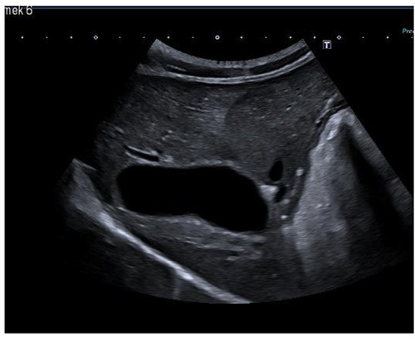
2D B-Mode
2-dimensional real-time or real-time gray-scale
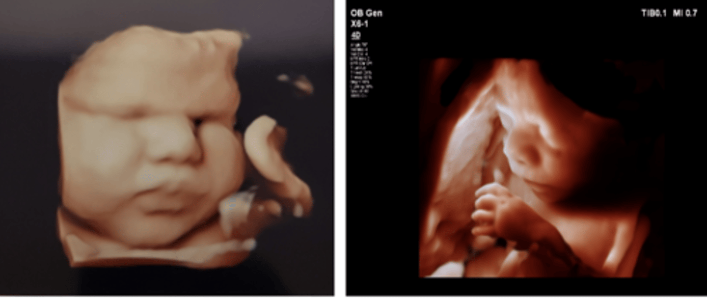
3D/4D Volume Imaging
3-dimensional/4-dimensional or volume imaging
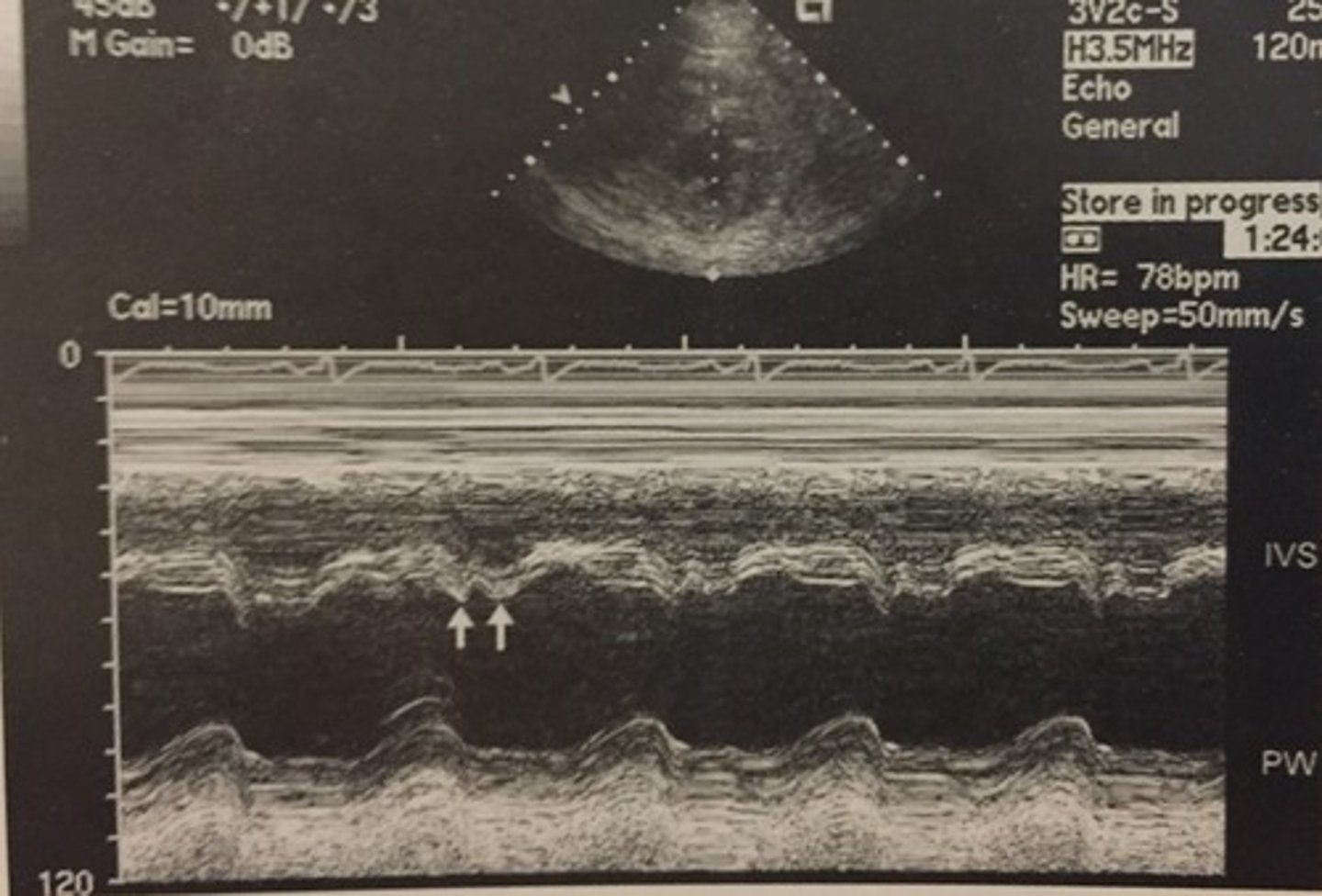
M-Mode
Motion mode
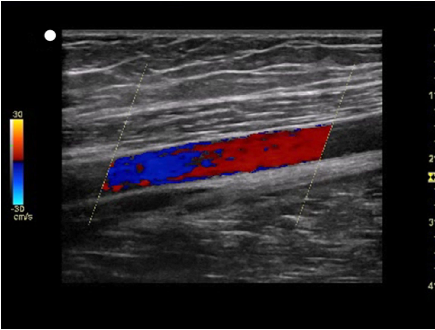
Color Doppler
Demonstrates presence of blood flow with color
Spectral Doppler
Estimates blood flow velocity
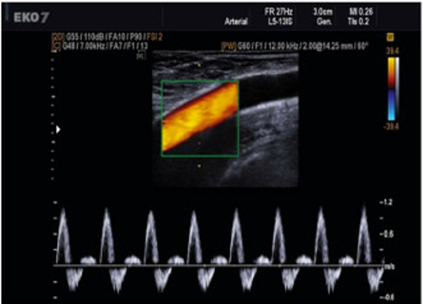
Sagittal Orientation
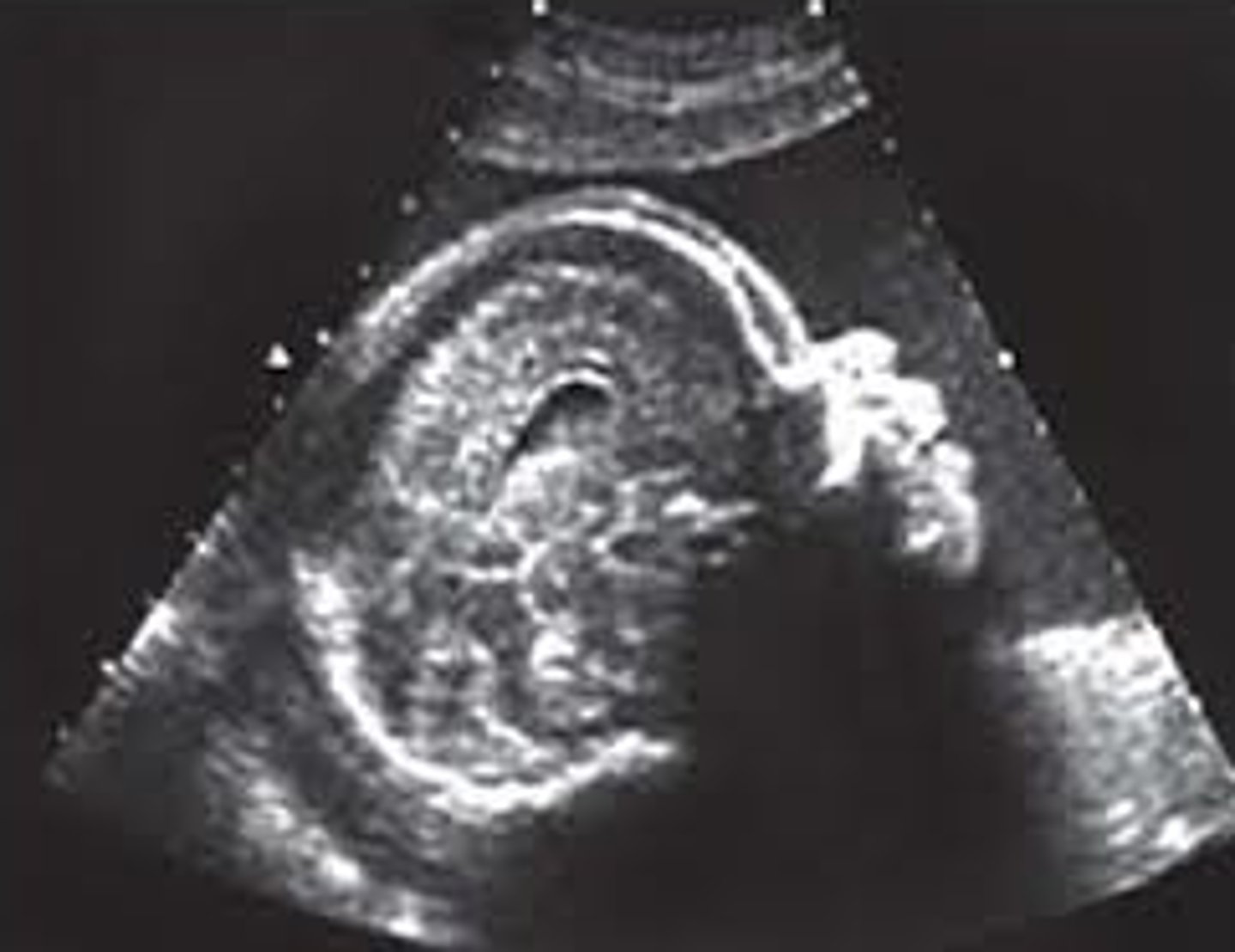
Transverse Orientation
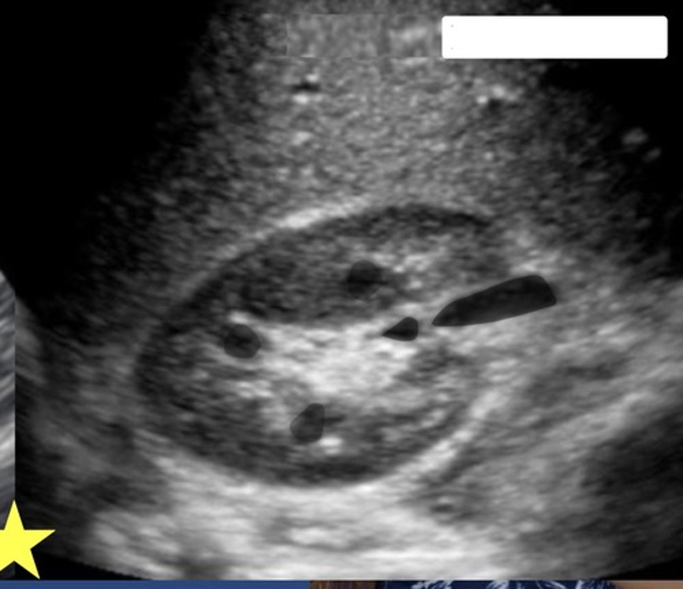
Coronal Orientation
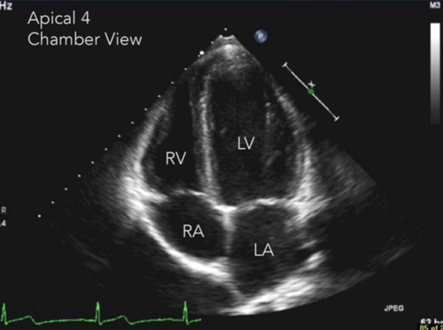
Echogenic
Capable of producing echoes
Anechoic
Without echoes
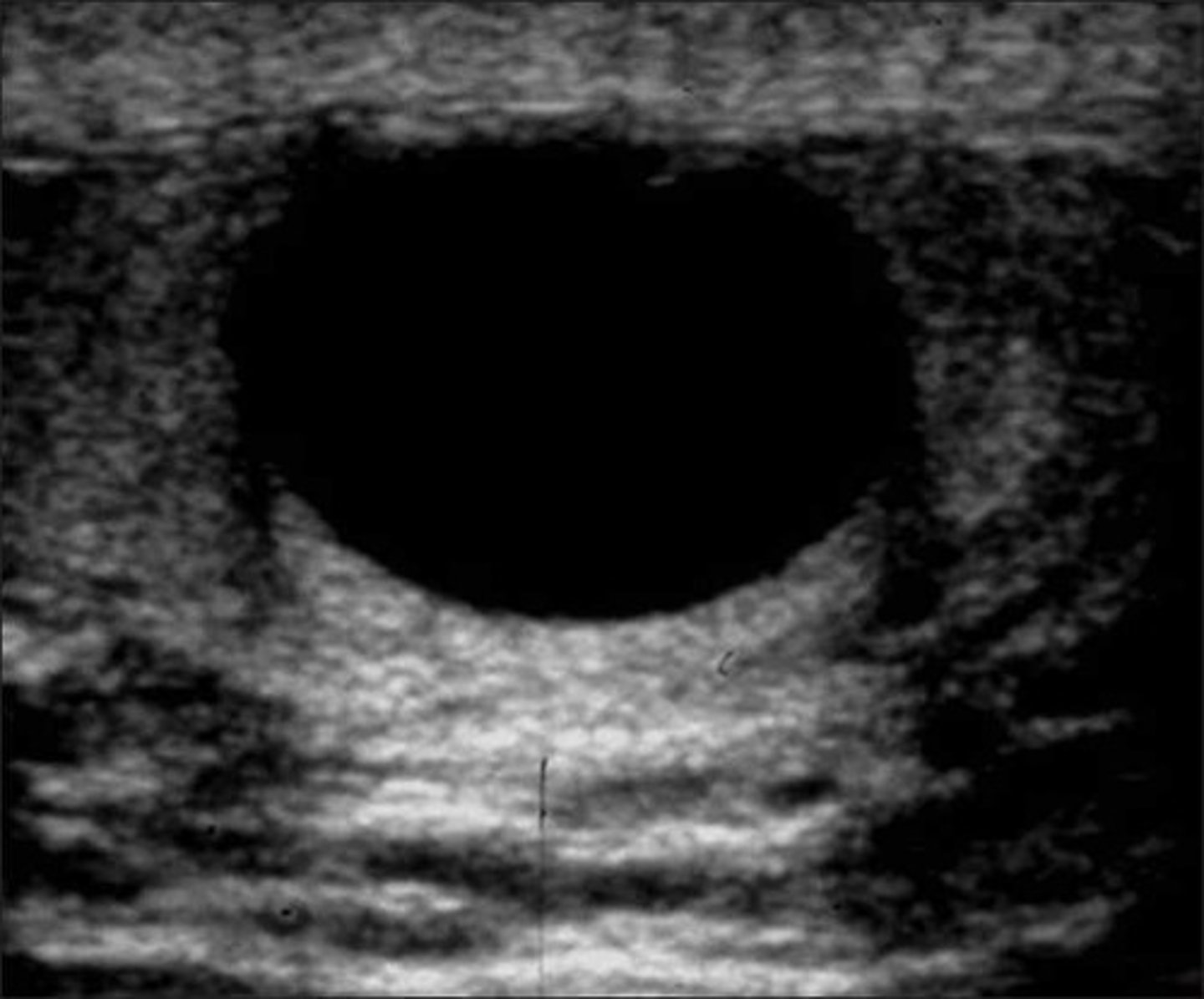
Hypoechoic
Low-level echoes
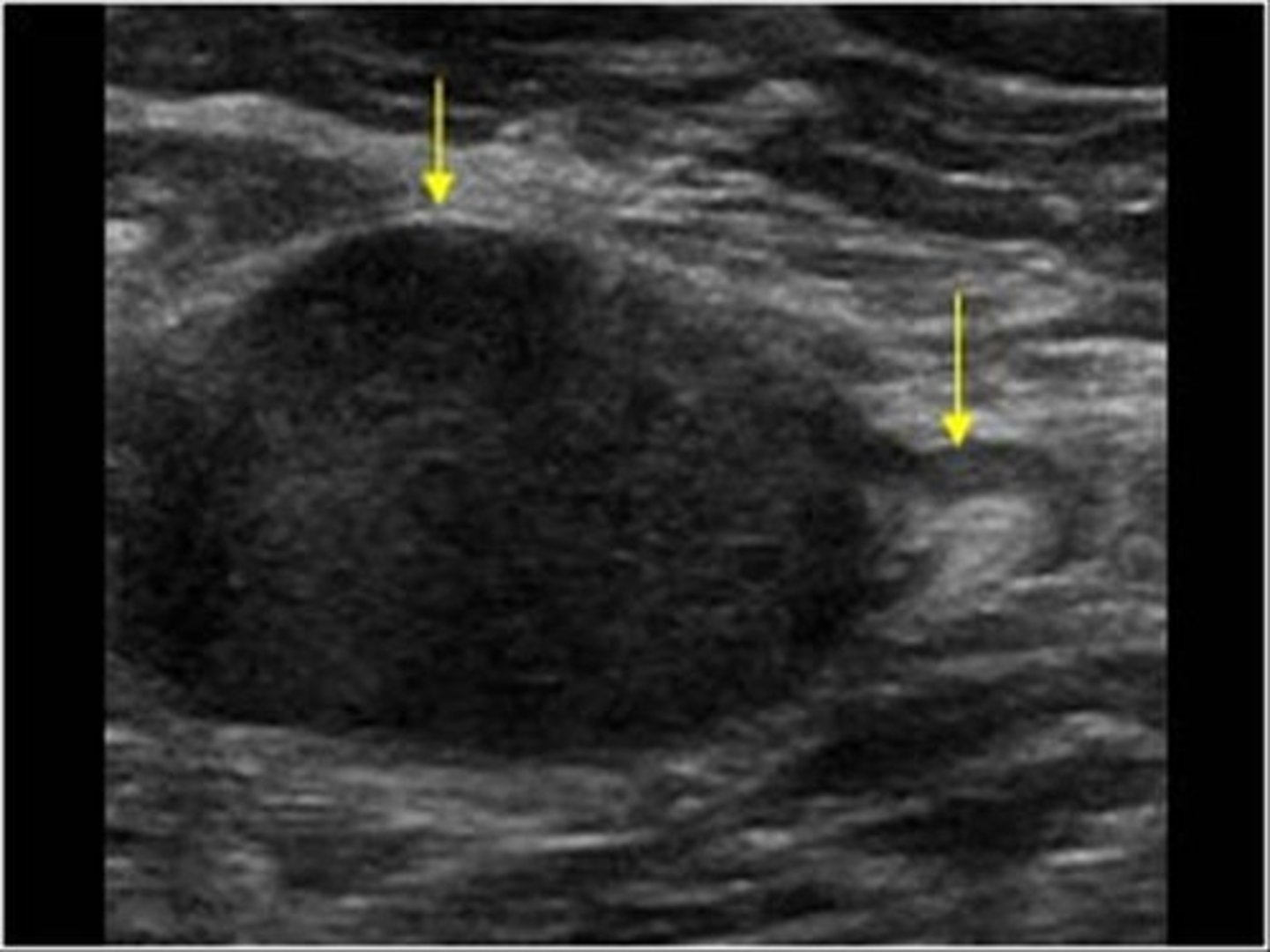
Hyperechoic
High-level echoes, bright, white
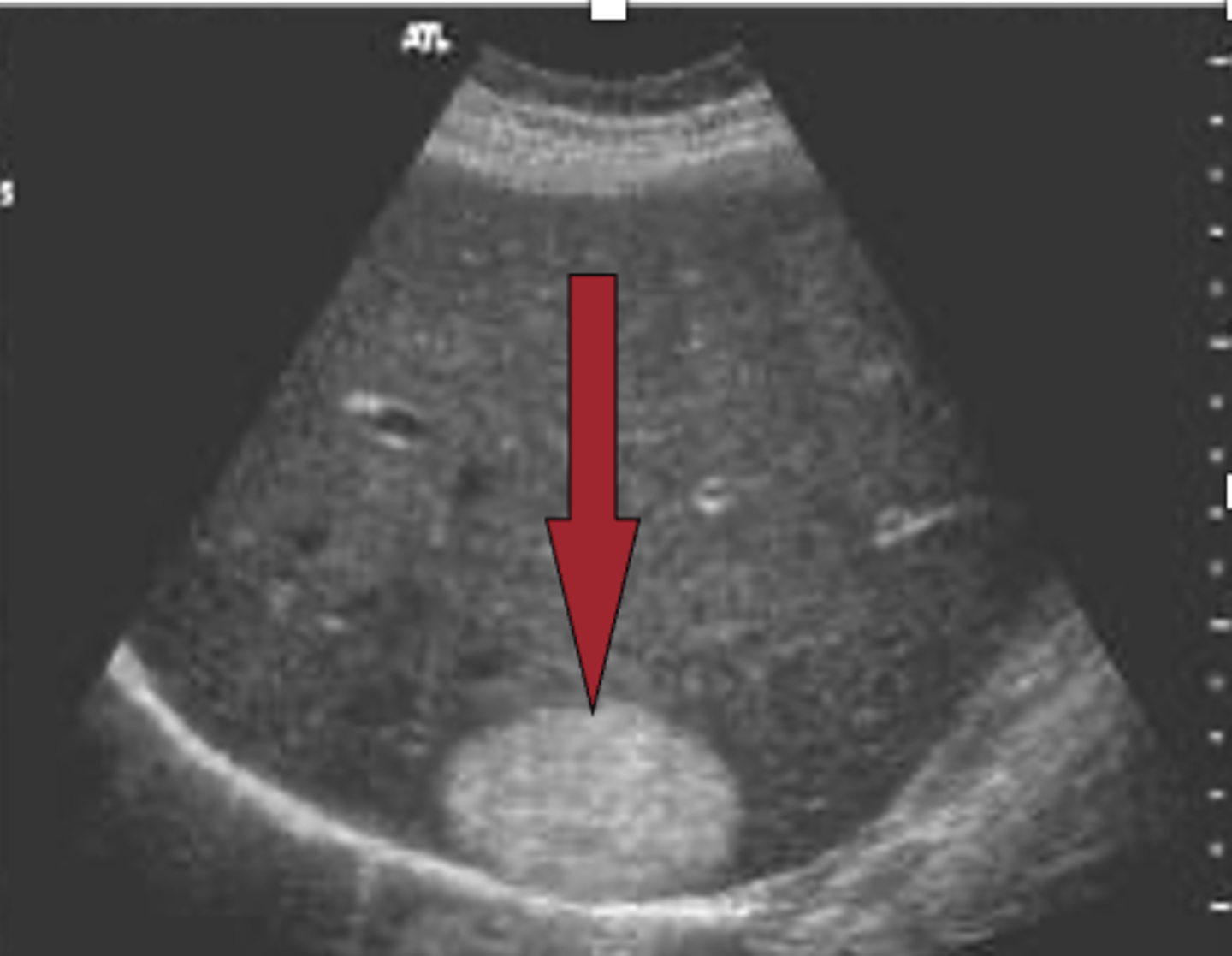
Homogeneous
Smooth texture
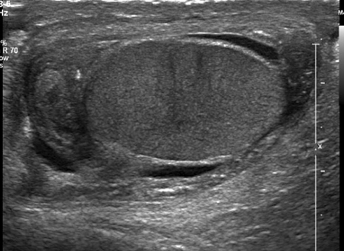
Inhomogeneous
Not uniform/smooth texture
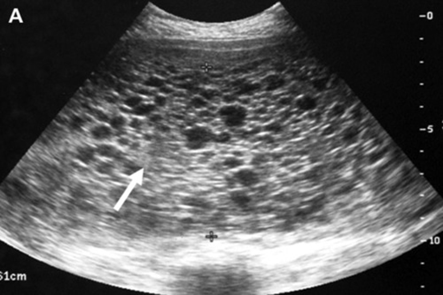
Shadowing
A structure absorbs/blocks sound
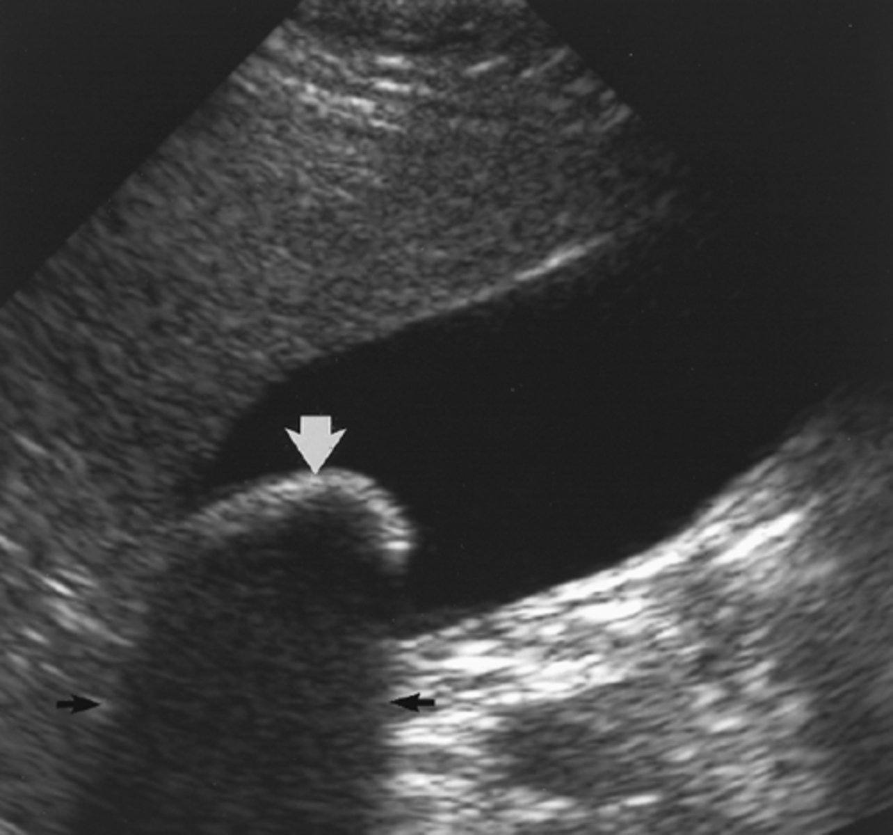
Enhancement
Increased echogenicity beneath low attenuating structures
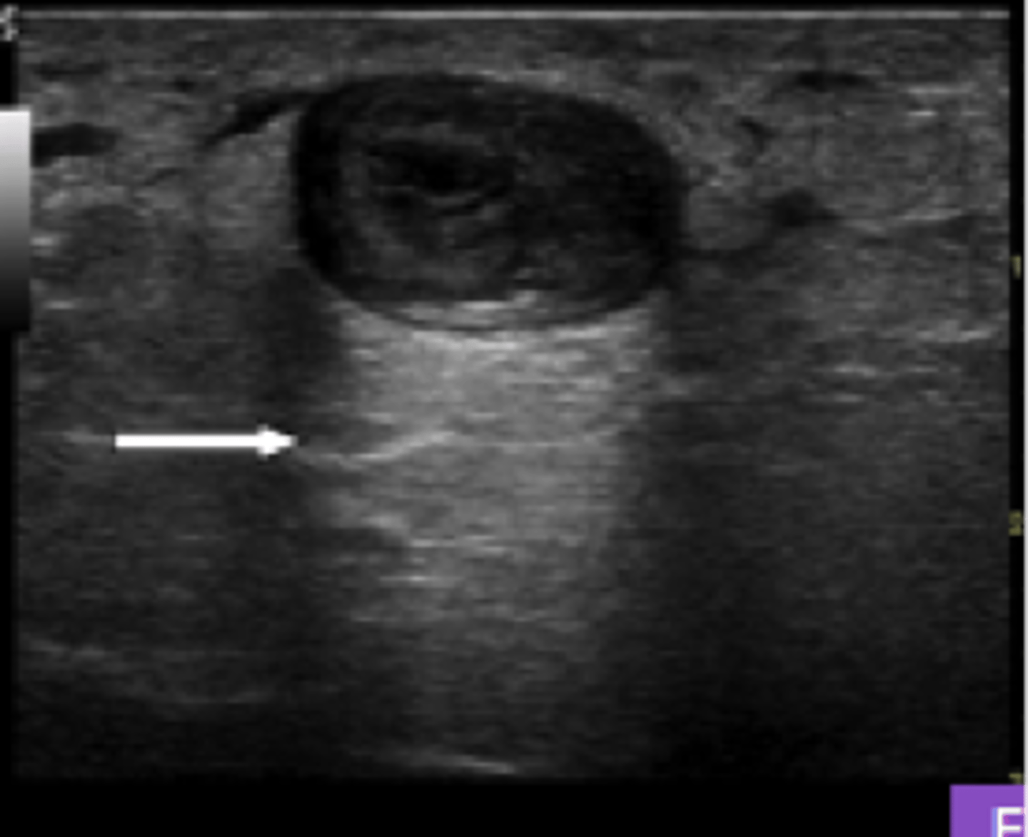
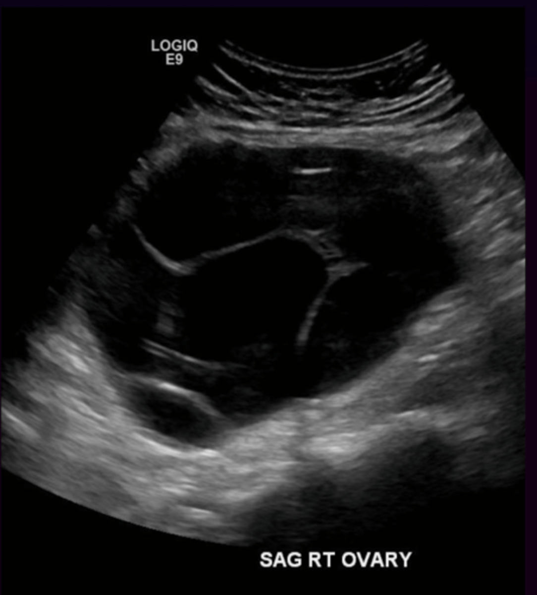
Cystic
Anechoic, smooth borders, enhancement
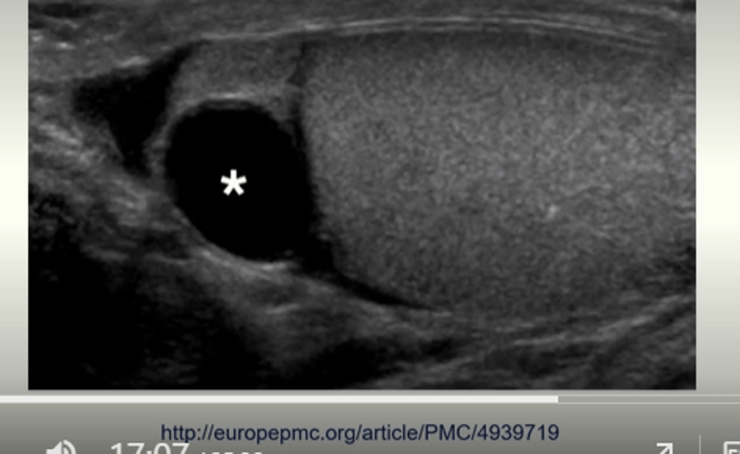
Complex
Echogenic and anechoic components
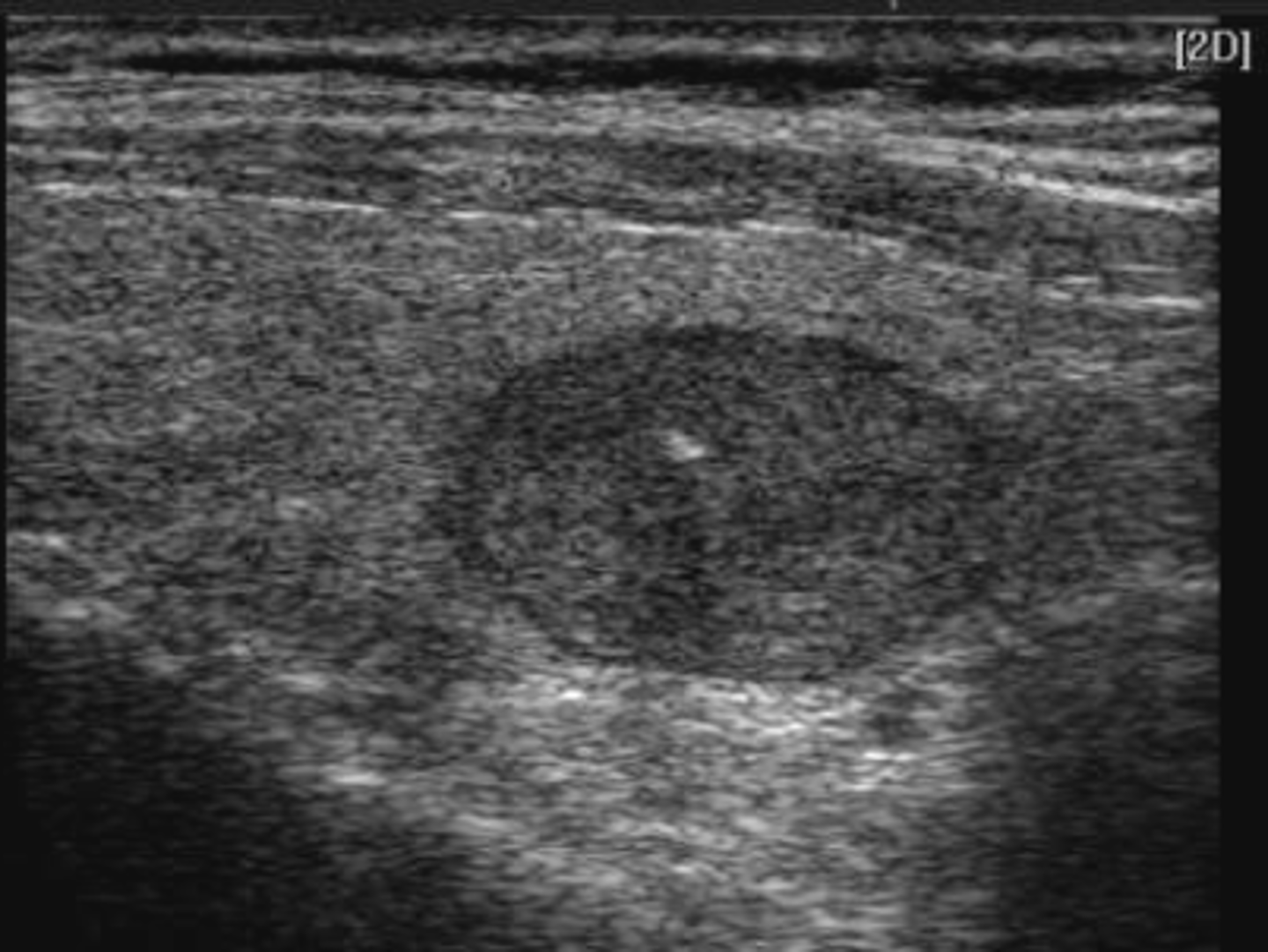
Solid
Echogenic (hypoechoic, medium or hyperechoic)
Structures that are likely to cause shadowing on sonographic images
Calcium deposits, stones, bones, & foreign objects
May occur when gas or air gets between the transducer and your target
Structures that are likely to cause enhancement on sonographic images
Cysts and bladder
May occur behind the gallbladder.
Usually doesn't occur behind blood vessels