Imaging Exam 2 Quizzes
1/19
There's no tags or description
Looks like no tags are added yet.
Name | Mastery | Learn | Test | Matching | Spaced |
|---|
No study sessions yet.
20 Terms
How often are the topics within the ACR Appropriateness Criteria reviewed and updated?
a) Every five years
b) Continually, due to advances in technology and new research
c) Only when major technological advancements occur
d) Annually by a legislative committee
b) Continually, due to advances in technology and new research
A 56-year-old woman fell, unobserved, at home, during a seizure and dislocated her right shoulder. She is currently under a physician’s care for seizure management but still suffers occasional seizures. Following the seizure her husband drove her to the emergency department where she was able to walk in and was cognitively intact. In the ED her shoulder was radiographed and found to be negative for fracture. The glenohumeral joint was re-located under sedation, and the arm was immobilized in a sling. A laceration on the scalp was sutured. She was discharged with medications for pain relief and muscle relaxants, instructed to follow up with her primary care physician, and referred to physical therapy for post shoulder dislocation management. At physical therapy, three days later, she presents cradling her right arm in the sling and holding her head rotated toward her arm. She is unable to actively move her head back to midline. Her entire right upper quadrant is painful to touch. According to Canadian Cervical Spine Rules, should her neck be radiographed?
Yes
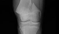
Refer to the figure. The _______is fractured in the most common epiphyseal injury pattern, a Salter–Harris ________:
a) femur; type 2
b) tibia; type 1
c) femur; type 1
d) tibia; type 2
a) femur; type 2
Radiologic hallmarks of rheumatoid arthritis include:
a) Asymmetrical joint space narrowing, sclerotic subchondral bone, and pseudocysts
b) Asymmetrical joint space narrowing and articular erosions
c) Osteophyte formation and osteoporosis
d) Concentric joint space narrowing, erosions of subchondral bone, and periarticular rarefaction
d) Concentric joint space narrowing, erosions of subchondral bone, and periarticular rarefaction
What is the digital representation of the ACR Appropriateness Criteria called, which can be integrated with electronic medical records?
a) ACR Guidelines
b) Image Wisely
c) ACR Select
d) Clinical Decision Rules
c) ACR Select
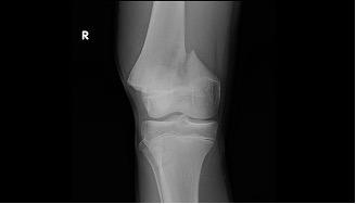
Refer to the figure. Radiographic assessment of a fractured bone must include:
a) The routine radiographic exam for the most distal joint
b) An AP view of the contralateral limb to assess normal values
c) Two views at right angles to each other
d) The routine radiographic exam for the most proximal joint
c) Two views at right angles to each other
Radiologic hallmarks of degenerative joint disease include:
a) Asymmetrical joint space narrowing and articular erosions
b) Asymmetrical joint space narrowing, sclerotic subchondral bone, and pseudocysts
c) Osteophyte formation and osteoporosis
d) Concentric joint space narrowing, sclerotic subchondral bone, and periarticular rarefaction
b) Asymmetrical joint space narrowing, sclerotic subchondral bone, and pseudocysts
Which of the following scenarios best describes how a physical therapist might utilize the ACR Appropriateness Criteria, according to the information presented?
a) To directly order advanced imaging studies such as MRI or CT scans for their patients
b) To save resources by bypassing communication with the physician and recommending treatment based solely on the ACR guidelines
c) To understand the most appropriate initial imaging for a specific musculoskeletal condition and discuss these findings with the referring physician
d) To independently determine a patient's diagnosis without consulting with a physician
c) To understand the most appropriate initial imaging for a specific musculoskeletal condition and discuss these findings with the referring physician
A 16-year-old male soccer player has a mid-air collision during a match and falls to the ground. He has immediate knee pain and is unable to touch his foot to the ground after being brought to stand by teammates. He sits out the rest of the match with ice on his knee. Following the match, he is able to limp unassisted to his car, but has to sit stretched out on the back seat as he cannot bend the knee sufficiently to get into the passenger side seat. His parent is asking whether he should go to the emergency department for radiographs or wait to see if he’s better in the morning. According to the Ottawa Knee Rules, should he have the knee radiographed?
Yes
A 68-year-old man presents to a direct access clinic after being involved in a parking lot fender- bender one week ago. He complains of neck pain that is constant when up he is up and relieved when lying down. He denies paresthesia. Active range of motion of his neck is limited in all directions by pain. He is tender to palpation along his cervical paraspinal soft tissues. According to NEXUS, should radiographs be ordered?
Yes
Match the type of cervical injury with its clinical feature.
Avulsion fractures
Compression fractures
Fracture dislocations
Disc herniation
Avulsion fractures: Fractures at spinous processes due to ligament failure
Compression fractures: Decreased disc height and vertebral body collapse
Fracture dislocations: Misalignment of vertebrae indicating instability
Disc herniation: Protrusion of disc material into the spinal canal
What imaging technique is most effective in showing acute soft tissue injuries in the cervical spine?
a) CT scan
b) MRI
c) Ultrasound
d) X-ray
b) MRI
What condition is commonly responsible for cervical myelopathy in chronic cases?
a) Rotator cuff injuries
b) Degenerative changes in the cervical spine
c) Inflammatory diseases
d) Acute trauma
b) Degenerative changes in the cervical spine
Match the shoulder structure with its corresponding pathology.
Rotator cuff
Glenoid labrum
Subacromial space
Humeral head
Rotator cuff: Tears leading to pain and dysfunction.
Glenoid labrum: Tears associated with instability.
Subacromial space: Narrowing leading to impingement.
Humeral head: Avascular necrosis from injury.
Match the type of wrist fracture with its characteristics.
Scaphoid fracture
Colles fracture
Boxer's fracture
Avulsion fracture
Scaphoid fracture: Often obscured on initial radiographs, common after falls.
Colles fracture: Distal radius fracture with dorsal angulation due to falls.
Boxer's fracture: Fracture of the fifth metacarpal from punching.
Avulsion fracture: Results from ligament or tendon pull on bone.
T/F: The Cobb measurement is used to assess the curvature of the spine in the frontal plane during scoliosis evaluation.
True
Match the imaging technique with its purpose in evaluating shoulder injuries.
AP radiograph
MRI
CT scan
Scapular Y view
AP radiograph: Shows glenohumeral joint space and detects fractures.
MRI: Utilized for detailed visualization of rotator cuff and labrum.
CT scan: Provides detailed assessment of complex fracture patterns.
Scapular Y view: Helps visualize the scapula and humeral head alignment.
Which imaging modality is considered the gold standard for diagnosing rotator cuff tears?
a) CT scan
b)Ultrasound
c) X-ray
d) MRI
d) MRI
What type of cervical injury is typically characterized by anterior compression and often occurs in older adults with osteoporosis?
a) Compression fractures
b) Fracture dislocations
c) Avulsion fractures
d) Facet dislocations
a) Compression fractures
Match the imaging technique with its purpose in cervical spine trauma.
Lateral view radiograph
CT scan
MRI
Cross table lateral radiograph
Lateral view radiograph: Assesses vertebral alignment and soft tissue abnormality
CT scan: Displays detailed bone structure
MRI: Visualizes neural tissues and edema
Cross table lateral radiograph: Used for supine patients