BFCP S03 + S04
1/80
Earn XP
Description and Tags
S03: Intro Cell + Tools of Cell Bio; S04: Cytoskeleton
Name | Mastery | Learn | Test | Matching | Spaced |
|---|
No study sessions yet.
81 Terms
T/F: all cells contain the same copies of the genome with minor exceptions
T
If all cells have the same genome, how do they perform different functions?
They differentiate based on how they use genetic instructions/cell signaling
What is the least complex eukaryote that we commonly use as a model organism?
Yeast
Why are yeast a good model organism?
Homologous proteins: similar in yeast and humans
Easy to grow and manipulate
Nematode C. elegans significance
First multicellular organism to have its complete genome sequence determined
Zebrafish are ideal for ____ studies because ______
developmental studies because their embryos develop outside of the mother and are transparent
Significance of cell culture
Is it usually one or multiple cells being harvested?
Allows for a controlled environment in vitro
Usually single type of cell
Flow cytometry purpose
Provides quantitative information on individual cells
Whether the target is present and at what quantity
Label cells with fluorochrome for a target molecule and allow them to flow through
All cells will scatter light
The cells with the target molecule scatter light AND emit fluorescence
Flow cytometry
Fluorescence activated cell sorter (FACS) purpose
To separate cells with a specific characteristic from a mixture using “gating”
Cells with a specific target will have fluorescence
Each cell droplet is given a negative charge proportional to the amount of target of interest present
More target of interest = more fluorescence = more negative electrical charge
Magnetic plates will sort cells
Cells can be “gated” and software can sort out the gated cells
Fluorescence activated cell sorter (FACS)
Cell lysis purpose
To study cells at the subcellular and molecular level
Rupture cells
Centrifuge the cell components by size and density
Heaviest precipitates first
Cell lysis
Antibodies are made up of WBCs called ______ and are (very specific/not specific) to their antigen.
B-cells
very specific
How can we produce antibodies of interest in animals?
Inject foreign protein of interest into animal
Animal creates antibodies against protein of interest
Method of extracting polyclonal antibodies
Harvest animal blood serum with a mixed combination of all antibodies produced
To make monoclonal antibodies, take ______ from the animal. Fuse these with a _____ to create a hybridoma.
To make monoclonal antibodies, take B-cells from the animal. Fuse these with a tumor cell lineH to create a hybridoma.
Hybridoma
A fused B cell and tumor cell that continuously produces monoclonal antibodies
T/F: light microscopy can be done on live cells
T
Brightfield light microscopy
Light is transmitted straight through the specimen
Phase-contrast light microscope
Makes highly transparent objects more visible
Differential-interference-contrast light microscope
Exaggerates differences in density
Using light and 2 sets of filters to excite fluorochromes and visualize their wavelength
Fluorescence microscopy
Explain the following methods of tagging molecules of interest in fluorescence microscopy. Indicate if they can be done live:
Genetic tagging
Epitope tagging
Immunolabeling with antibodies
Genetic: adding GFP to proteins of interest and tracking LIVE
Epitope: adding an epitope tag to a protein and detecting it via antibody binding to epitope (usually fixed)
Immunolabeling: fluorescence secondary antibody binds to primary antibody which binds to target (only fixed)
Laser scanning confocal microscope
Fixed or live?
Using lasers to focus on a very specific point and combining data to create a 3D structure. Usually on fixed cells.
T/F: electron microscopy can be done on live cells
F - only fixed samples
Transmission electron microscopy
Beams of electrons go THROUGH the target to see ultrastructural details
Scanning electron microscopy
Beam of electrons onto the sample surface
What is the benefit of electron microscopy > light microscopy?
Can reveal much smaller structures and in greater detail
What are 3 methods of following protein dynamics?
Fluorescence resonance energy transfer (FRET)
Fluorescence recovery after photobleaching (FRAP)
Photoactivation
Tag 2 proteins with different fluorescents
Excite protein X with the appropriate laser and observe if protein Y emits light
Purpose: to determine if 2 proteins interact
Fluorescence resonance energy transfer (FRET)
Tag protein of interest
Use a laser to bleach the fluorescence
Watch how other labeled proteins diffuse throughout this area by tracking the reappearance of fluorescence
Purpose: to understand the protein’s kinetic parameters
Fluorescence recovery after photobleaching (FRAP)
Photoactivate protein of interest with a wavelength of light
Watch how that fluoresced target protein move around/away from that spot into other regions
Purpose: to understand the target protein’s kinetic parameters
Photoactivation
3 parts of the cytoskeleton from smallest → largest, and identify if they are polar or nonpoar
Microfilaments (actin filaments): polar
Intermediate filaments: nonpolar
Microtubules: polar
Which part of the cytoskeleton is the toughest and most durable, having the greatest tensile strength against mechanical stress?
Intermediate filaments
Where are intermediate filaments found?
Throughout the cytoplasm - around/inside the nucleus and extending to the cell periphery
Which cytoskeleton part is found underlying and strengthening the nuclear envelope?
Intermediate filaments
Which cytoskeleton part is responsible for limiting the extension of cell stretching and pulling?
Intermediate filaments
Intermediate filaments have a ______-like shape
rope-like
The structure and composition of intermediate filaments
Long fibrous proteins that form tetramers. 8 tetramers form a long rope
The following are examples of which cytoskeleton part: keratins, desmins, vimentin, neurofilaments, nestin, nuclear lamins
Intermediate filaments
Keratin is an example of ________ and is found in ____ cells.
Keratin is an example of an intermediate filament and is found in epithelial cells.
Lines and strengthens the inside surface of the nuclear envelope
Nuclear lamina
The nuclear lamina is made up of ___________.
intermediate filaments
How are IFs stabilized and reinforced into bundles?
With accessory proteins like plectin
Epidermolysis bullosa simplex (EBS)
Mutations in keratin genes that encode IFs cause the skin to be highly vulnerable to mechanical injury
Which cytoskeleton part acts as an intracellular “highway” to move cell components?
Microtubules
Explain the dynamic instability of microtubules
They can rapidly disassemble in one location and reassemble in another
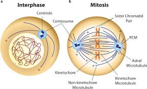
Microtubules grow out from ______
centrosomes
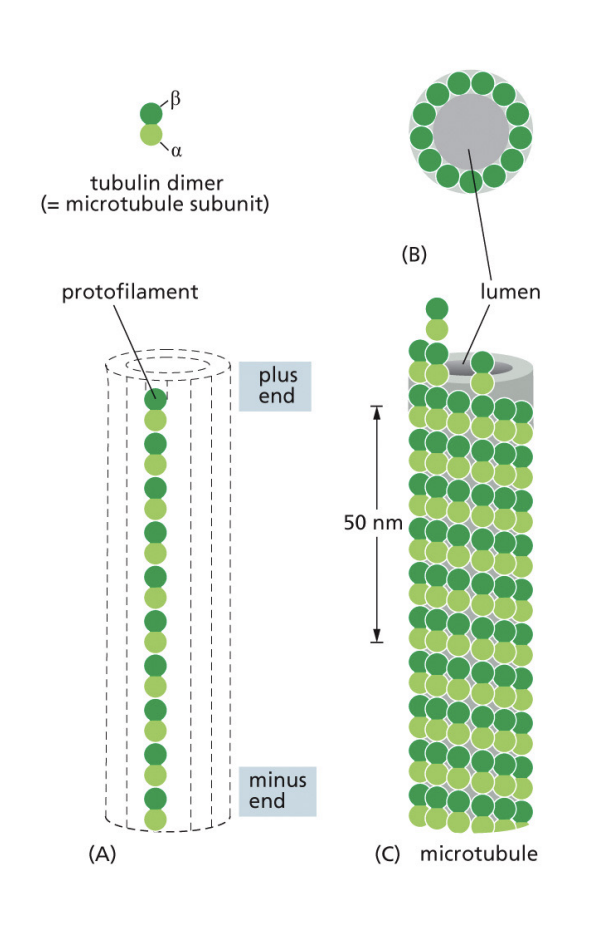
Microtubules are composed of dimers of ___ (+/-) and ____ (+/-) tubulins. Each microtubule has __ protofilaments.
Microtubules are composed of dimers of a (-) and b (+) tubulins. Each microtubule has 13 protofilaments.
The centrosome is composed of 2 ____ surrounded by pericentriolar material. It is located (close/far away) from the nucleus, except when the cell is in mitosis.
2 centrioles
close
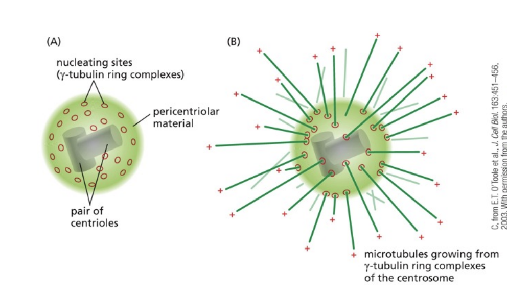
Microtubules grow from the __________ ring complex on centrosomes that serve as the nucleation site for the growth of microtubules.
y-tubulin ring complex

Centrosome function
Controls the production and location of microtubules
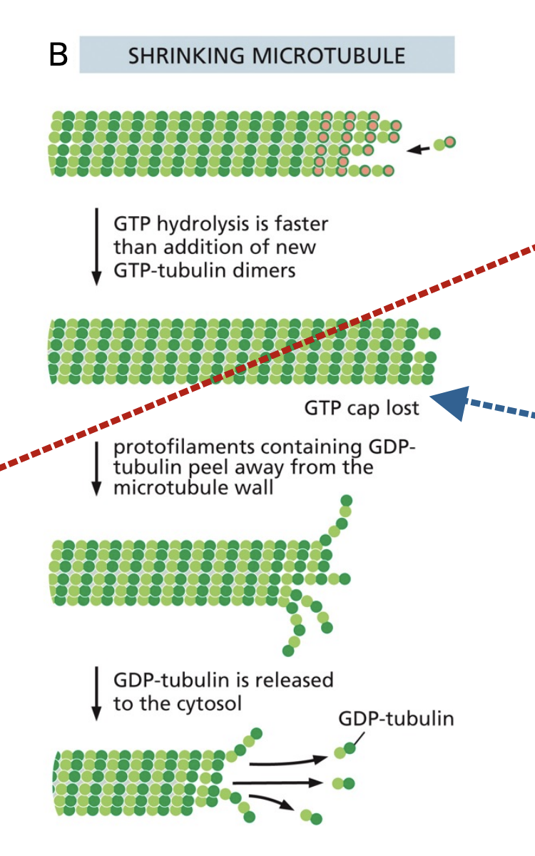
Explain how microtubules are built using GTP
Tubulin dimers that are bound to GTP are added to the + end of a growing microtubule. Shortly after addition, they are hydrolyzed into GDP.

GTP cap
The end of a microtubule, where the dimers are still attached to provide stability to the molecule
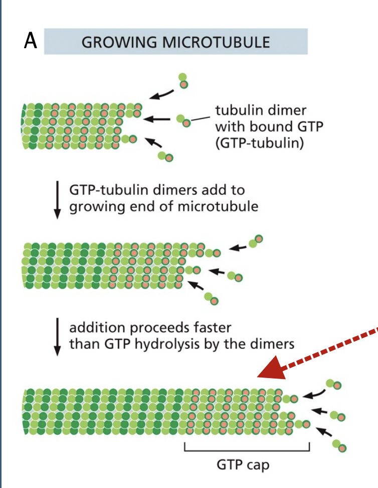
What would happen if there was no GTP cap on a microtubule?
Rapid depolymerization due to the instability of GDP
Microtubule-associated proteins (MAP)
Regulates the organization of microtubules
Prevents depolymerization when the GTP cap is lost by providing stability
Motor proteins use energy from _______ to travel along _____
ATP hydrolysis
microtubules
Transport of 2 microtubule motor proteins: kinesin and dynein
Kinesin: moves towards the + end, away from the middle/centrosome
Dynein: move towards the - end, towards the middle/centrosome
Kinesins and dyneins have 2 ___ on one end that attach to ____ and hydrolyze ____.
Kinesins and dyneins have 2 globular heads on one end that attach to microtubules and hydrolyze ATP.
Kinesins and dyneins have 1 ___ that interacts with _____.
Kinesins and dyneins have 1 tail that interacts with cargo.
The propulsion of a globular head of a motor protein forward is driven by _____
The binding of ATP on the other head
Kinesins tend to move the organelle _____ outward, while dyneins tend to move the organelle ____ inward.
Kinesin moves the ER
Dynein moves the Golgi
Effects of colchicine treatment
Causes microtubules to disassemble, changing the locations of the ER and golgi
Cilia and flagella are composed of ______ and use ____ to drive their movement.
Cilia and flagella are composed of microtubules and use dynein to drive their movement.
The definition and structure of the axoneme
9+2 structure: 9 outer doublets of microtubules, and 2 single microtubules in the middle
Found in cilia and flagella
The microtubule doublets in cilia/flagella ___ to allow for movement, while linker proteins ______
bend
linker proteins keep the doublets paired while sliding
T/F: dynein “walks” along cilia/flagella to move them
F: we don’t want the microtubule doublets to separate, so the microtubules bend instead
What powers the locomotion of cilia and flagella?
ATP hydrolysis
What do microtubule-specific drugs target?
Prevention of microtubule polymerization/depolymerization
Which cytoskeleton part is responsible for cell movement and the flexibility of cell shape?
Microfilaments
Actin filaments are also called ______
microfilaments
Where are microfilaments located?
Around the cell, but mostly in the cell cortex
Structure of microfilaments
Actin monomers polymerize to form actin filmanets
Microtubule polymerization involved the hydrolization of _____, while microfilament polymerization involves the hydrolization of _____.
Microtubule polymerization involved the hydrolization of GTP, while microfilament polymerization involves the hydrolization of ATP.
What is actin treadmilling?
When ATP-bound actin is added to an actin filament on the + end at the same rate as ADP-bound actin is being removed from the - end.
Actin-binding proteins function
What controls these proteins?
Where and when actin filaments form and grow
Controlled by extracellular signals
Actin combining with ____ allows it to form the sarcomere structure and contract muscles.
myosin
Actin filaments concentrated in the cell cortex allow for _______.
support of the outer surface and mechanical strength
Explain how cell crawling depends on actin using lamellipodia and filopodia, and myosin
-Actin polymerization on the protruding end allows the cell to push forward
-Lamellipodia forms protrusions on the plasma membrane to move the cell forward
-Filopodia detects cues to guide the cell
-Contraction ofm yosin on the rear end of the cell draws the cell forward
Microvilli are made up of ________.
actin filaments