EXSC 418 Week #2: Superficial and Deep Back; Posterior Neck
 Call Kai
 Learn
Learn Practice Test
 Spaced Repetition
 Match
Match Flashcards
 Knowt Play
1/97

Earn XP
Description and Tags
Vocabulary flashcards covering skeletal landmarks, muscle origins, insertions, actions, innervations, and blood supplies for EXSC 418 Week 2.
Name | Mastery | Learn | Test | Matching | Spaced | Call with Kai | Chat |
|---|
No analytics yet
Send a link to your students to track their progress
98 Terms

Vertebrochondral Ribs
Ribs 8-10.


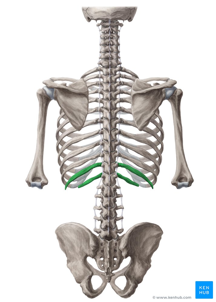
Vertebral Ribs
Ribs 11-12.


Accessory Nerve (Cranial Nerve XI) (Yellow)
Nerve that provides innervation to the trapezius muscle. (TEAL)


Thoracodorsal Nerve
Nerve that provides innervation to the latissimus dorsi muscle.

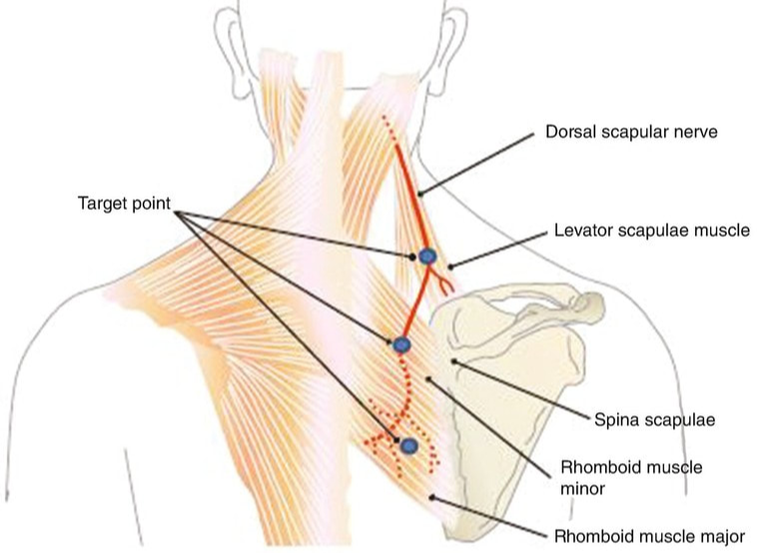
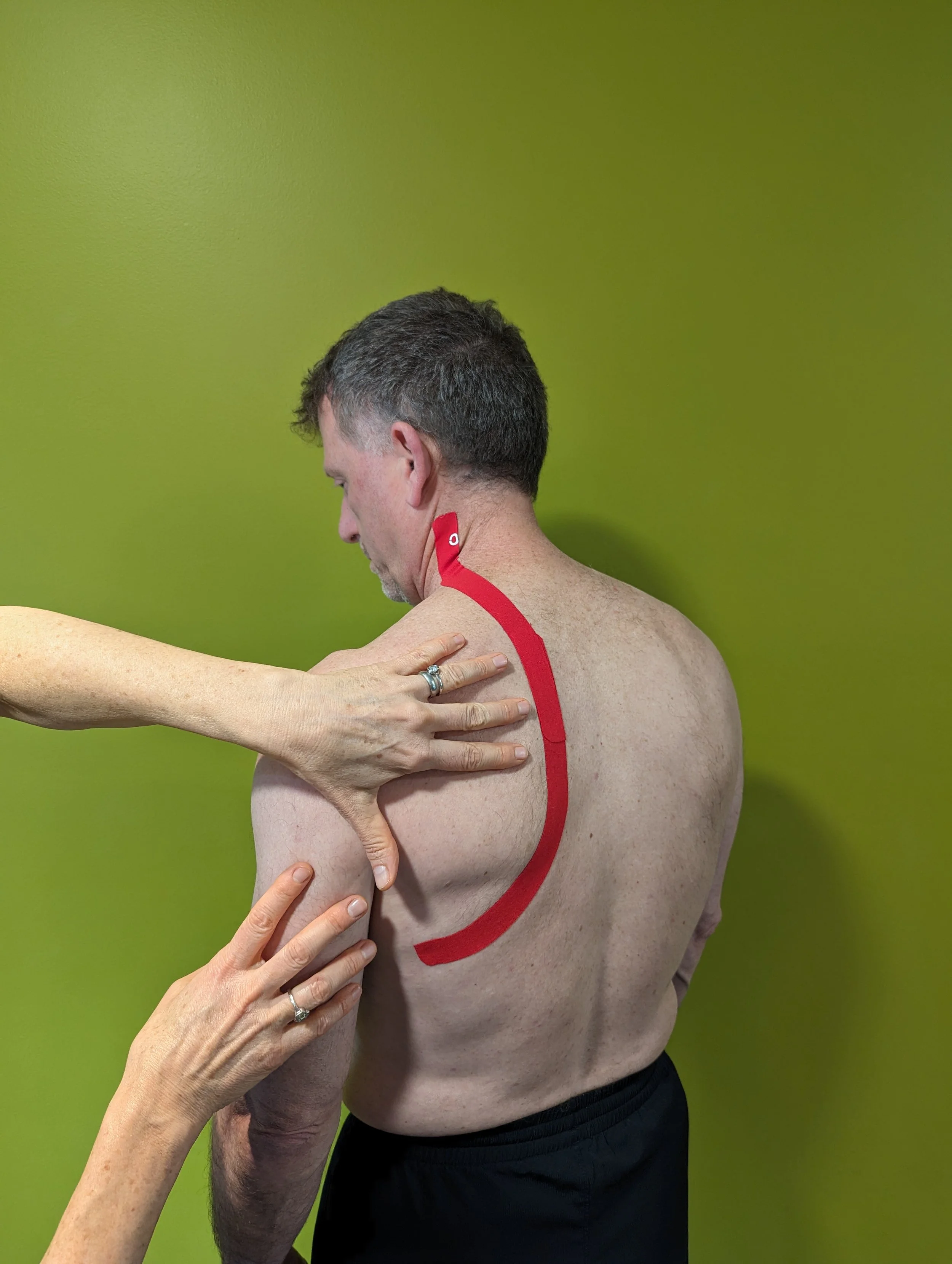
Dorsal Scapular Nerve
Nerve that provides innervation to the levator scapulae, rhomboideus major, and rhomboideus minor muscles.

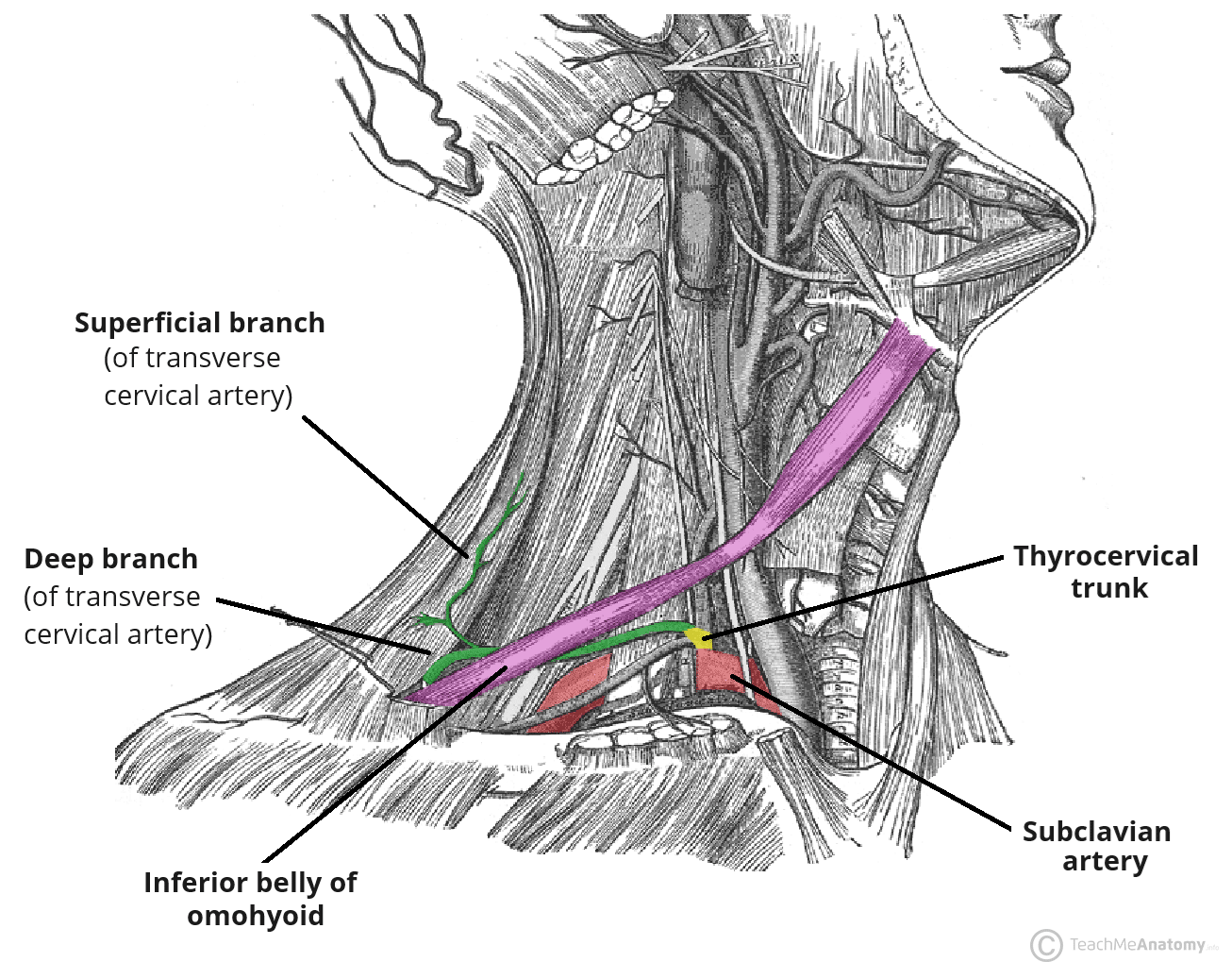
Transverse Cervical Artery
Artery providing blood supply to the trapezius muscle.

Thoracodorsal Artery
Artery providing blood supply to the latissimus dorsi muscle.


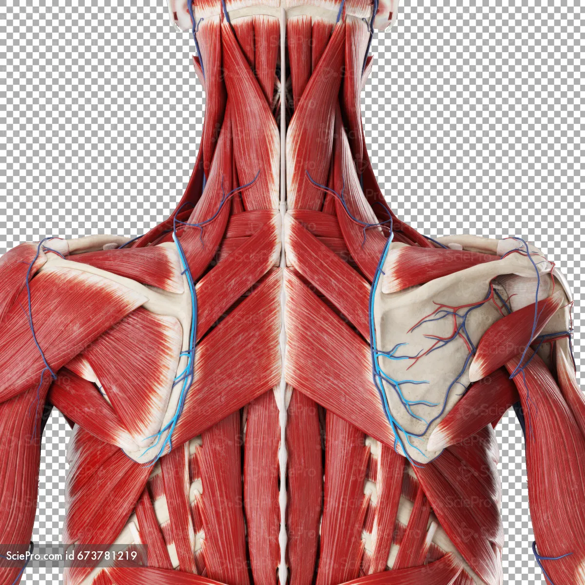
Dorsal Scapular Artery
Artery providing blood supply to the levator scapulae, rhomboid major, and rhomboid minor muscles. (BLUE medial side of scaps)

External Occipital Protuberance
Big bump in middle-bottom of occipital bone….. Prominent median elevation located on the outer surface of the occipital bone that serves as an attachment site for the ligamentum nuchae and trapezius.

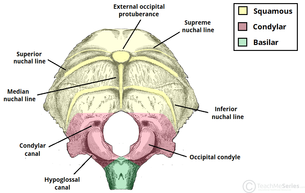
Nuchal Lines (*2 sets, superior and inferior*)
Ridges running laterally on the posterior surface of the occipital bone that serve as insertion points for neck and back muscles.


Temporal Bone - Mastoid Process
Conical projection located on the inferior posterior portion of the temporal bone, serving as an attachment site for muscles such as the splenius capitis.


Vertebrosternal Ribs
Ribs 1 through 7 (true ribs) that attach directly to the sternum via their own costal cartilages.

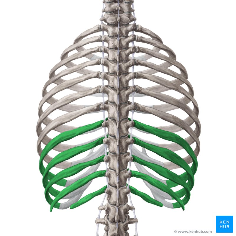
Vertebrochondral Ribs
Ribs 8 through 10 (false ribs) whose costal cartilages attach indirectly to the sternum by fusing with the cartilage of the rib directly above them.


Vertebral Ribs
Ribs 11 and 12 (floating ribs) that have no anterior attachment to the sternum or other costal cartilages.

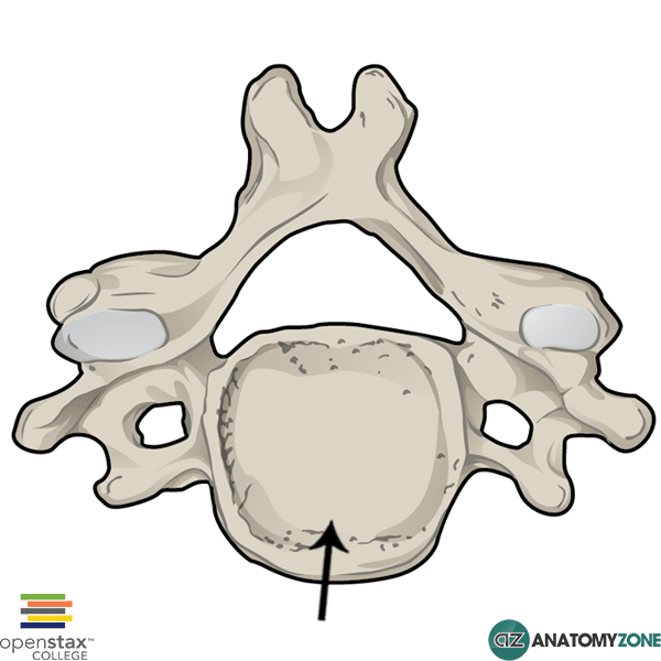
Body
Thick, cylindrical anterior portion of a vertebra responsible for supporting weight.
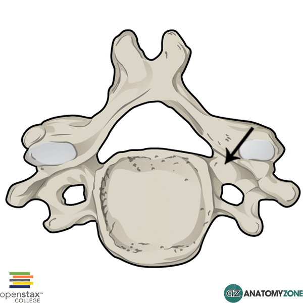
Pedicle
Short, thick pillar extending posteriorly from the vertebral body to form the lateral wall of the vertebral arch.


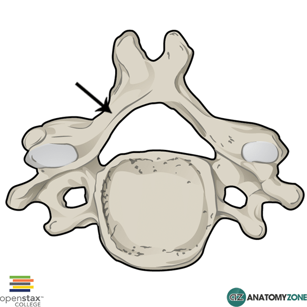
Lamina
Broad, flat plate extending medially from the pedicle to fuse at the posterior midline of the vertebral arch.

Spinous Process of Vertebra
Posterior projection extending from the fusion of the two laminae, serving as a site for muscle and ligament attachment.


Transverse Process of Vertebra
Lateral projection extending from the junction of the pedicle and lamina on each side of a vertebra.


Vertebral Foramen
Central opening enclosed by the body and vertebral arch that houses and protects the spinal cord.


Intervertebral Foramen
Lateral opening created between the pedicles of adjacent vertebrae for the passage of spinal nerves.


Intervertebral Disc
Fibrocartilaginous pad sandwiched between adjacent vertebral bodies to absorb shock and allow movement.


Sacral Promontory
Anteriorly projecting superior border of the body of the S1 sacral vertebra.
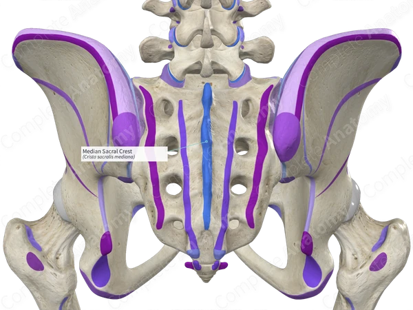
Median Sacral Crest (blue in center)
Central ridge formed on the posterior surface of the sacrum by the fusion of sacral spinous processes.


Sacral Canal
Hollow pathway running through the length of the sacrum, forming the caudal continuation of the vertebral canal.

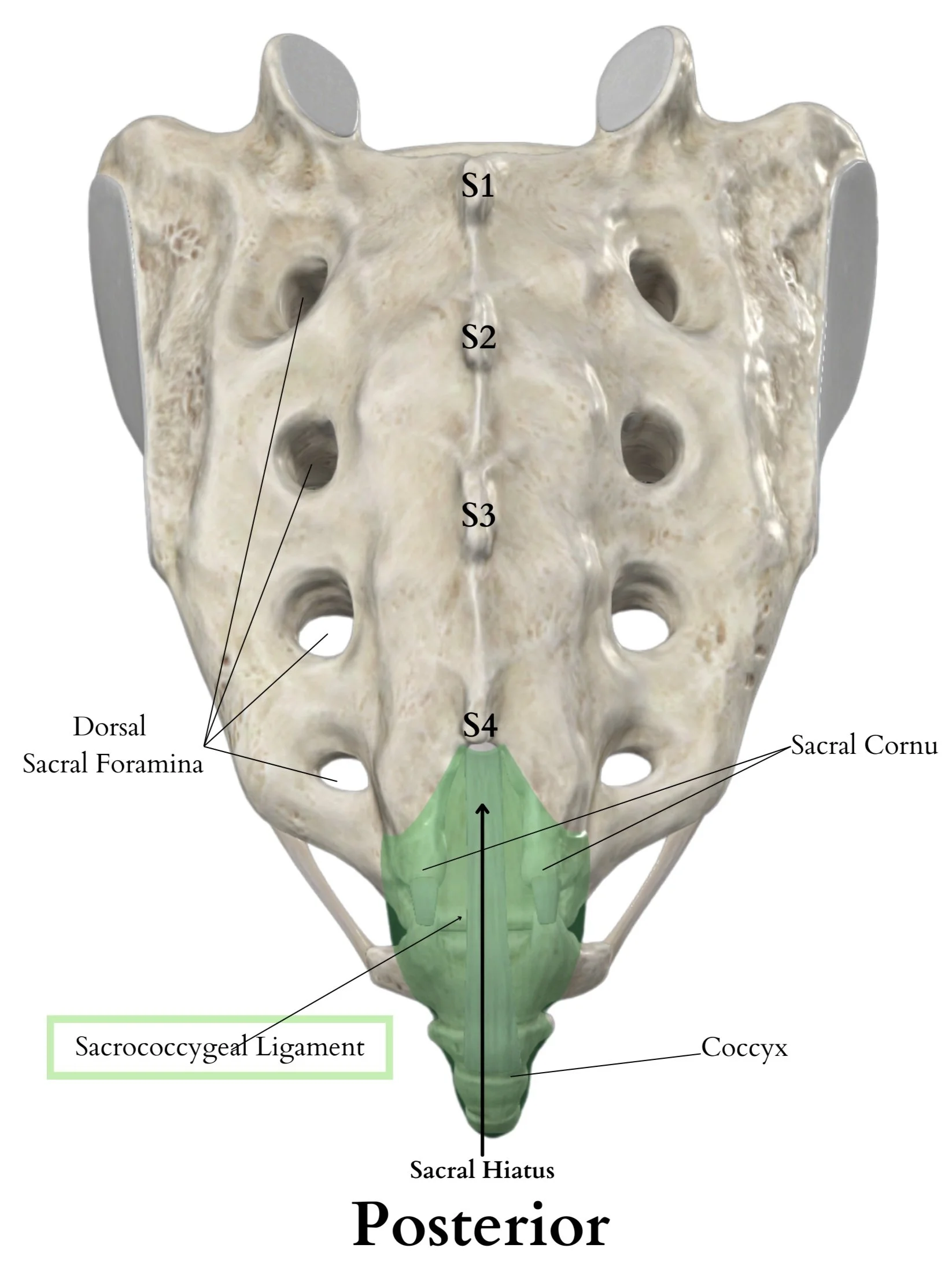
Sacral Hiatus
Inferior opening at the caudal end of the sacral canal resulting from the failure of the S5 laminae to fuse.
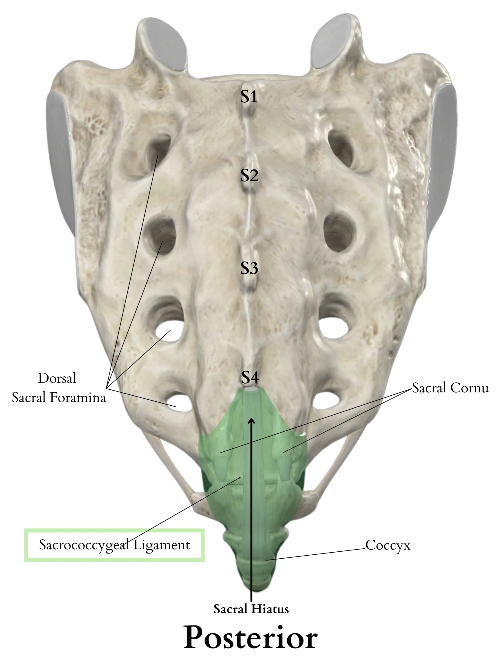
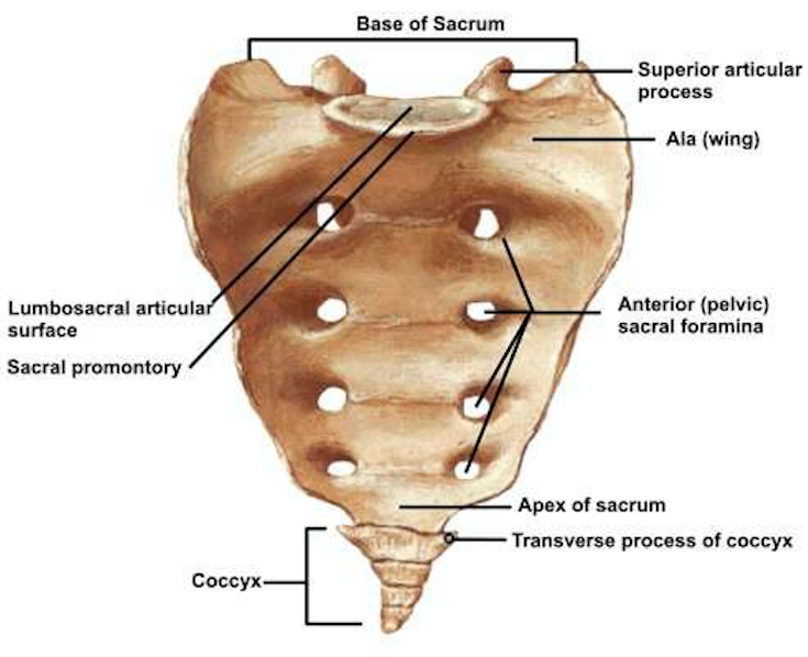
Anterior and Posterior Sacral Foramina
Paired openings on the sacrum (Front and Back) allowing passage for the anterior and posterior rami of sacral spinal nerves.


Coccyx - Apex
Tapered, inferior-most extremity of the coccyx.


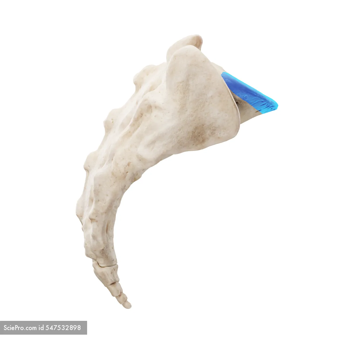
Coccyx - Base
Broad superior surface of the coccyx that articulates with the apex of the sacrum. (RED is just the whole coccyx)

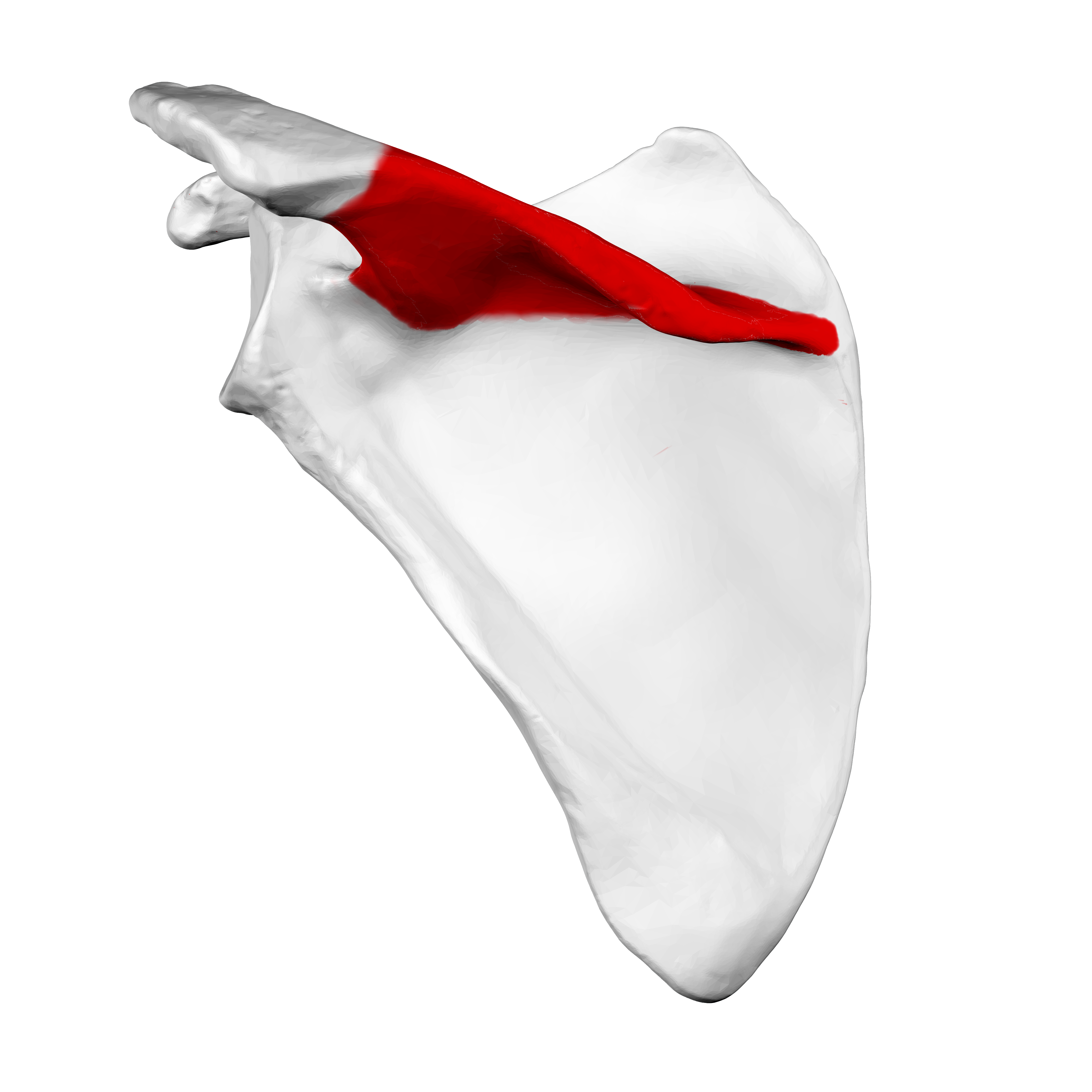
Scapula - Spine (Posterior View)
Prominent transverse ridge running across the posterior surface of the scapula. (posterior view)


Scapula - Acromion
Expanded lateral extremity of the scapular spine that articulates with the clavicle.

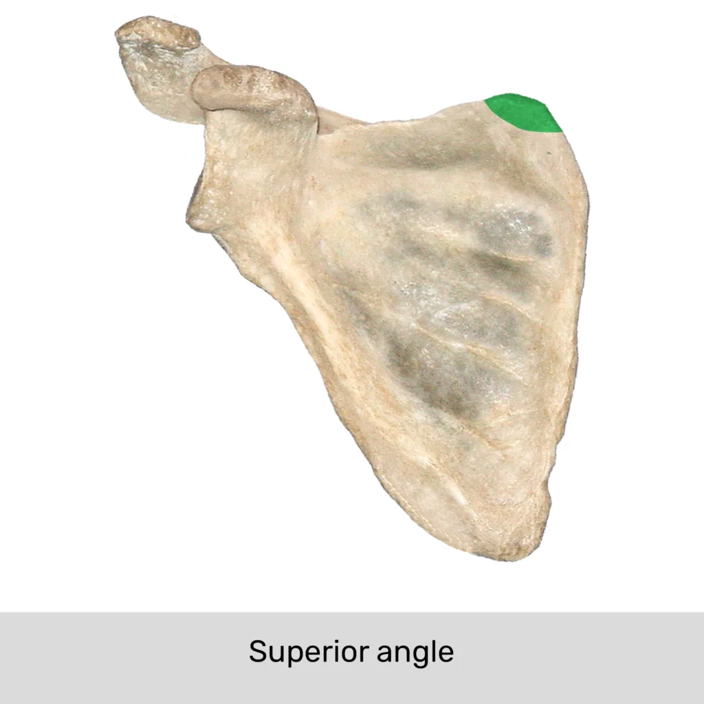
Scapula - Superior Angle
Junction formed by the meeting of the superior and medial borders of the scapula.
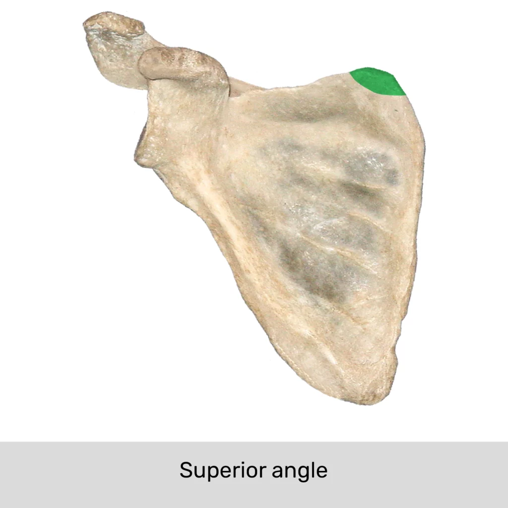

Scapula - Medial Border
Long vertical margin of the scapula facing the vertebral column (also called the vertebral border).

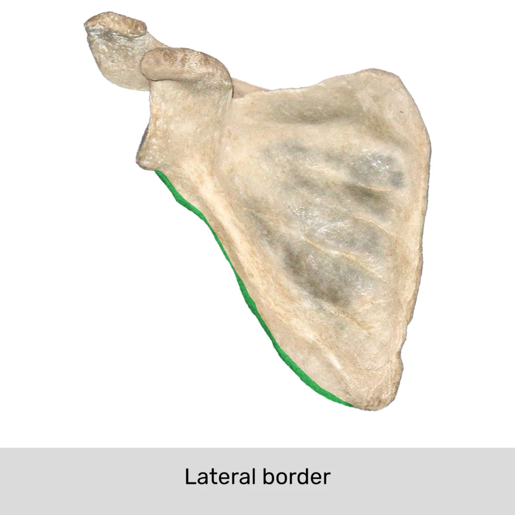
Scapula - Lateral Border
Thick edge of the scapula facing the axilla (also called the axillary border).
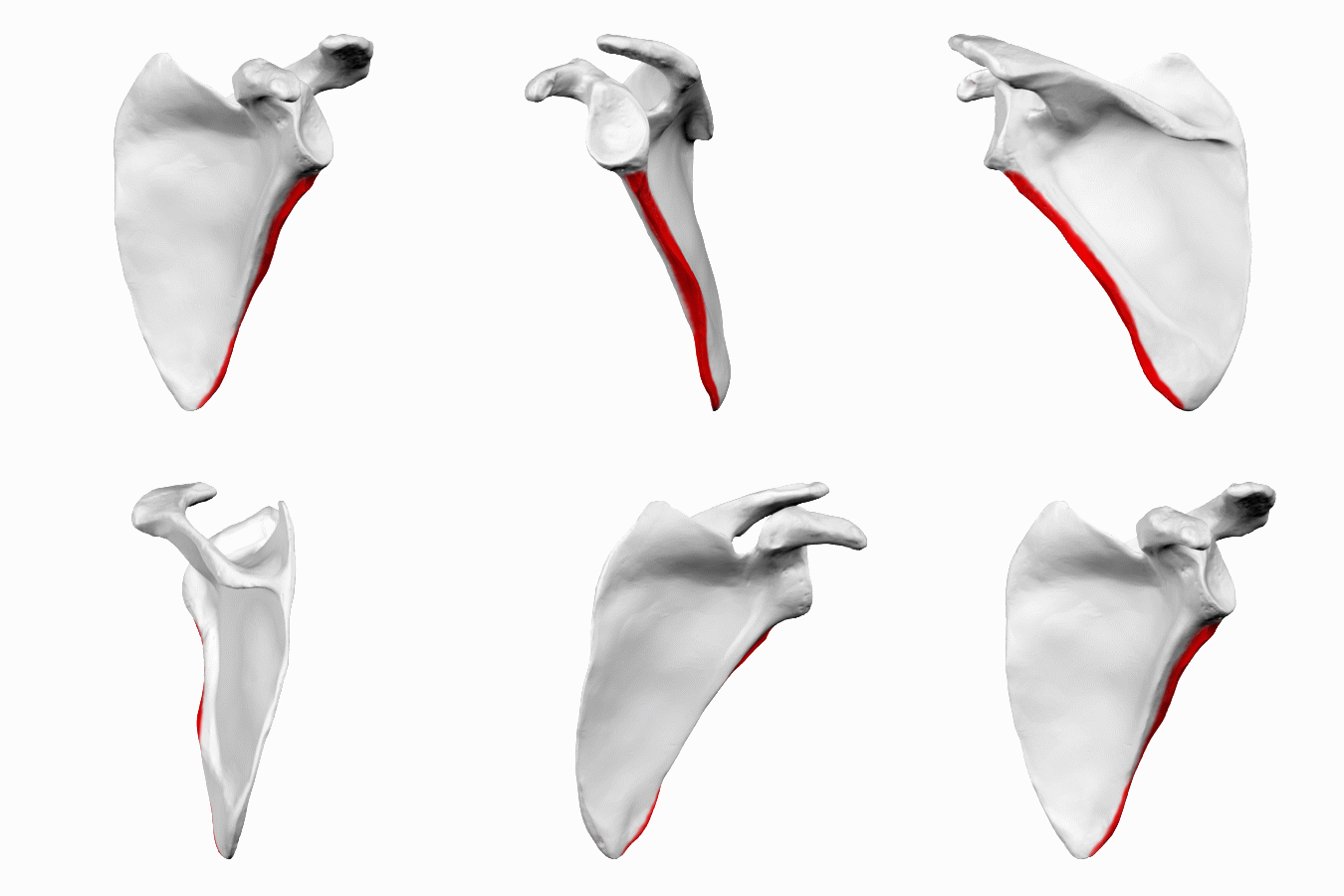

Humerus - Lesser Tubercle
Anterior projection located on the proximal humerus medial to the intertubercular groove.
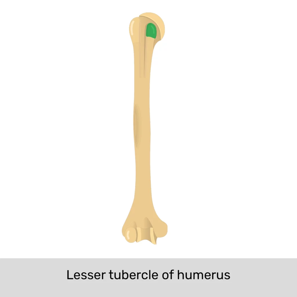

Humerus - Intertubercular Groove
Deep depression running vertically on the proximal humerus between the greater and lesser tubercles.


Ilium - Iliac Crest
Superior margin of the ilium bone serving as an origin and insertion point for back and abdominal muscles.


Trapezius - Origin
External occipital protuberance of occipital bone, ligamentum nuchae (C1-C6), and spinous processes of C7 through T12.

Trapezius - Insertion
Spine of scapula, acromion process of scapula, and lateral 1/3 of clavicle.

Trapezius - Actions
Upper fibers: elevation and upward rotation of scapula
Middle fibers: retraction of scapula
Lower fibers: depression and upward rotation of scapula.

Trapezius - Innervation
Accessory nerve (Cranial Nerve XI).

Trapezius - Blood Supply
Transverse cervical artery.

Latissimus Dorsi - Origin
Spinous processes of T7 through L5 and posterior iliac crest of ilium.

Latissimus Dorsi - Insertion
Crest of lesser tubercle of humerus and floor of intertubercular groove of humerus.

Latissimus Dorsi - Actions
Extension, medial rotation, and adduction of the humerus.

Latissimus Dorsi - Innervation
Thoracodorsal nerve.

Latissimus Dorsi - Blood Supply (BLUE circle)
Thoracodorsal artery.

Levator Scapulae - Origin
Transverse processes of C1 through C4 vertebrae.

Levator Scapulae - Insertion
Superior angle of the scapula.

Levator Scapulae - Actions
Elevation of scapula and downward rotation of scapula.

Levator Scapulae - Innervation
Dorsal scapular nerve.

Levator Scapulae - Blood Supply (whole red is dorsal scapular artery and branches, top part supplies blood to Lev. Scap)
Dorsal scapular artery.

Rhomboideus Major - Origin
Spinous processes of T2 through T5 vertebrae.

Rhomboideus Major - Insertion
Medial (vertebral) border of scapula inferior to the base of the spine.

Rhomboideus Major - Actions
Retraction, elevation, and downward rotation of the scapula. (Same as Minor)

Rhomboideus Major - Innervation
Dorsal scapular nerve.

Rhomboideus Major - Blood Supply
Dorsal scapular artery.

Rhomboideus Minor - Origin
Spinous processes of C7 and T1 vertebrae, and lower ligamentum nuchae.

Rhomboideus Minor - Insertion
Medial (vertebral) border at the base of the spine of the scapula.

Rhomboideus Minor - Actions (same as major)
Retraction, elevation, and downward rotation of the scapula. (minor)
Dorsal scapular nerve.
Rhomboideus Minor - Innervation (same as major)

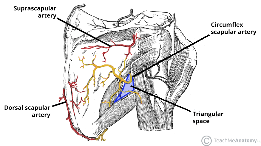
Dorsal scapular artery.
Rhomboideus Minor - Blood Supply (same as major)
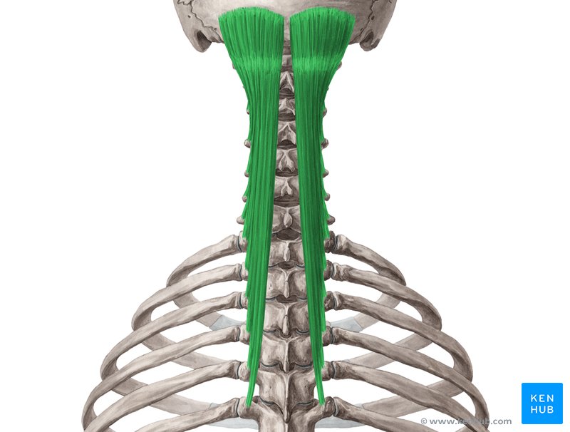
Splenius Capitis - Origin
Lower half of ligamentum nuchae and spinous processes of C7-T4 vertebrae.

Splenius Capitis - Insertion
Mastoid process of temporal bone and occipital bone inferior to lateral nuchal line.

Splenius Capitis - Actions
Extension of head and neck, lateral flexion of head and neck, and ipsilateral rotation of head.

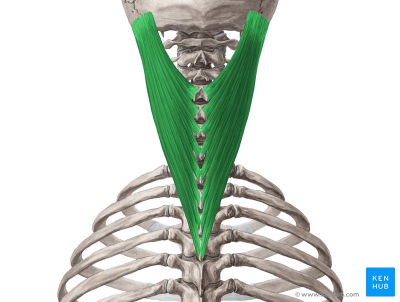
Splenius Capitis - Innervation
Dorsal rami of middle cervical spinal nerves.
Splenius Capitis - Blood Supply
Occipital artery.


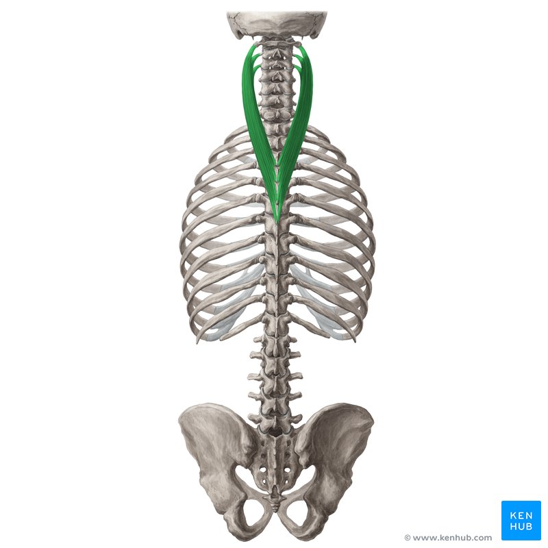
Splenius Cervicis - Origin
Spinous processes of T3-T6 vertebrae.

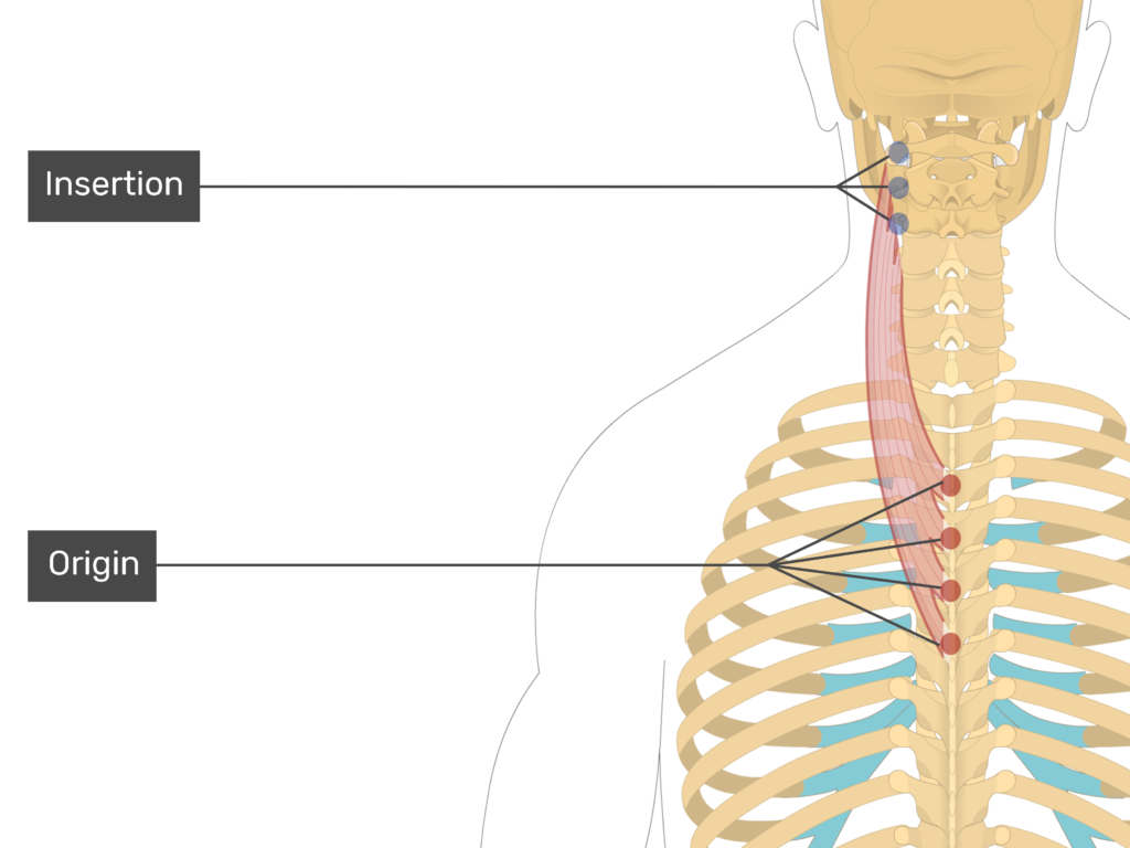
Splenius Cervicis - Insertion
Transverse processes of C1-C3 vertebrae (anterior to Levator Scapulae).

Splenius Cervicis - Actions
Extension of head and neck, lateral flexion of head and neck, and ipsilateral rotation of head.
Splenius Cervicis - Innervation
Dorsal rami of spinal nerves.
Splenius Cervicis - Blood Supply
Occipital artery.


Semispinalis Group - Muscular Components
Composed of semispinalis capitis, semispinalis cervicis, and semispinalis thoracis.
Semispinalis Group - Origin
Transverse processes of C7 through T12 vertebrae.
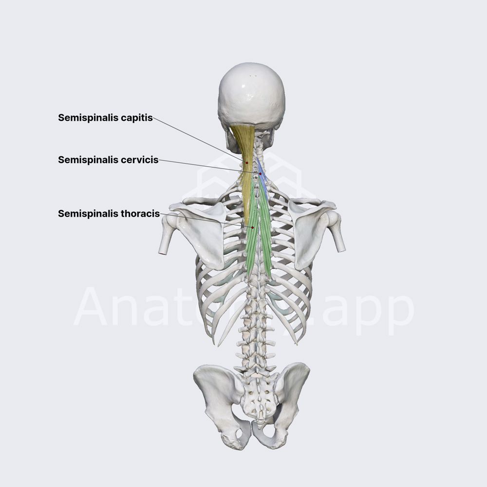
Semispinalis Group - Insertion
Capitis: between superior and inferior nuchal lines of occipital bone; Cervicis & Thoracis: vertebral spines 4-6 vertebrae above origin.

Semispinalis Group - Actions
Extension of head and neck, and contralateral rotation of head and vertebral column.

Semispinalis Group - Innervation
Dorsal rami of spinal nerves.

Semispinalis Group - Blood Supply
Occipital artery.
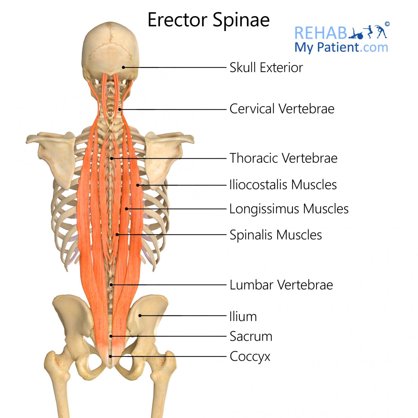
Erector Spinae Group - Muscular Components
Composed of spinalis (medial), longissimus (intermediate), and iliocostalis (lateral).
I LIKE SPINACH <3

Erector Spinae Group - Origin
Transverse and spinous processes of T10-L5, sacrum, and posterior iliac crest of ilium.

Erector Spinae Group - Insertion
Spinous processes of thoracic vertebrae, transverse processes of cervical/thoracic vertebrae, ribs, occipital bone, and temporal bone.

Erector Spinae Group - Actions
Extension of vertebral column, lateral flexion of vertebral column, head, and neck.

Erector Spinae Group - Innervation
Dorsal rami of spinal nerves C1 through S5.

Erector Spinae Group - Blood Supply
Deep cervical artery, posterior intercostal arteries, subcostal arteries, and lumbar artery.

Serratus Posterior Superior - Origin
Spinous processes of C7-T3 vertebrae and ligamentum nuchae.

Serratus Posterior Superior - Insertion
Superior borders of ribs 1 through 4.

Serratus Posterior Superior - Actions
Elevates upper ribs (draws upper ribs up and back).

Serratus Posterior Superior - Innervation
Ventral rami of T1-T4 spinal nerves (intercostal nerves).

Serratus Posterior Superior - Blood Supply
Posterior intercostal arteries 1 through 4.

Serratus Posterior Inferior - Origin
Spinous processes of T11-L2 vertebrae via thoracolumbar fascia.

Serratus Posterior Inferior - Insertion
Inferior borders of ribs 8 through 12.

Serratus Posterior Inferior - Actions
Depresses lower ribs (draws lower ribs down and back).

Serratus Posterior Inferior - Innervation
Ventral rami of T9-T12 spinal nerves (intercostal nerves).

Serratus Posterior Inferior - Blood Supply
Lowest posterior intercostal artery, subcostal artery, and first two lumbar arteries.
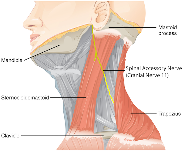
Cranial Nerve XI (Accessory Nerve) Target
Provides motor innervation specifically to the trapezius muscle.
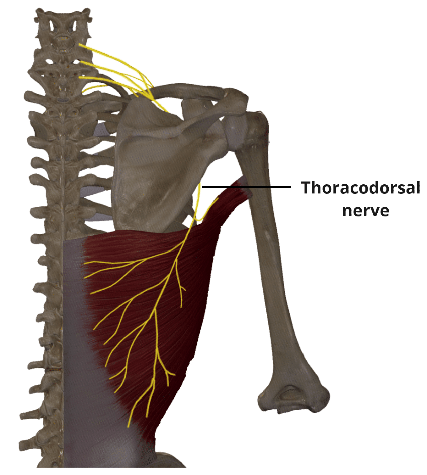
Thoracodorsal Nerve Target
Provides motor innervation specifically to the latissimus dorsi muscle.
Dorsal Scapular Nerve Targets
Provides motor innervation to levator scapulae, rhomboideus major, and rhomboideus minor.
Transverse Cervical Artery Target
Provides primary blood supply to the trapezi