Ch. 11 - Nervous Tissue Lecture
1/65
There's no tags or description
Looks like no tags are added yet.
Name | Mastery | Learn | Test | Matching | Spaced |
|---|
No study sessions yet.
66 Terms
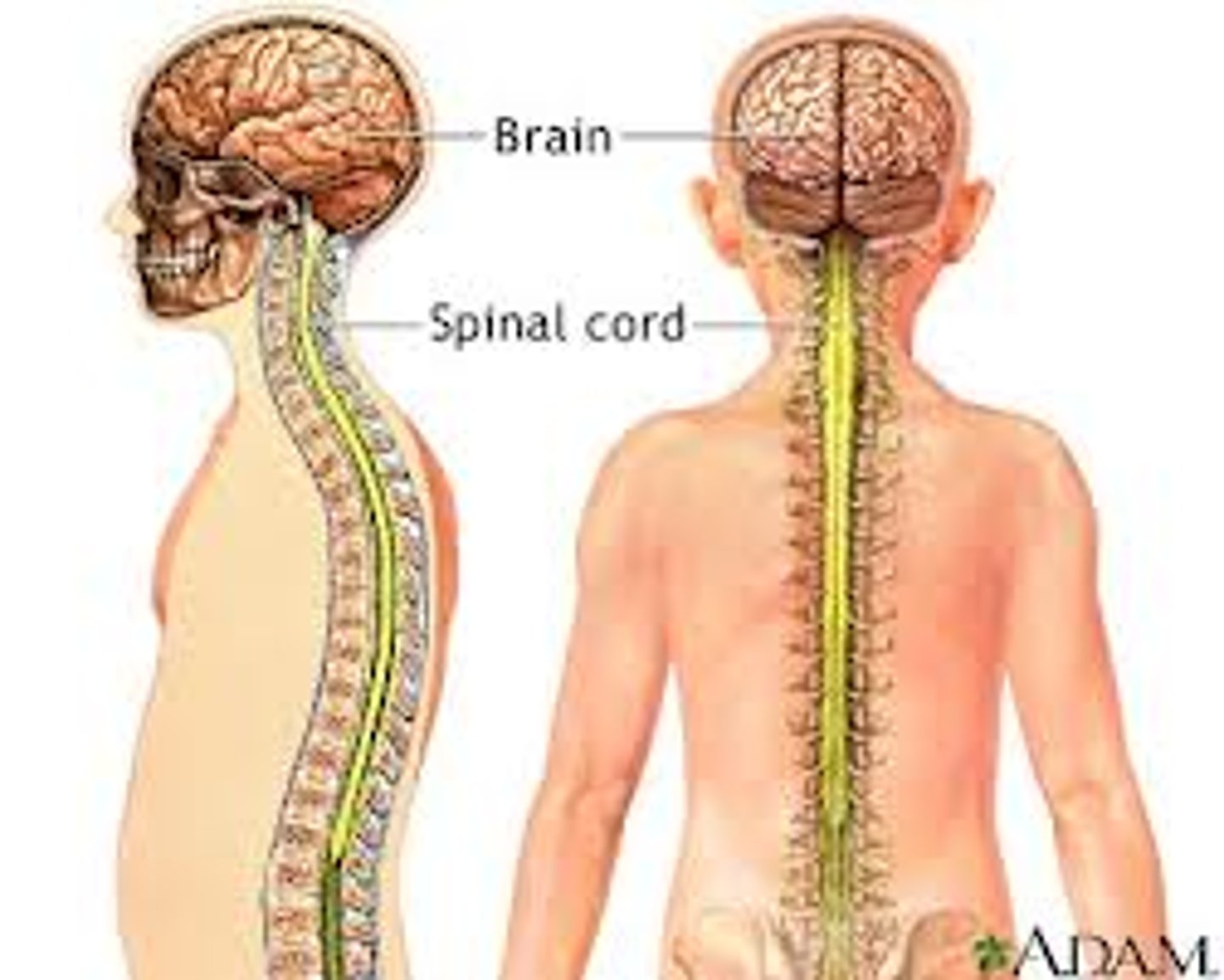
Nervous system structures
Brain
cranial nerves (12 pairs)
spinal cord
spinal nerves (31 pairs)
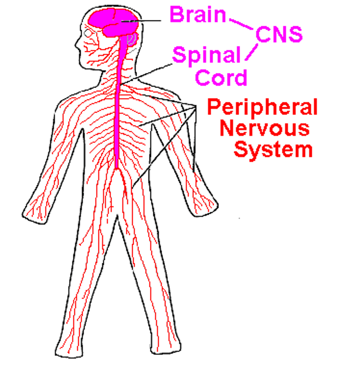
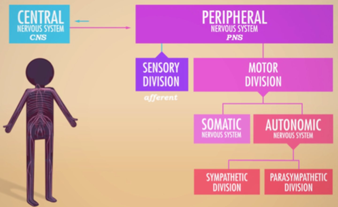
3 parts of the nervous system
Central Nervous system
Peripheral Nervous system
Enteric Nervous system
Central Nervous System (CNS)
brain + spinal cord.
Doesn’t include cranial and spinal nerves.
Processes information and integrates/coordinates both sensory and motor commands
Peripheral Nervous System (PNS)
Contains nerves (bundles of axons) ganglion (Collections of cell bodies, grey matter)
Two subsections
Sensory (afferent) and motor (efferent)
Sensory PNS
Brings information to CNS
General sensory receptors (Pain, touch, pressure, temperature)
Special sensory organs (Smell, taste, sight, sound)
Motor PNS
receives motor commands from CNS
2 subsections of PNS:
Somatic (voluntary movement such as movement of skeletal muscle)
Autonomic (involuntary movement such as smooth and cardiac muscle)
Enteric Nervous System (ENS)
focuses on the digestion of food. Known as the nervous system of the gut
Overview of nervous system function (steps)
Sensory receptors receive info, detect changes in internal/external environment
Information travels to CNS
Information gets processed in CNS
Development of motor commands in motor division of PNS
Effectors respond to motor commands
Nervous tissue cells
neurons and neuroglia

Neurons
electrically excitable-action potentials
unique functions of NS
Neuroglia
Support, nourish, and protect neurons
Divide throughout lifetime
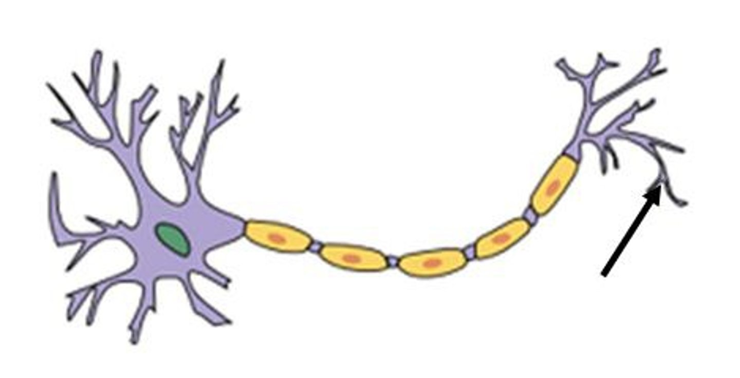
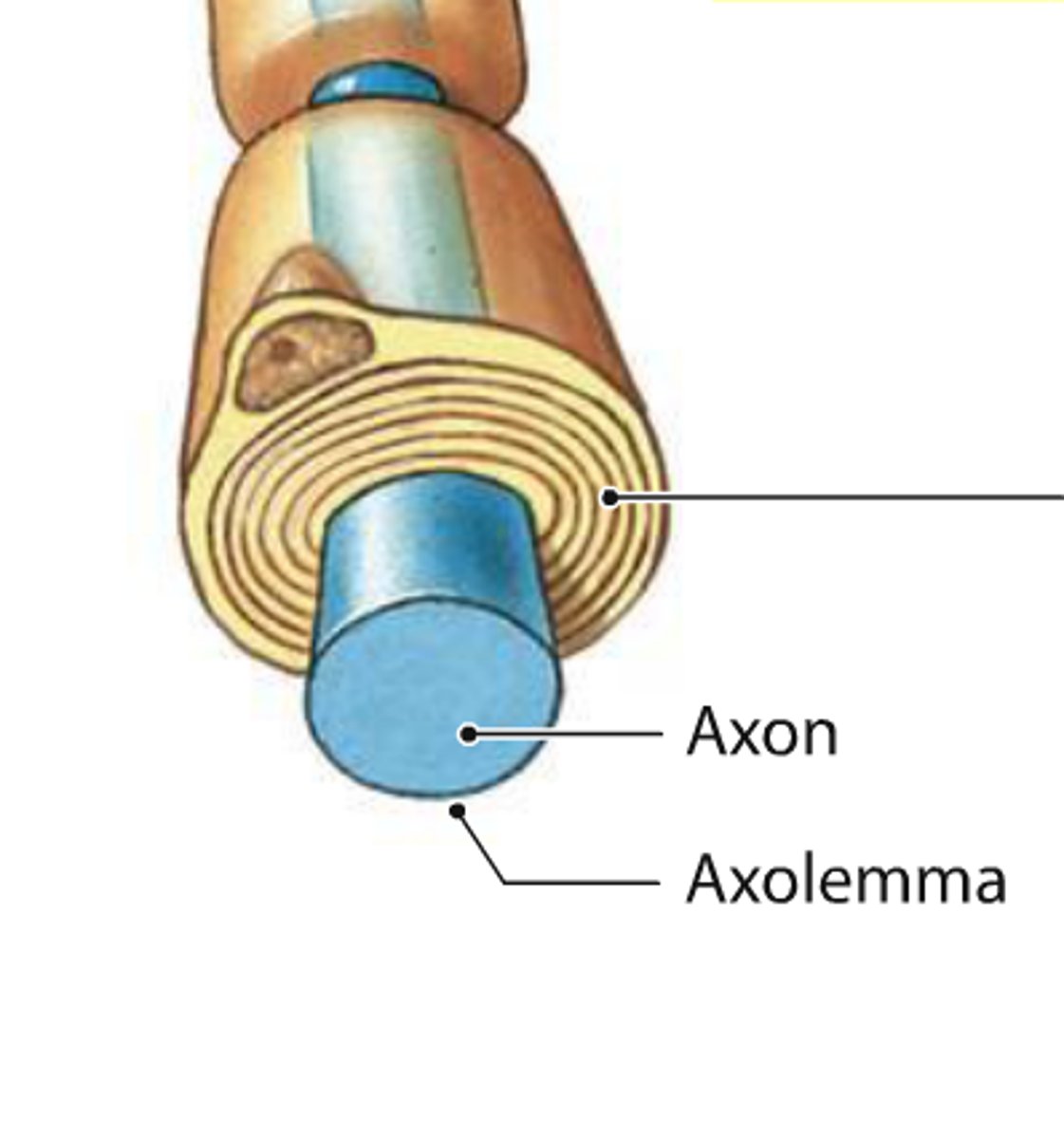
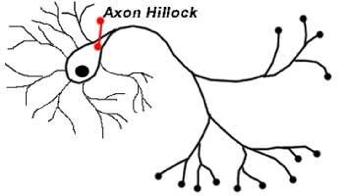
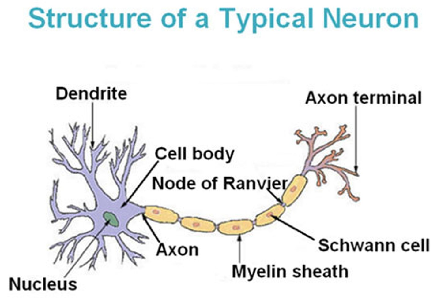
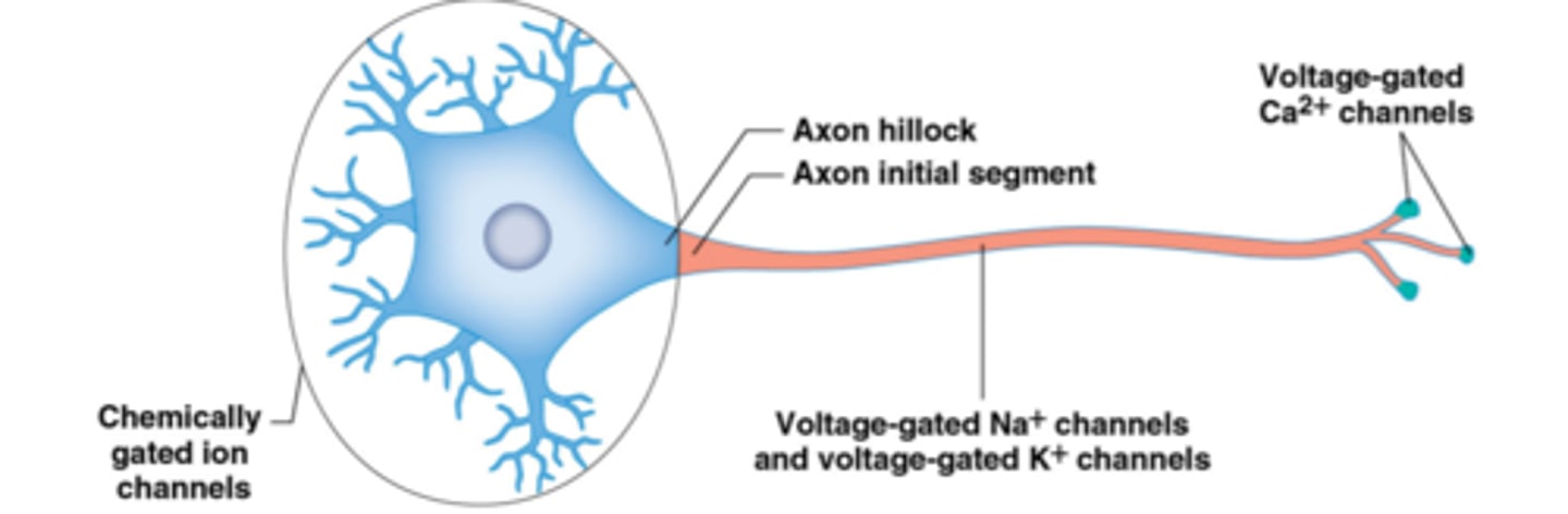
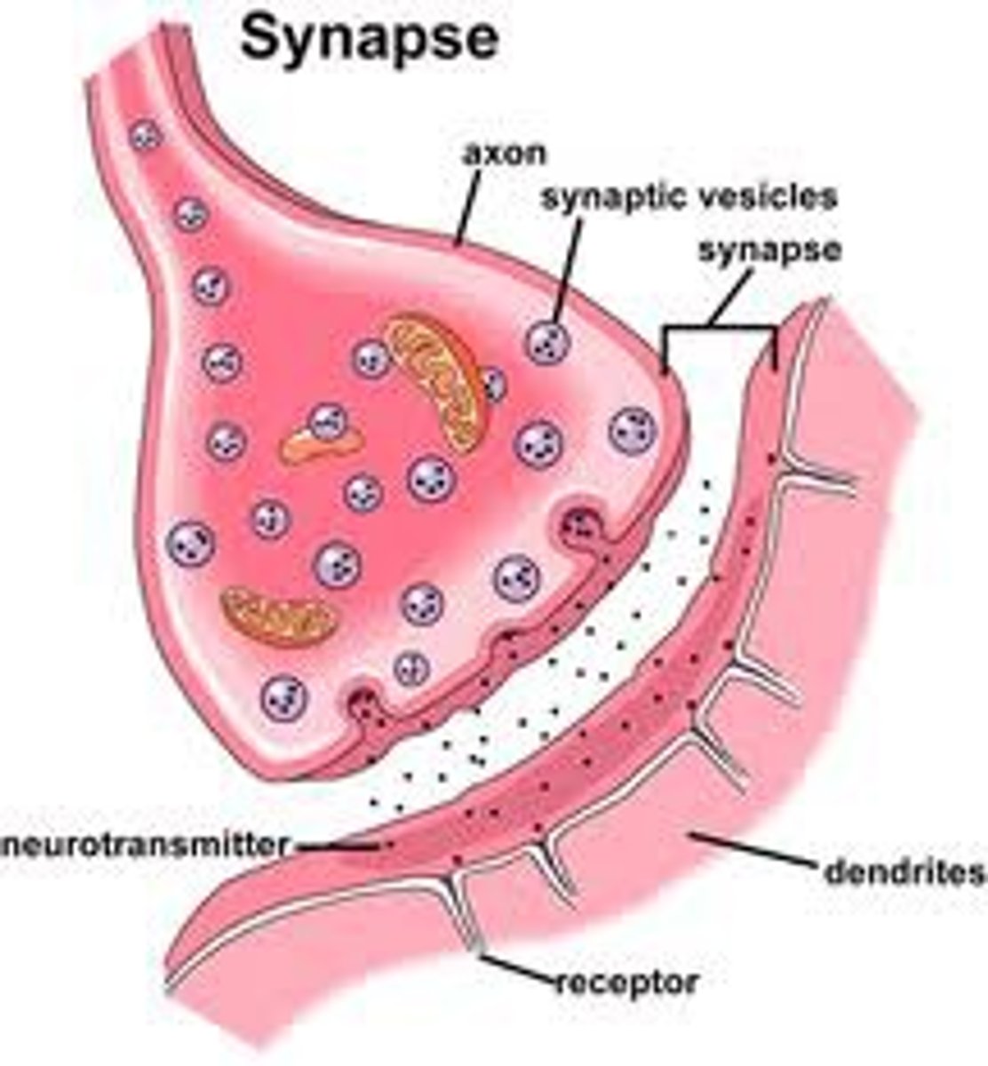
Axon
Carries info to other cells
Regions:
Axon hillock
Axolemma
Axoplasm
Axon terminals
Axon terminals
Branches at the end of the axon that contain tiny pouches, or sacs, called synaptic vesicles.
axoplasm
cytoplasm of axon
Axolemma
plasma membrane of axon
axon hillock
Cone shaped region where axon joins cell body
Where action potential begins if it decides to carry it out
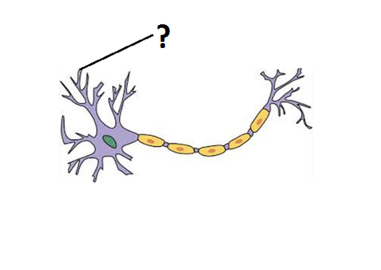
Dendrites
Receive stimuli from environment/other neurons
highly branched = dendritic spines
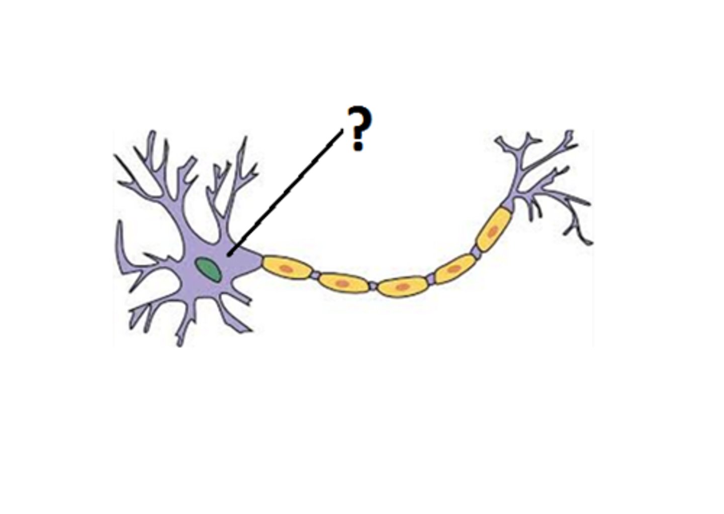
Cell body (Soma)
nucleus
cytoplasm
includes lysosomes, mitochondria, Golgi complex
Nissl bodies = rough ER
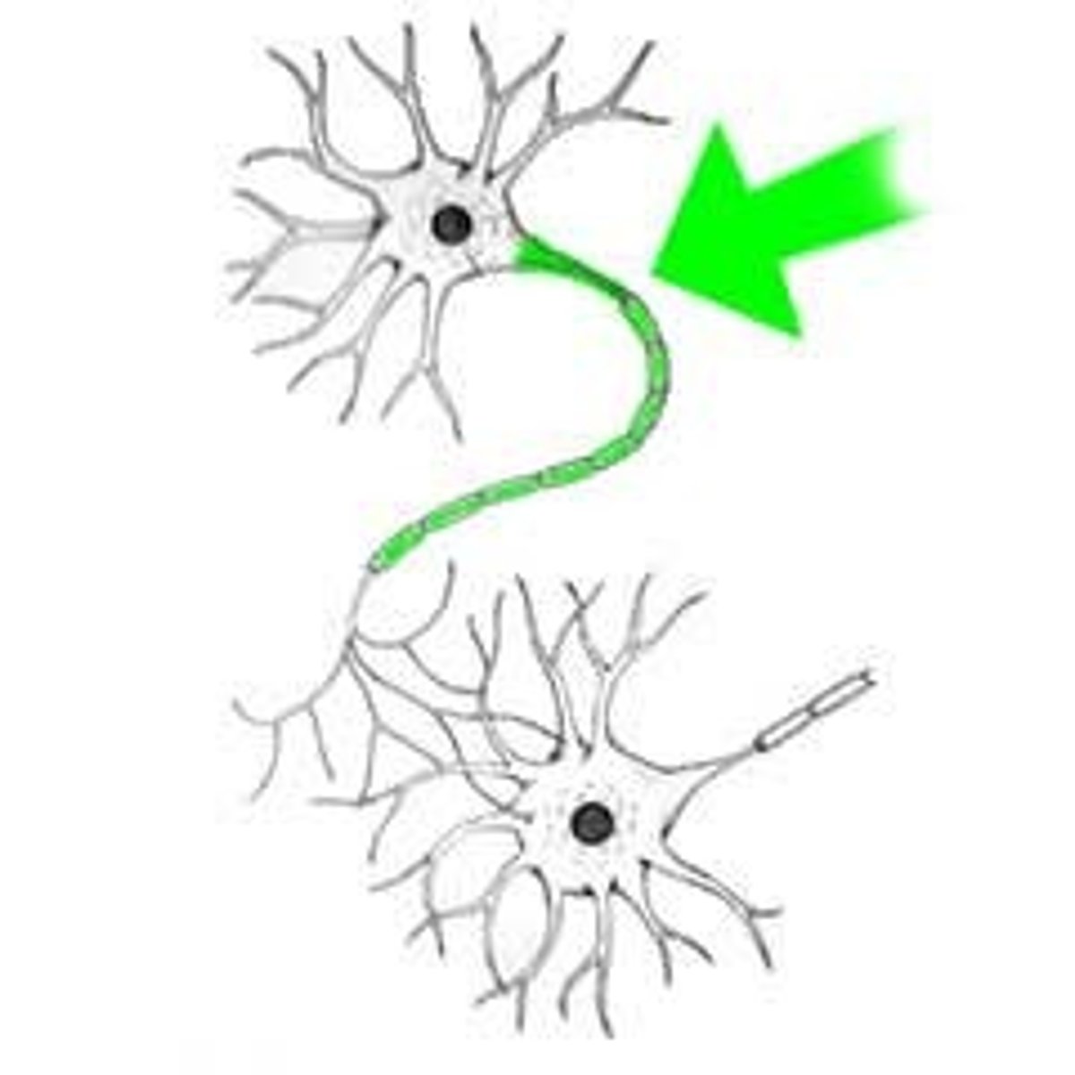
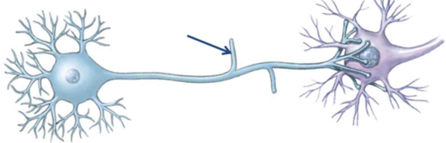
Synapse
Where neuron (presynaptic cell) communicates with another cell (postsynaptic cell)
Neurotransmitters are most common method of communication
collateral branch
single neuron branches to 1+ cell
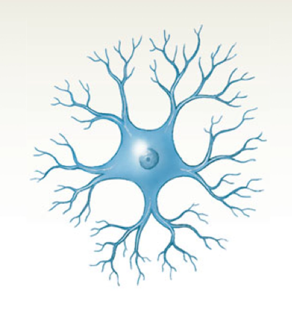
anaxonic neuron
small neurons
lack axonal distinguishing features
in brain + special sense organs
functions poorly understood
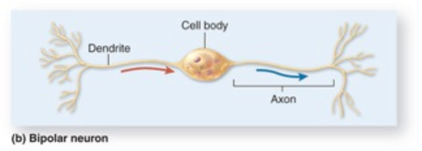
bipolar neurons
2 distinct processes
1 dendritic process
1 axon.
These are small, rare
in special sense organs
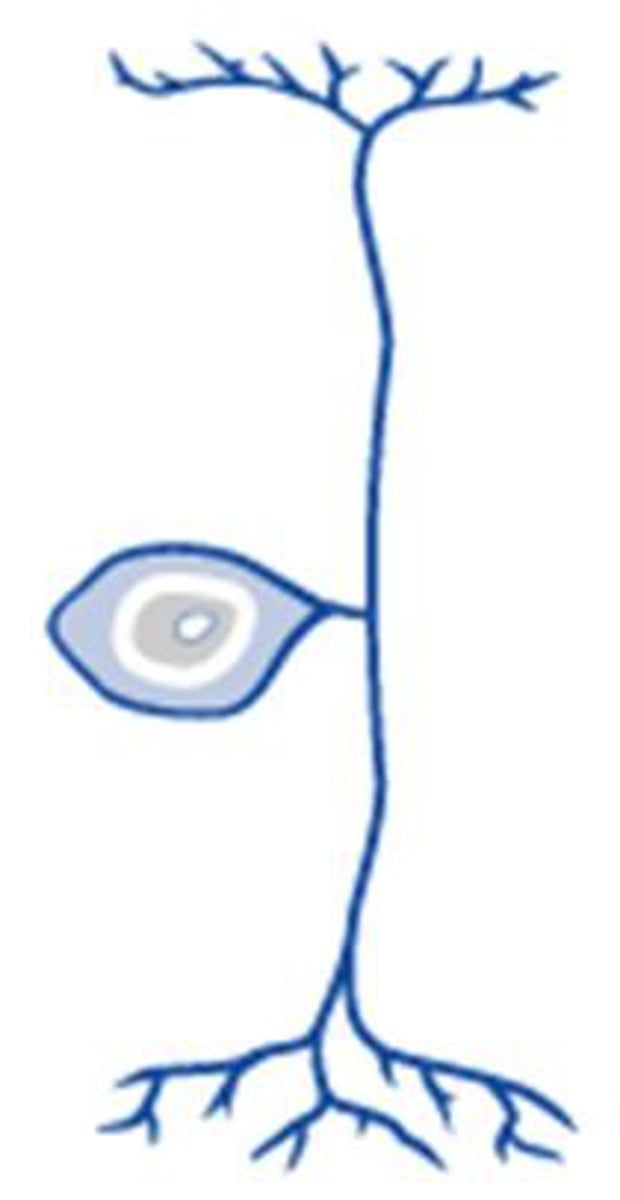
Unipolar neurons
Dendrites + axon are continuous (fused)
Most neurons in PNS
can be over 1 meter long
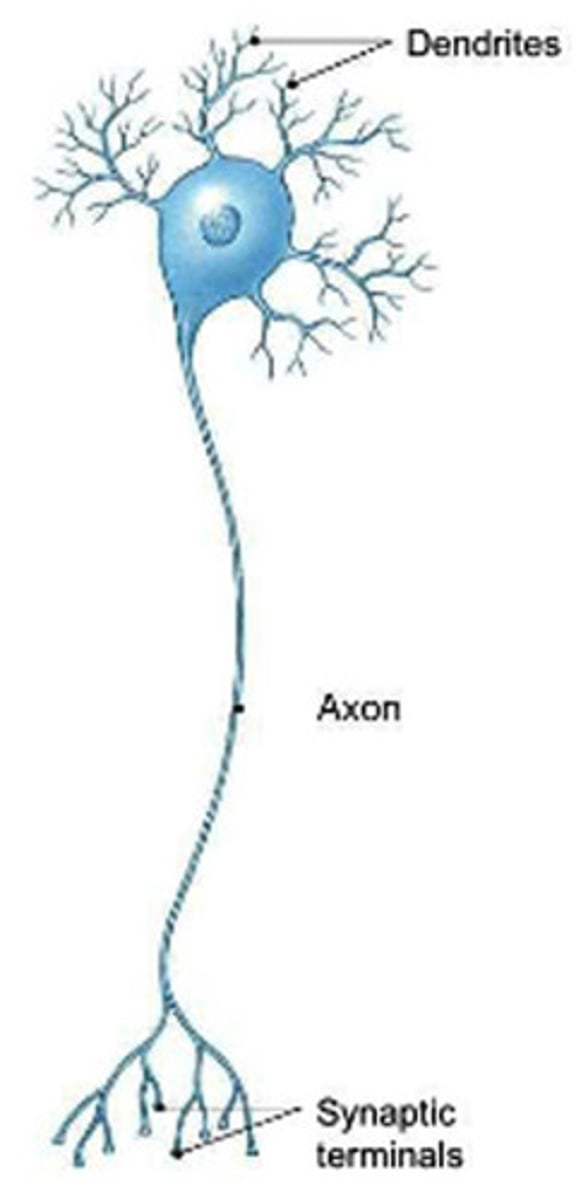
Multipolar neurons
2+ dendrites + 1 axon
Most neurons in CNS
make up all motor neurons to skeletal muscles
may be long
sensory (afferent) neurons
Receiving stimuli and sends information to CNS
Mostly Unipolar
Contain Sensory ganglia (collection of neuron cell bodies in PNS)
Somatic sensory neurons: outside world
Visceral sensory neurons: internal systems
Different receptors on sensory neuronsi:
interoceptors; internal organs
proprioceptors; position/movement of skeletal muscles and joints
exteroceptors: external environment
interneuron
In CNS
between sensory and motor neurons.
Receive info from PNS & CNS
responsible for higher functions (memory and learning)
Motor (efferent) neurons
Send signals from CNS to muscles + glands (effectors)
2 types:
Somatic motor neurons → skeletal muscles
voluntary
cell body in CNS
Visceral motor neuron → smooth, cardiac muscle, glands, adipose tissue
CNS neuroglia
Support, protect, nourish neurons.
smaller than neurons, more numerus
~1/2 volume of nervous system
no action potential
cell division in mature nervous system
myelinated axons in CNS
axons w/ myelin sheaths (CNS white matter)
internodes - myelin-wrapped areas
Nodes of Ranvier
gaps between internodes
unmyelinated axon in CNS
axons without myelin sheaths
CNS grey matter
cell bodies, dendrites, unmyelinated axons
PNS neuroglia
2 types:
Schwann cell: produce myelin in PNS (A in image).
Satellite cells: surround peripheral cell bodies
regulate environment around neurons (B in image)
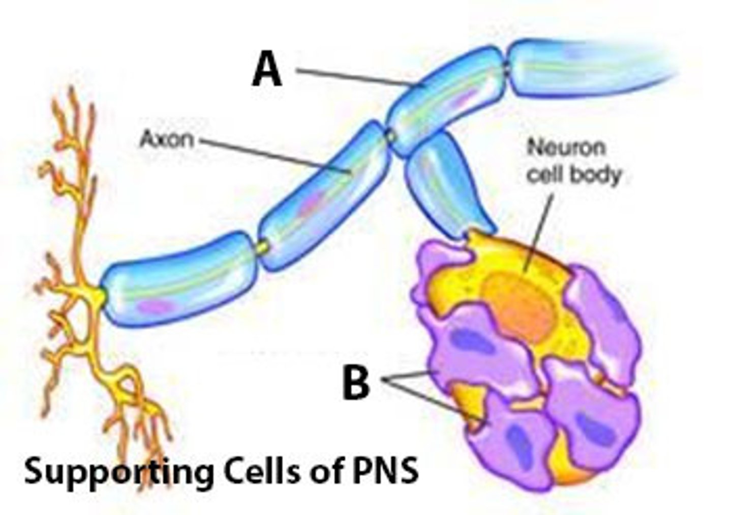
Myelinated axons in PNS
Formed by myelinating Schwann cells
A single Schwann cell wraps a single internode
Unmyelinated axons in PNS
single nonmyelinating Schwann cell wraps around segments of a group of axons
No nodes of ranvier
Axon injury and repair in CNS
limited because…
many more axons involved
astrocytes make scar tissue that can block axon growth in damaged area
astrocytes release chemicals blocking axon regrowth
Membrane potential
From unequal charge distribution across membrane
Due to…
difference in membrane permeability
active transport mechanisms
Contributors to resting membrane potential
High Na+ concentration in extracellular fluid
High K+ concentration in cytosol
Maintaining Resting membrane potential
Leak channels:
passive
allow K+ to leave, Na+ to enter
Sodium-potassium pump:
active
ejects 3 Na+ ions for every 2 K+ ions that are brought back into the cell
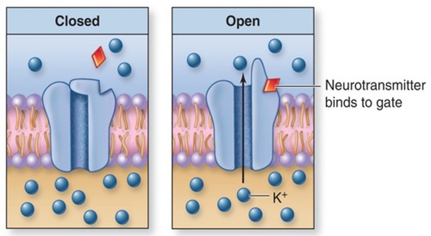
chemically gated channels
ligand-gated ion channels
Open when they bind specific chemicals
Ex: neurotransmitters, hormones, ions
Most abundant on dendrite and cell bodies
Voltage-gated channels
Changes in membrane potential open the gate
sodium channels w/ 2 gates - activation gate, inactivation gate
Na+ and K+ channels along axon
Ca2+ channels along axon terminal
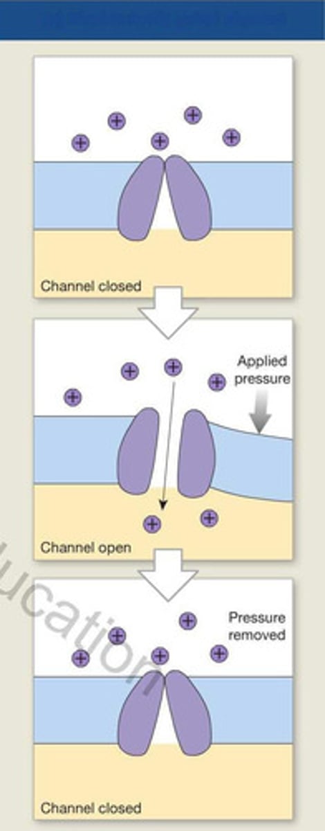
Mechanically gated channels
Open in response to physical distortion of membrane surface
(stretch, pressure, touch, vibration)
Graded potentials
stimulus causes ligand/mechanically gated ion channel to open
@ resting potential, chemically gates channels are closed
membrane expose to chemical that opens chemically gates Na channels
gates open, Na+ enters
shift from resting potential to + value
Sodium ions entering move away from channels (attract to - charges along inner surface)
local current
changes in membrane potential
When a chemical stimulus opens sodium ion channels, depolarization occurs (Na+ comes into cell) making the membrane potential more positive (excitatory response)
When a chemical stimulus opens a potassium channel, potassium leaves the cell, making membrane potential more negative (inhibitory response)
Pain
degree of depolarization decreases with distance from stimulus
The change of membrane potential is proportional to the stimulus size
Potentials and their channel type
depends on leak channels
Graded potentials depends on chemically gated channels
Action potentials depend on voltage gated channels
excitatory (depolarizing)
inhibitory (hyperpolarizing)
Action potentials
All-or-none
irreversible
once formed, always the same amplitude
rapid (1 ms)
reverse membrane potential, then restores it
short and long distance communication
runs along axon
steps of action potential generation
1. Depolarization to threshold
2. Activation of Na+ channels, rapid depolarization
3. Inactivation of Na+ channels and activation of K+ channels
4. Potassium on channels close, return to resting membrane potential
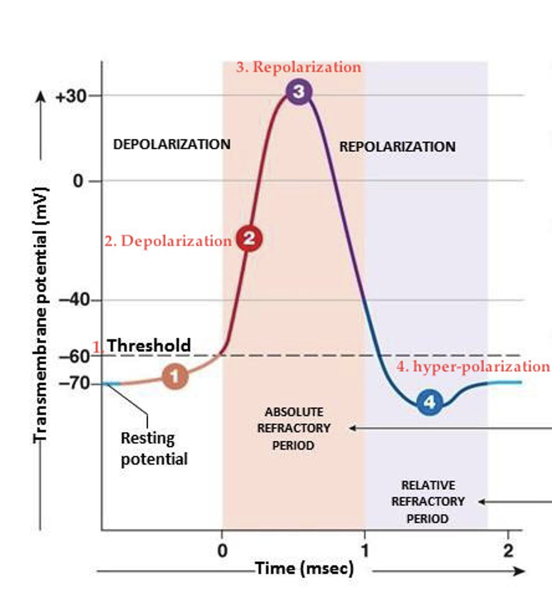
abolute refractory period
the time following an action potential which a new action potential cannot be initiated (can't respond to another stimulus)
relative refractory period
A period after firing when a neuron is returning to its normal polarized state and will fire again only if the incoming message is much stronger than usual
propogation of the action potential
transmission of an action potential down an axon
2 types:
Continuous propagation: action potential appears to move step by step through the entire axon
Occurs in unmyelinated axons, much SLOWER
Saltatory propagation: in myelinated axons, depolarizes only at nodes
faster, speed varies w/ axon diameter
Synapses
Information is transferred from neuron to neuron or from neuron to an effector cell.
2 types:
1. Chemical synapse
2. Electrical synapse
Chemical synapses
Most abundant type of synapse
neurotransmitter
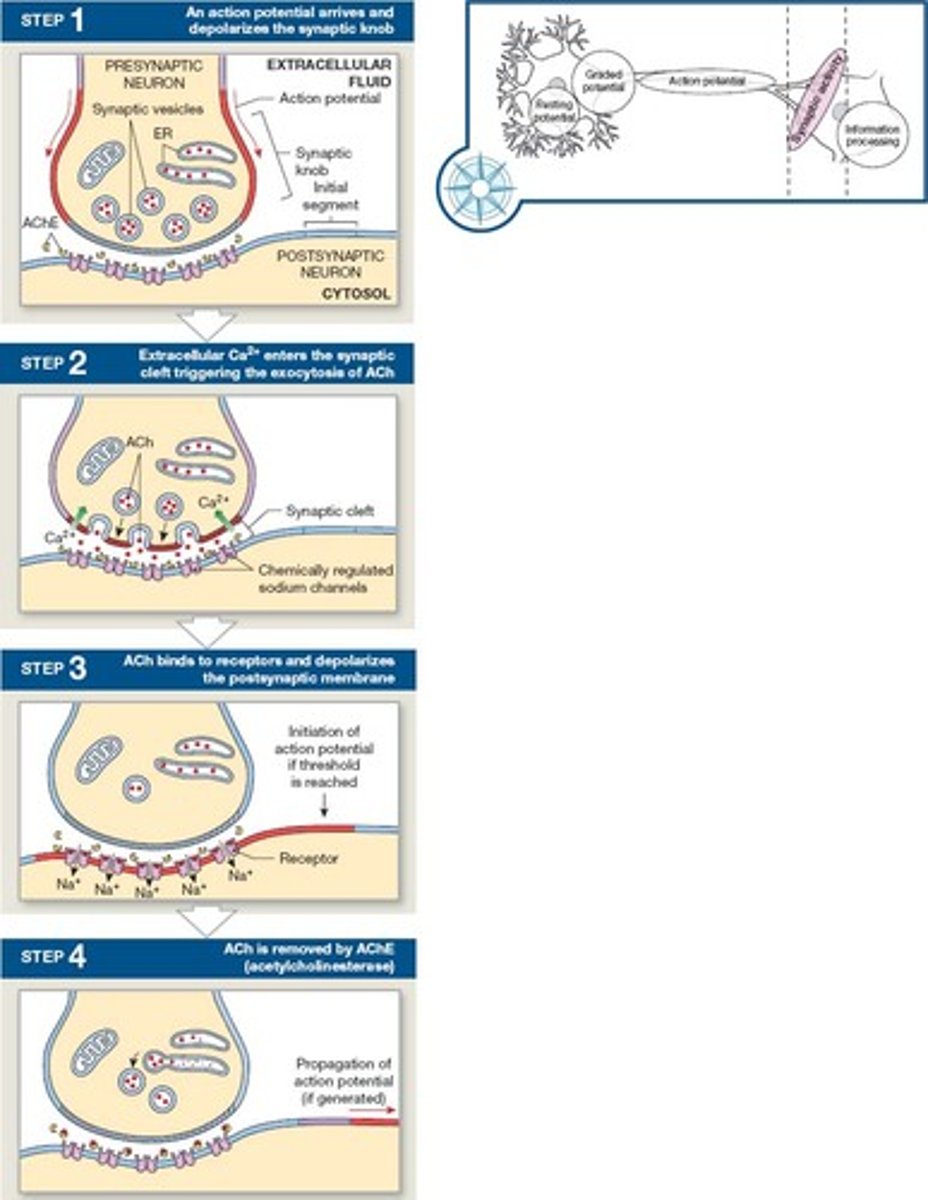
Cholinergic synapses - release acetylcholine
most common
steps of cholinergic synapse
1. Axon terminal depolarized by arriving action potential.
2. Depolarization opens voltage gated calcium channels
3. ACh diffuses across synaptic cleft, binds to chemically gated Na+ channel receptors on postsynaptic membrane
4. Effects on the postsynaptic membrane are temporary
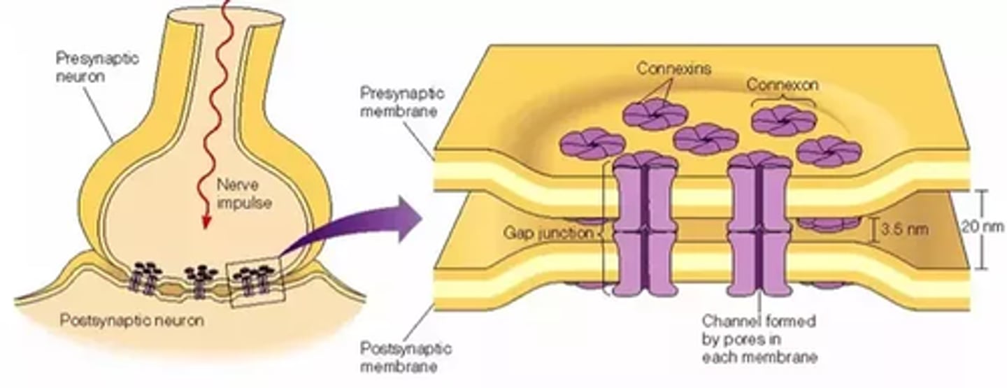
Electrical synapses
contain tunnels called gap junctions
connect pre and postsynaptic neurons
rare
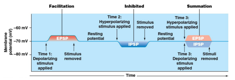
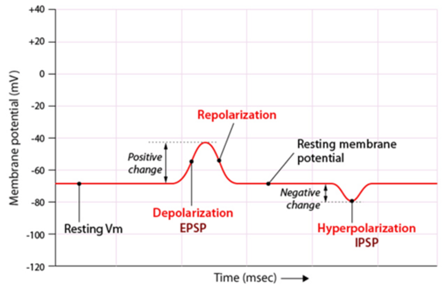
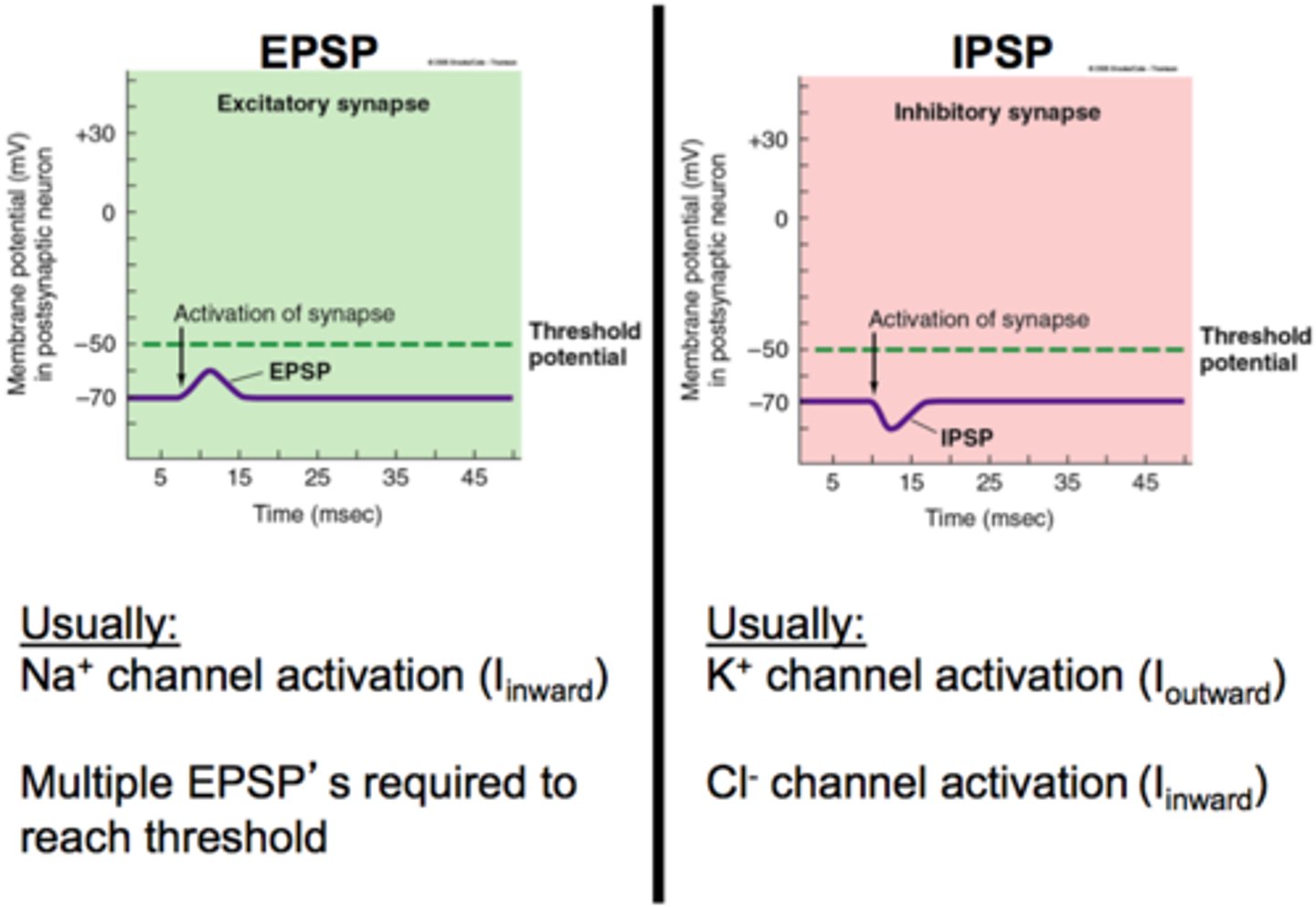
postsynaptic potential
graded potential in postsynaptic membrane in response to a neurotransmitter
1. Excitatory postsynaptic potential (EPSP):
graded depolarization, shifts membrane potential closer to threshold
2. Inhibitory postsynaptic potential (IPSP):
Graded hyperpolarization, shifts membrane potential farther away from threshold
autonomic nervous system
(involuntary movement such as smooth and cardiac muscle)
2 Subsections
Sympathetic: Stress, “fight or flight”
Parasympathetic: Relaxed, “rest and digest”
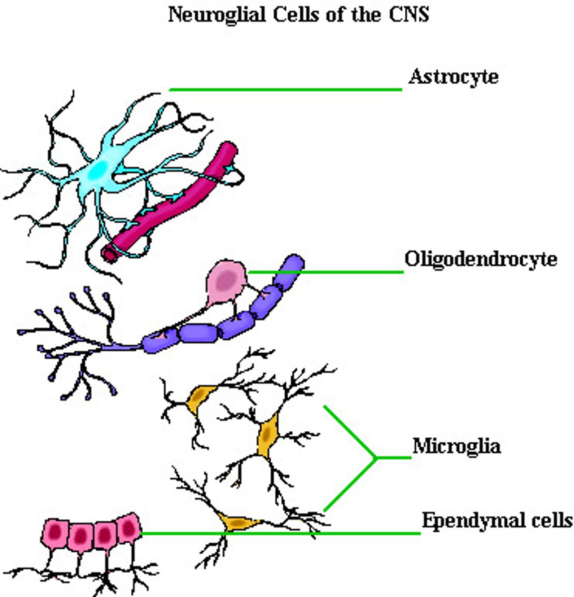
Ependymal cells
lines central canal (spinal cord) + ventricles (brain)
produce, circulate, monitor cerebrospinal fluid (CSF)
Microglia
engulf invading microbes
clear cell debris + waste products
Astrocytes
form blood-brain barrier
maintain ion, nutrient, gas concentration
take up excess neurotransmitters
Oligodendrocytes
support fibers in CNS
produce myelin
resting membrane potential
potential of neuron to send signals
-70 mV
inside more negative than outside
contributors to resting membrane potential
extracellular fluid (ECF) - high Na+, Cl-
cytosol - high K+, Pr-
graded potential
temporary local change
decreases with distance
from stimulus
action potential
electrical event
triggered by sufficiently large graded potential
synaptic activity
presynaptic - releases neurotransmitters
postsynaptic - bind neurotransmitter to receptors
changes permeability
produce graded potentials
summation
integration of effects of graded potentials
collective effects of both EPSPs and IPSPs
net effect may be no change in membrane potential