ST Imaging Final Image Study Questions

0.0(0)
Studied by 0 people
call kaiCall Kai
Locked
learnLearn
examPractice Test
spaced repetitionSpaced Repetition
heart puzzleMatch
flashcardsFlashcards
GameKnowt Play
Card Sorting

1/53

encourage image

There's no tags or description

Looks like no tags are added yet.

Last updated 3:48 PM on 9/2/26
Name
Mastery
Learn
Test
Matching
Spaced
Call with Kai
Chat

No analytics yet

Send a link to your students to track their progress

54 Terms

1
New cards

d. fibrous dysplasia

Based on the appearance of the right 4th posterior rib lesion what is the best differential diagnosis:

a. osteochondroma

b. Ewings sarcoma

c. metastasis

d. fibrous dysplasia

<p>Based on the appearance of the right 4th posterior rib lesion what is the best differential diagnosis:</p><p>a. osteochondroma </p><p>b. Ewings sarcoma </p><p>c. metastasis </p><p>d. fibrous dysplasia</p>
2
New cards

a. superior sulcus tumor

The prominent lesion in the right lung field is best described as:

a. superior sulcus tumor

b. right middle lobe atelectasis

c. pneumonia granuloma

d. hamartoma

<p>The prominent lesion in the right lung field is best described as:</p><p>a. superior sulcus tumor </p><p>b. right middle lobe atelectasis </p><p>c. pneumonia granuloma </p><p>d. hamartoma</p>
3
New cards

c. retrocardiac space

What anatomical space is shown by the arrow:

a. retrovertebral space

b. retrosternal space

c. retrocardiac space

d. subdiaphragmatic space

<p>What anatomical space is shown by the arrow:</p><p>a. retrovertebral space </p><p>b. retrosternal space </p><p>c. retrocardiac space </p><p>d. subdiaphragmatic space</p>
4
New cards

c. eventration

The finding along the right hemidiaphragm is termed:

a. interstitial lung disease e

b. epicardial fat pad

c. eventration

d. hiatal hernia

<p>The finding along the right hemidiaphragm is termed:</p><p>a. interstitial lung disease e</p><p>b. epicardial fat pad </p><p>c. eventration</p><p>d. hiatal hernia</p>
5
New cards

b. False

T/F: A pleural effusion is present:

a. True

b. False

<p>T/F: A pleural effusion is present:</p><p>a. True</p><p>b. False</p>
6
New cards

c. Schwannoma

Which of the following is NOT a good differential for conditions at this location:

a. Lymphoma

b. Thymoma

c. Schwannoma

d. Goiter

<p>Which of the following is NOT a good differential for conditions at this location:</p><p>a. Lymphoma </p><p>b. Thymoma </p><p>c. Schwannoma </p><p>d. Goiter</p>
7
New cards

a. air bronchogram sign

What name sign is present in the right lung abnormality confirming airspace disease:

a. air bronchogram sign

b. meniscus sign

c. silhouette sign

d. luftsichel sign

<p>What name sign is present in the right lung abnormality confirming airspace disease:</p><p>a. air bronchogram sign </p><p>b. meniscus sign </p><p>c. silhouette sign</p><p>d. luftsichel sign</p>
8
New cards

b. False

T/F: The lesion indicated by the arrows silhouettes the right heart border:

a. True

b. False

<p>T/F: The lesion indicated by the arrows silhouettes the right heart border:</p><p>a. True</p><p>b. False</p>
9
New cards

b. mass

What pattern of lung disease is shown:

a. airspace

b. mass

c. consolidation

d. interstitial

<p>What pattern of lung disease is shown:</p><p>a. airspace </p><p>b. mass </p><p>c. consolidation </p><p>d. interstitial</p>
10
New cards

c. meniscus sign

What name sign is shown in the image above:

a. batwing sign

b. cat under the rug sign

c. meniscus sign

d. air bronchogram sign
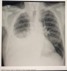
<p>What name sign is shown in the image above:</p><p>a. batwing sign </p><p>b. cat under the rug sign </p><p>c. meniscus sign</p><p>d. air bronchogram sign</p>
11
New cards

d. RML

Based on the image what lobe is collapsed:

a. RLL

b. RUL

c. LLL

d. RML

<p>Based on the image what lobe is collapsed:</p><p>a. RLL </p><p>b. RUL </p><p>c. LLL</p><p>d. RML</p>
12
New cards

a. left lower lung zone

A large pulmonary lesion is present How would you best describe its location:

a. left lower lung zone

b. left upper lung zone

c. right lower lung zone

d. right upper lung zone

<p>A large pulmonary lesion is present How would you best describe its location:</p><p>a. left lower lung zone </p><p>b. left upper lung zone </p><p>c. right lower lung zone </p><p>d. right upper lung zone</p>
13
New cards

d. mass

What pattern of lung disease does the lesion fit into:

a. interstitial

b. airspace

c. reticular

d. mass

<p>What pattern of lung disease does the lesion fit into:</p><p>a. interstitial </p><p>b. airspace </p><p>c. reticular </p><p>d. mass</p>
14
New cards

a. posterior mediastinum

Based on the position of the lesion on frontal and lateral projections this lesion is localized to what location:

a. posterior mediastinum

b. middle mediastinum

c. superior mediastinum

d. anterior mediastinum

<p>Based on the position of the lesion on frontal and lateral projections this lesion is localized to what location:</p><p>a. posterior mediastinum </p><p>b. middle mediastinum </p><p>c. superior mediastinum </p><p>d. anterior mediastinum</p>
15
New cards

c. hilum

What anatomical location are the arrows pointing to:

a. upper lung zone

b. cardiac silhouette

c. hilum

d. aortic arch

<p>What anatomical location are the arrows pointing to:</p><p>a. upper lung zone </p><p>b. cardiac silhouette </p><p>c. hilum </p><p>d. aortic arch</p>
16
New cards

a. tall thin female

What is the typical patient presentation for this radiographic finding:

a. tall thin female

b. old obese female

c. old obese male

d. tall thin male

<p>What is the typical patient presentation for this radiographic finding:</p><p>a. tall thin female </p><p>b. old obese female </p><p>c. old obese male </p><p>d. tall thin male</p>
17
New cards

a. pectus excavatum

The finding indicated by the arrow is termed:

a. pectus excavatum

b. anterior mediastinal mass

c. sternum fracture

d. pectus carinatum

<p>The finding indicated by the arrow is termed:</p><p>a. pectus excavatum </p><p>b. anterior mediastinal mass </p><p>c. sternum fracture </p><p>d. pectus carinatum</p>
18
New cards

c. CT lung window

What imaging modality and window is shown:

a. MRI T2

b. MRI T1

c. CT lung window

d. CT bone window

<p>What imaging modality and window is shown:</p><p>a. MRI T2 </p><p>b. MRI T1 </p><p>c. CT lung window </p><p>d. CT bone window</p>
19
New cards

c. right rib 9

What rib demonstrates acute fracture:

a. left rib 9

b. right rib 3

c. right rib 9

d. left rib 7

<p>What rib demonstrates acute fracture:</p><p>a. left rib 9 </p><p>b. right rib 3 </p><p>c. right rib 9 </p><p>d. left rib 7</p>
20
New cards

b. pectus excavatum

The patient has:

a. sternum fracture

b. pectus excavatum

c. pectus carinatum

d. anterior mediastinal mass

<p>The patient has:</p><p>a. sternum fracture </p><p>b. pectus excavatum </p><p>c. pectus carinatum </p><p>d. anterior mediastinal mass</p>
21
New cards

a. Any of the above

Based on the image what does the patient have?

a. Any of the above

b. hydrothorax

c. chylothorax

d. hemothorax

<p>Based on the image what does the patient have? </p><p>a. Any of the above </p><p>b. hydrothorax </p><p>c. chylothorax </p><p>d. hemothorax</p>
22
New cards

b. Thoracic aneurysm

Based on the image what is NOT an appropriate differential:

a. Lymphoma

b. Thoracic aneurysm

c. Metastasis

d. Goiter
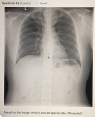
<p>Based on the image what is NOT an appropriate differential:</p><p>a. Lymphoma </p><p>b. Thoracic aneurysm</p><p>c. Metastasis </p><p>d. Goiter</p>
23
New cards

a. Congestion

Based on the pattern of lung disease shown which of the following is NOT one of the classic clinical presentations:

a. Congestion

b. Cough

c. Crackles

d. Clubbing of nails

<p>Based on the pattern of lung disease shown which of the following is NOT one of the classic clinical presentations:</p><p>a. Congestion </p><p>b. Cough</p><p>c. Crackles </p><p>d. Clubbing of nails</p>
24
New cards

d. Superior sulcus tumor

Based on the chest wall findings which of the following should NOT be considered a differential:

a. Old rib fracture

b. Intercostal schwannoma

c. Pleural fibroma

d. Superior sulcus tumor

<p>Based on the chest wall findings which of the following should NOT be considered a differential:</p><p>a. Old rib fracture </p><p>b. Intercostal schwannoma </p><p>c. Pleural fibroma </p><p>d. Superior sulcus tumor</p>
25
New cards

b. Snowman sign

c. Pituitary macroadenoma

Please choose the TWO most appropriate findings/conditions for this 50-year-old male with headaches and loss of vision.

a. Hydrocephalus

b. Snowman sign

c. Pituitary macroadenoma

d. Chiari II malformation

e. Spinal dysraphism

<p>Please choose the TWO most appropriate findings/conditions for this 50-year-old male with headaches and loss of vision. </p><p>a. Hydrocephalus </p><p>b. Snowman sign </p><p>c. Pituitary macroadenoma </p><p>d. Chiari II malformation </p><p>e. Spinal dysraphism</p>
26
New cards

c. Optic chiasm

With the patient's history of blindness, which structure is likely involved in the patient's condition?

a. Cranial nerve II

b. Corpus colosum

c. Optic chiasm

d. Cranial nerve III
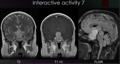
<p>With the patient's history of blindness, which structure is likely involved in the patient's condition? </p><p>a. Cranial nerve II </p><p>b. Corpus colosum </p><p>c. Optic chiasm </p><p>d. Cranial nerve III</p>
27
New cards

c. Neurosurgical referral

What is the best next step for this patient?

a. EENT referral

b. Chiropractic referral

c. Neurosurgical referral

d. Ophthalmologist referral

<p>What is the best next step for this patient? </p><p>a. EENT referral </p><p>b. Chiropractic referral </p><p>c. Neurosurgical referral </p><p>d. Ophthalmologist referral</p>
28
New cards

a. Infection

d. Prevertebral soft tissue swelling

Please choose the TWO most appropriate findings/conditions for this 30-month-old female with noisy breathing and growing neck mass.

a. Infection

b. Zenker diverticulum

c. Esophageal varices

d. Prevertebral soft tissue swelling

e. Trauma
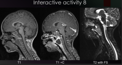
<p>Please choose the TWO most appropriate findings/conditions for this 30-month-old female with noisy breathing and growing neck mass. </p><p>a. Infection </p><p>b. Zenker diverticulum </p><p>c. Esophageal varices </p><p>d. Prevertebral soft tissue swelling </p><p>e. Trauma</p>
29
New cards

a. Yes

This is an emergency referral.

a. Yes

b. No

<p>This is an emergency referral. </p><p>a. Yes </p><p>b. No</p>
30
New cards

a. Yes

Would this lead to difficulty swallowing?

a. Yes

b. No
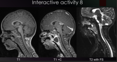
<p>Would this lead to difficulty swallowing? </p><p>a. Yes </p><p>b. No</p>
31
New cards

c. Calcifications in hilar region

d. Pericardial fat pad

Please choose the TWO best findings/conditions for this chest radiograph of a 30 year old male. Select 2 correct answer(s)

a. Absent heart syndrome

b. Bilateral tenting of the diaphragm

c. Calcifications in hilar region

d. Pericardial fat pad

e. Tracheal deviation

<p>Please choose the TWO best findings/conditions for this chest radiograph of a 30 year old male. Select 2 correct answer(s) </p><p>a. Absent heart syndrome </p><p>b. Bilateral tenting of the diaphragm </p><p>c. Calcifications in hilar region </p><p>d. Pericardial fat pad </p><p>e. Tracheal deviation</p>
32
New cards

b. PA at 72" FFD

What are the standard patient position and x-ray tube position for a chest radiograph?

a. PA at 40" FFD

b. PA at 72" FFD

c. AP at 40" FFD

d. AP at 72" FFD

<p>What are the standard patient position and x-ray tube position for a chest radiograph? </p><p>a. PA at 40" FFD </p><p>b. PA at 72" FFD </p><p>c. AP at 40" FFD </p><p>d. AP at 72" FFD</p>
33
New cards

a. No further action necessary

Based on the radiographic findings, how would this patient best be managed?

a. No further action necessary

b. Blood evaluation for lymphoma

c. Consult with oncologist

d. Referral to pulmonologist

<p>Based on the radiographic findings, how would this patient best be managed? </p><p>a. No further action necessary </p><p>b. Blood evaluation for lymphoma </p><p>c. Consult with oncologist </p><p>d. Referral to pulmonologist</p>
34
New cards

a. Fluid in the pleural space

d. Obscured right heart and diaphragm border

Please choose the TWO best findings/conditions for this 25 year old male with chest pain and difficulty breathing. Select 2 correct answer(s)

a. Fluid in the pleural space

b. Abdominal contents within right lung space

c. Large tumor of the diaphragm

d. Obscured right heart and diaphragm border

e. Interstitial lung disease

<p>Please choose the TWO best findings/conditions for this 25 year old male with chest pain and difficulty breathing. Select 2 correct answer(s) </p><p>a. Fluid in the pleural space </p><p>b. Abdominal contents within right lung space </p><p>c. Large tumor of the diaphragm </p><p>d. Obscured right heart and diaphragm border </p><p>e. Interstitial lung disease</p>
35
New cards

d. Chest CT

What imaging modality is the best follow up?

a. Apical lordotic chest radiograph

b. Abdominal ultrasound

c. Abdominal CT

d. Chest CT
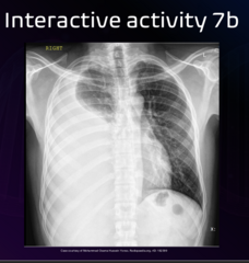
<p>What imaging modality is the best follow up? </p><p>a. Apical lordotic chest radiograph </p><p>b. Abdominal ultrasound </p><p>c. Abdominal CT </p><p>d. Chest CT</p>
36
New cards

a. Emergency referral to the hospital

Given the patient symptoms and radiographic findings, what is the best next course of action?

a. Emergency referral to the hospital

b. Referral to primary care physician

c. Trial of chiropractic care

d. No follow-up necessary
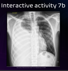
<p>Given the patient symptoms and radiographic findings, what is the best next course of action? </p><p>a. Emergency referral to the hospital </p><p>b. Referral to primary care physician </p><p>c. Trial of chiropractic care </p><p>d. No follow-up necessary</p>
37
New cards

d. right middle

What lung lobe is highlighted in green?

a. lingula

b. right lower

c. left lower

d. right middle

<p>What lung lobe is highlighted in green?</p><p>a. lingula </p><p>b. right lower </p><p>c. left lower </p><p>d. right middle</p>
38
New cards

a. air bronchogram sign

The left lung demonstrates opacification. The radiating air densities noted superimposed over the region of increased density is termed:

a. air bronchogram sign

b. target sign

c. bulging fissure sign

d. silhouette sign
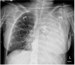
<p>The left lung demonstrates opacification. The radiating air densities noted superimposed over the region of increased density is termed: </p><p>a. air bronchogram sign </p><p>b. target sign </p><p>c. bulging fissure sign </p><p>d. silhouette sign</p>
39
New cards

c. right lung apex

A pulmonary mass is present. Identify the location.

a. left lung apex

b. right lower lung zone

c. right lung apex

d. left lower lung zone

<p>A pulmonary mass is present. Identify the location. </p><p>a. left lung apex </p><p>b. right lower lung zone </p><p>c. right lung apex </p><p>d. left lower lung zone</p>
40
New cards

a. Anterior mediastinum

Where is the lesion located?

a. Anterior mediastinum

b. Lung parenchyma

c. Retrocardiac space

d. Posterior mediastinum

<p>Where is the lesion located? </p><p>a. Anterior mediastinum </p><p>b. Lung parenchyma </p><p>c. Retrocardiac space </p><p>d. Posterior mediastinum</p>
41
New cards

a. Pleural effusion

What is the pertinent finding?

a. Pleural effusion

b. Pneumothorax

c. Anterior mediastinal mass

d. Lymphoma

<p>What is the pertinent finding? </p><p>a. Pleural effusion </p><p>b. Pneumothorax </p><p>c. Anterior mediastinal mass </p><p>d. Lymphoma</p>
42
New cards

b. Mass-like lesion

d. Posterior mediastinum density

Please select the TWO most appropriate findings/conditions in this adult patient. Select 2 correct answer(s)

a. Eventration of the diaphragm

b. Mass-like lesion

c. Airspace disease

d. Posterior mediastinum density

e. Atelectasis

<p>Please select the TWO most appropriate findings/conditions in this adult patient. Select 2 correct answer(s) </p><p>a. Eventration of the diaphragm </p><p>b. Mass-like lesion </p><p>c. Airspace disease </p><p>d. Posterior mediastinum density </p><p>e. Atelectasis</p>
43
New cards

b. CT with contrast

What is the imaging imaging modality that would best characterize this finding/condition?

a. No further imaging needed

b. CT with contrast

c. Bone scan

d. US with contrast
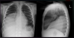
<p>What is the imaging imaging modality that would best characterize this finding/condition? </p><p>a. No further imaging needed </p><p>b. CT with contrast </p><p>c. Bone scan </p><p>d. US with contrast</p>
44
New cards

d. Referral to hospital for surgical consult

What is the best next course of action?

a. Referral to hepatologist

b. Referral to vascular surgeon

c. Raise your hand

d. Referral to hospital for surgical consult

<p>What is the best next course of action? </p><p>a. Referral to hepatologist </p><p>b. Referral to vascular surgeon </p><p>c. Raise your hand </p><p>d. Referral to hospital for surgical consult</p>
45
New cards

c. Mass-like calcification

e. Extra pleural sign

Please choose the TWO best findings/conditions in the following image. Select 2 correct answer(s)

a. Left heart border silhouetting

b. Left upper lung zone opacification

c. Mass-like calcification

d. Consolidation

e. Extra pleural sign

<p>Please choose the TWO best findings/conditions in the following image. Select 2 correct answer(s) </p><p>a. Left heart border silhouetting </p><p>b. Left upper lung zone opacification </p><p>c. Mass-like calcification </p><p>d. Consolidation </p><p>e. Extra pleural sign</p>
46
New cards

d. pleural or extrapleural lesion

What differential diagnosis is most reasonable?

a. pneumonia

b. anterior mediastinal mass

c. pulmonary mass

d. pleural or extrapleural lesion

<p>What differential diagnosis is most reasonable?</p><p>a. pneumonia </p><p>b. anterior mediastinal mass </p><p>c. pulmonary mass </p><p>d. pleural or extrapleural lesion</p>
47
New cards

b. Local infection of the pleura

What would be the most likely associated finding or patient history?

a. Rib fracture

b. Local infection of the pleura

c. Blunt trauma to the left chest wall

d. Drug packing

<p>What would be the most likely associated finding or patient history? </p><p>a. Rib fracture </p><p>b. Local infection of the pleura </p><p>c. Blunt trauma to the left chest wall </p><p>d. Drug packing</p>
48
New cards

b. Hyperlucency in the left upper lung zone

d. Triangular density in the left lower lung zone

Please select the TWO most appropriate answers based on the image presented of this 70 year old female with cough and dyspnea. Select 2 correct answer(s)

a. Blunted costophrenic angles

b. Hyperlucency in the left upper lung zone

c. Round density in the right middle lung zone

d. Triangular density in the left lower lung zone

e. Air bronchogram sign in right middle lung zone
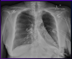
<p>Please select the TWO most appropriate answers based on the image presented of this 70 year old female with cough and dyspnea. Select 2 correct answer(s) </p><p>a. Blunted costophrenic angles </p><p>b. Hyperlucency in the left upper lung zone </p><p>c. Round density in the right middle lung zone </p><p>d. Triangular density in the left lower lung zone </p><p>e. Air bronchogram sign in right middle lung zone</p>
49
New cards

b. Chest CT without contrast

Which imaging modality is best to further characterize this patients condition?

a. MRI with contrast

b. Chest CT without contrast

c. A nuclear imaging perfusion/ventilation study

d. PET scan of Chest
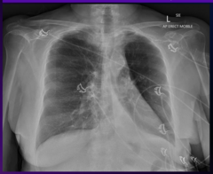
<p>Which imaging modality is best to further characterize this patients condition? </p><p>a. MRI with contrast </p><p>b. Chest CT without contrast </p><p>c. A nuclear imaging perfusion/ventilation study </p><p>d. PET scan of Chest</p>
50
New cards

c. Emergency hospital referral

What is the best next course of action for this patient?

a. Referral to chiropractor

b. Referral to physical therapy

c. Emergency hospital referral

d. Referral cardiologist

<p>What is the best next course of action for this patient? </p><p>a. Referral to chiropractor </p><p>b. Referral to physical therapy </p><p>c. Emergency hospital referral </p><p>d. Referral cardiologist</p>
51
New cards

c. atelectasis

Given the radiographic findings, what diagnosis is likely?

a. viral pneumonia

b. cardiomegaly

c. atelectasis

d. pulmonary mass

<p>Given the radiographic findings, what diagnosis is likely? </p><p>a. viral pneumonia </p><p>b. cardiomegaly </p><p>c. atelectasis </p><p>d. pulmonary mass</p>
52
New cards

a. Airspace involvement

b. Pneumonia

Please select the TWO most appropriate answers based on the image presented of this 50 year old female with shortness of breath, fever, cough, and chest pain. Select 2 correct answer(s)

a. Airspace involvement

b. Pneumonia

c. Mass Interstitial involvement

d. Enlarged cardiac silhouette

<p>Please select the TWO most appropriate answers based on the image presented of this 50 year old female with shortness of breath, fever, cough, and chest pain. Select 2 correct answer(s) </p><p>a. Airspace involvement </p><p>b. Pneumonia </p><p>c. Mass Interstitial involvement </p><p>d. Enlarged cardiac silhouette</p>
53
New cards

d. Labs

Which of the following can best help narrow your differential for this patient?

a. Ultrasound

b. PET scan

c. Lateral radiograph

d. Labs

<p>Which of the following can best help narrow your differential for this patient? </p><p>a. Ultrasound </p><p>b. PET scan </p><p>c. Lateral radiograph </p><p>d. Labs</p>
54
New cards

b. A course of anti-biotics

What is the best next course of action for this patient?

a. Referral to pulmonologist

b. A course of anti-biotics

c. Referral to chiropractor

d. Referral to orthopedist

<p>What is the best next course of action for this patient? </p><p>a. Referral to pulmonologist </p><p>b. A course of anti-biotics </p><p>c. Referral to chiropractor </p><p>d. Referral to orthopedist</p>



