42 Diseases and Disorders of the Lower Respiratory Tract II
 Call Kai
 Learn
Learn Practice Test
 Spaced Repetition
 Match
Match Flashcards
 Knowt Play
1/51

There's no tags or description
Looks like no tags are added yet.
Name | Mastery | Learn | Test | Matching | Spaced | Call with Kai | Chat |
|---|
No analytics yet
Send a link to your students to track their progress
52 Terms
Exercise induced pulmonary hemorrhage
This is described as "alveolo-capillary membrane rupture" in horses, where the blood comes from pulmonary circulation and stress on the alveolo-capillary membrane
a. locomotor forces
b. pulmonary capillary hypertension
c. rheologic properties of blood (how materials flow and deform under stress, like viscosity and elasticity)
f. subatmospheric pleural pressure
Select the top risk factors for exercise induced pulmonary hemorrhage in horses:
a. locomotor forces
b. pulmonary capillary hypertension
c. rheologic properties of blood (how materials flow and deform under stress, like viscosity and elasticity)
d. laminitis
e. degenerative suspensory ligament disorder
f. subatmospheric pleural pressure
Exercise induced pulmonary hemorrhage
This is a disorder of horses that run at high speeds (like thoroughbreds and standardbreds), where the more intense the exercise, the greater the proportion of horses with this disorder
Dorsal area between the vertebrae and angle of the diaphragm is the most common area for hemorrhage
Where is exercise induced pulmonary hemorrhage seen in horses on histopathology?
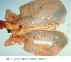
BILATERAL
Exercise induced pulmonary hemorrhage normally shows *unilateral or bilateral* epistaxis
Because 80-90% bleed into their lungs
Bilateral epistaxis may be seen in horses with Exercise induced pulmonary hemorrhage. However, some don't exhibit any signs besides poor performance. Why might you not see the blood?
Endoscopy 30-120 minutes post exercise and graded on a scale of 1-4 for how much blood is in the trachea (1 is normal, 4 is severe pooling)
How do you diagnose Exercise induced pulmonary hemorrhage?
RBC and erythrophages, maybe hemosiderophages if this has been going on over 21 days
Cytology is even more sensitive than endoscopy for Exercise induced pulmonary hemorrhage diagnosis. What do you see on BALF?
Unilateral
Ethmoidal hematomas and guttural pouch mycosis tend to cause *unilateral or bilateral* epistaxis
you want to *decrease* pressure in the pulmonary capillaries and *increase* alveolar inspiratory pressure
To prevent stress rupture of the capillaries (which causes exercise induced pulmonary hemorrhage), you want to *increase or decrease* pressure in the pulmonary capillaries and *increase or decrease* alveolar inspiratory pressure
Furosemide (except not europe)
What medications allow you to decrease pressure in the pulmonary capillaries to prevent stress rupture of the capillaries, and thus exercise induced pulmonary hemorrhage?
In the upper resp. tract, you can use flair strips and correct dynamic collapse
What can you do to the upper respiratory tract to increase alveolar inspiratory pressure (aka make it less negative) to prevent stress rupture of the capillaries, and thus exercise induced pulmonary hemorrhage?
This is a "flair strip" used to increase alveolar inspiratory pressure
What is this and what does it do to prevent stress rupture of the capillaries to prevent exercise induced pulmonary hemorrhage?
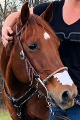
Use bronchodilators, corticosteroids, and reduce environmental dust
What can you do to the lower respiratory tract to increase alveolar inspiratory pressure (aka make it less negative) to prevent stress rupture of the capillaries, and thus exercise induced pulmonary hemorrhage?
FALSE = one episode will lead to more episodes that become worse overtime. you will need to have a conversation with the owner about retiring the horse early
TRUE OR FALSE: episodes of exercise induced pulmonary hemorrhage are not related in horses, and having one episode does not predispose to another
Bronchopneumonia and pleuropneumonia
What infectious lower airway disease affects adult horses to cause pneumonia?
Anaerobes
Pneumonia may be caused by several different pathogens in horses: *gram positives, negatives, and anaerobes*. Which of these types has the LEAST favorable prognosis?
Streptococcus equi ssp. zooepidemicus
note: it is an important distinction that streptococcus equi ssp equi, if present, IS NOT commensal and causes strangles, but streptococcus equi ssp. zooepidemicus IS commensal and causes pneumonia
This gram positive pneumonia causative agent in horses is present in NORMAL flora and is a beta hemolytic streptococcus
Prolonged transportation
What predisposes a horse to pneumonia?
Because these conditions allow for colonization of the lung by opportunistic bacteria
Weakened immune defenses and massive numbers of bacteria allow pneumonia to happen in horses WHY?
LOWERED HEAD POSTURE
What posture is essential for muco-ciliary clearance in horses?
Because lowered head posture allows muco-ciliary clearance in horses. So, if the head is restrained and this cannot happen, the head restraint is the single most predisposing factor for development of pneumonia associated with long distance transport
We know prolonged transport predisposes a horse to pneumonia. But why is this?
Pneumonia in horses
What is this the pathophysiology of:
infiltration with inflammatory cells --> damage to epithelium --> flood of inflammatory cells/debris --> exudative pleuropneumonia --> fibrinopurulent pleuropneumonia --> fibrin deposits
Early intervention
What is the most crucial part of stopping pneumonia in horses?
DECREASED breathing sounds and MUFFLED sounds on percussion
With pneumonia, a horse will have *increased or decreased* breathing sounds in the lower lungs and *muffled or exaggerated* sounds on percussion.
They have pain in the chest cavity and will try and keep their elbows away from their body
What does it mean that horses have "pleurodynia" with pneumonia as a clinical sign?
Transtracheal wash
Which is BEST for pneumonia diagnosis in horses? *transtracheal wash or BALF*?
Pneumonia = lung lobe consolidation
What are you seeing on this equine ultrasound and what is your diagnosis?
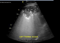
Pneumonia = consolidation (middle left bright white part of screen) and fluid
What are you seeing on this equine ultrasound and what is your diagnosis?
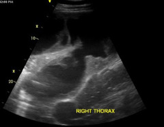
Pneumonia = upper lung only visible, lower lung covered with fluid
What are you seeing on this equine radiograph and what is your diagnosis?
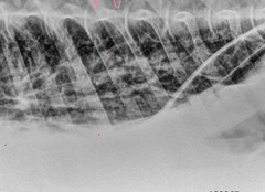
ANTIBIOTICS = penicillin (+), gentamicin (-), metronidazole (anaerobes)
What is the BIGGEST treatment for pneumonia in horses?
Minimum 10 days or more than that until signs resolve
How long do you treat horses with pneumonia using antibiotics?
To dec. volume causing respiratory distress or if there is evidence of sepsis = do BEFORE fibrin is present
Antibiotics are the MAIN treatment of pneumonia in horses. However, you may also do pleural drainage on top of this. Why?
Overall good = 90% of horses survive pneumonia and 60% return to athletic performance
Overall, the prognosis is *good or guarded* for pneumonia in horses (unless hemorrhagic necrotizing pneumonia occurs)
Rhodococcus equi
What is the most common cause of pneumonia in foals under 6 months?
Gram POSITIVE, intracellular (alveolar macrophages)
Rhodococcus equi is a bacteria that is gram *negative or positive* and survives *intracellularly or extracellularly*
YES if you are immunocompromised
Is Rhodococcus equi zoonotic?
FOALS ONLY (like under 6 months)
What age of horse is the ONLY one susceptible to rhodococcus?
b. inhalation
The following are BOTH routes for rhodococcus to infect foals. However, which is the PRIMARY:
a. ingestion
b. inhalation
Rhodococcus equi = extensive pulmonary abscessation and chronic suppurative bronchopneumonia
A 4 month old foal presents with increased respiratory rate, decreased appetite, cough, tachypnea, and a mild fever. The foal eventually progresses to acute respiratory distress and death. This is the necropsy. Thoughts?
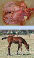
Rhodococcus equi
Hyperfibrinogenemia, serum amyloid A spike, and neutrophilic leukocytosis on hematology and biochemistry of a recently deceased 6 month old foal tips you off to what?
Rhodococcus equi
A 3 month old foal presents with alveolar "snowstorm" and ill defined regions of consolidation. There are nodular lung lesions and mediastinal lumphadenopathy. Thoughts
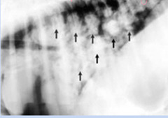
Rhodococcus equi
Radiographs showing a 1-4 month old foal with nodular lung lesions and mediastinal lymphadenopathy on radiographs. This is HIGHLY SUGGESTIVE and is basically WHAT until proven otherwise?
Rhodococcus equi
A 2 month old foal presents with focal areas of consolidation that look like comet tails on ultrasound and extra-pulmonary lesions. Thoughts?
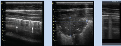
Rhodococcus equi
Clinical signs of lower respiratory in a foal, cytologic evidence of septic airway inflammation, and radiographic/ultrasound evidence of bronchopneumonia in conjunction with PCR amplification of Vap A are diagnoses for WHAT problem?
a. PCR with amplification of Vap A
c. clinical signs of lower respiratory disease in a foal
d. cytologic evidence of septic airway inflammation
f. radiographic/ultrasound evidence of bronchopneumonia
Select the diagnostic tests for rhodococcus equi:
a. PCR with amplification of Vap A
b. bronchoalveolar lavage
c. clinical signs of lower respiratory disease in a foal
d. cytologic evidence of septic airway inflammation
e. arthrocentesis evidence of septic arthritis
f. radiographic/ultrasound evidence of bronchopneumonia
Macrolides like azithromycin/clarithromycin + rifampin
What are the BEST TREATMENTS for rhodococcus equi in foals?
Hyperthermia, diarrhea in foals. Colitis in mares
Erythromycin is a macrolide but is considered last ditch for treatment of rhodococcus in foals. Why? What are some adverse effects?
Synergistic activity: dec. liklihood of resistance, prolonged half life, lipid soluble to penetrate cells, and oral bioavailability
Why do you use macrolides like clarithromycin/azithromycin in combination with rifampin for treatment of foals with Rhodococcus?
3-8 weeks (maybe even past resolution of clinical signs)
How long do you treat foals with rhodococcus with macrolides and rifampin?
Mares are coprophagic (eat feces), and so if the baby is treated with Erythromycin and the mare ingests it, it will disrupt her natural gut flora and allow overgrowth of Clostridium dificile
We said erythromycin may cause enterocolitis in mares if you treat her baby with it for rhodococcus. Why does this make sense?
Ultrasound
Which is the more common screening test for Rhodococcus equi in foals, *ultrasound or x-ray*