Chapter 9 Study Guide (The Muscular System)
1/77
There's no tags or description
Looks like no tags are added yet.
Name | Mastery | Learn | Test | Matching | Spaced | Call with Kai |
|---|
No analytics yet
Send a link to your students to track their progress
78 Terms
Skeletal Muscle Tissue
A type of muscular tissue that is striated, under voluntary control, multinucleated, and attached to bones.
Cardiac Muscle Tissue
A type of muscular tissue that is striated, under involuntary control, has a single nucleus, has intercalated discs, and is found in the heart.
Smooth Muscle Tissue
A type of muscular tissue that is non-striated, under involuntary control, has a single nucleus, and is found in hollow organs.
Produces Skeletal Movement: Contracting muscles cause movement. Muscles pull on tendons to move bones.
Stabilizes Body Position and Posture: Even while not moving, muscles contract to keep us upright. Constant muscular contraction prevents us from just collapsing.
Supports Soft Tissues: Supports organs in abdominopelvic cavity, since they don’t have much structure. The muscles line the cavity to protect internal tissues and organs.
Stores and Move Substances throughout the Body: Hollow organs, hollow sacs of muscle, store substances and move substances along. These organs have rings of muscular tissue called sphincters and they contract to prevent organs from loosing substance. Muscles provide voluntary control over swallowing, defecation, and urination.
Thermogenesis: Generates heat whenever muscles contract. Shivering, working out, breaks ATP and generates body heat. Maintains body temperature.
Functions of the Muscular Tissue
Excitability: Respond to stimulation.
Contractibility: Can shorten actively and exert pull or tension.
Extensibility: Can contract over a range of resting lengths.
Elasticity: Ability of muscle to return to original length after contraction.
Properties of Muscle Tissue
Skeletal Muscle Tissue
Connective Tissues: Areolar, dense irregular, adipose, elastic, etc.
Blood vessels: Blood flow is needed because muscles need nutrients to function and produce ATP.
Nerves: Helps muscles contract
Subcutaneous Layer: Insulates and protects. Areolar and adipose tissue.
Tissues and components of muscles that make them organs
Fascia
The dense irregular connective tissue that wraps around muscles. It supports muscles and organs, lines body cavities, and is around limbs. Provides pathway for blood vessels and nerves to access muscle tissue. Some will have stem cells inside to repair muscle tissues.
Fascicle
A bundle of muscle fibers. Vary in the number of muscle fibers inside.
Endomysium
The fascia around each muscle fiber. Lined with specialized cells called myosatelite cells, they surround every muscle fiber and repair muscle fibers. It binds muscle fiber to its neighbor and supports capillaries. Consists of delicate network of reticular fibers.
Perimysium
The fascia that surrounds the fascicles. Divide the muscle into internal compartments. Contain bundle of muscle fibers, fascicles. Also contains collagen and elastic fibers, blood vessels, and nerves.
Epimysium
The fascia that is outside (surrounds) the muscle. Dense irregular connective tissue surrounding entire skeletal muscle. Separates muscle from surrounding tissues and organs. Connected to deep fascia.
Tendon
The dense regular connective tissue that attaches muscle facia to bone periosteum. Attaches muscle to bone, cartilage, skin, or other muscles. Continuous with periosteum, makes strong bond so any contraction of muscle pulls on attached bone.
Aponeurosis
Broad and flat tendons. They are tendons that form thick and flattened sheets.
Fibromyalgia
A connective tissue disorder/disease that is the inflammation of tendons and muscles. Symptoms include pain, fatigue, and stiffness in joints. Causes are unknown but could be immune system related. Treatments include antibiotics, rest, and exercise.
Tendinitis
A connective tissue disorder/disease that is the inflammation of tendons due to repetitive use. Symptoms include pain in joints and tendons. The tendons don’t have much blood flow so it takes longer to heal. It is common in the elderly and athletes that use specific tendons repetitively. Treatment includes rest and the reduction of movement. The repetitive use causes microscopic tears to accumulate in the tendon over time.
Every skeletal muscle fiber is connected to one nerve.
One nerve may connect with multiple muscle fibers.
Nerves lie within connective tissues.
Nerves are bundle of axons that enter the epimysium, branch through perimysium, and enter endomysium to attach to muscle fibers.
Motor unit: One neuron and all the muscle fibers it is connected to.
Nerves stimulate contraction.
Nerve Supply of Muscles
Muscles require a lot of blood flow (nutrients, oxygen, waste removal).
Every time a muscle contracts, they consume oxygen, glucose, and make waste.
Blood vessels deliver oxygen and nutrients needed to produce energy in the form of ATP in skeletal muscles.
In the endomysium, arteries supply a large capillary network.
Capillaries are coiled.
Blood Supply of Muscles
Muscle Fiber
A muscle cell.
They span the entire length of their muscle.
During embryonic development, myoblasts fuse together to make one very long muscle fiber.
Long and cylindrical.
Striated: Organization of myofibrils and their molecular components give skeletal muscle a striated appearance.
Polynucleated (one nucleus for each myoblast that fuse together)
Muscles cannot reproduce.
Muscle Fiber Characteristics
Myoblasts
Muscle stem cells. Embryonic cells that fuse to form individual skeletal muscle fibers.
Myosatellite Cells
When myoblasts don’t fuse with muscle fibers but remain in adult skeletal muscle tissue as stem cells. When muscle is injured, these cells differentiate and assist in repairing and regenerating muscle.
Sarcolemma
The plasma membrane of the muscle fiber.
Transverse (T) Tubules
A structure of the muscle fiber that is an extension of the sarcolemma in the form of tubes. IT wraps around organelles. Deep indentations in sarcolemma surface form these. Extend into sarcoplasm. These structures and sarcolemma conduct electrical impulses called action potentials, to stimulate muscle fiber contraction.
Sarcoplasm
The cytoplasm of the muscle fiber. It is the space in the fibers. Anything inside sarcolemma. Contains a lot of proteins called myoglobin.
Myoglobin
Oxygen-storing protein in the sarcoplasm of a muscle fiber. Muscle fibers are able to store excess oxygen for when mitochondria need them. They are red in color because of the iron that is necessary to bind oxygen molecule, allows muscle cells to store oxygen.
Myofibrils
Contracting organelles in the muscle fiber. Contain mitochondria and glycogen granules scattered throughout to provide ATP to power contractions. Lie parallel and along the whole length of the muscle fiber. Made of cytoskeletal filaments (myofilaments). They are fine cylindrical fibers. The active shortening is responsible for muscle fiber contraction.
Sarcoplasmic Reticulum (SR)
A structure in the muscle fiber that stores calcium ions which contract muscle fibers. They are a series of tubes related to the smooth endoplasmic reticulum. There are enlarge areas at the ends of the tubes. Surrounds each myofibril and controls myofibril contraction. They are the storage and release site of calcium ions.
Terminal Cisternae
The enlarged areas at the ends of the tubes of the SR. Located on each side of T tubule. The tubules of SR enlarge, fuse, and form these expanded chambers.
Triads
A structure within the muscle fiber that consists of two terminal cisternae and 1 transverse (T) tubule in the middle of them.
Myofilaments
Protein filaments that are made of cytoskeleton and allow for contraction. There are two types: thin and thick. They are cytoskeletal microfilaments. Do not run the entire length of myofibril. They are short chopped up pieces. Inside the myofibrils they are held in compartments called a sarcomere.
Thin Filament
A type of filament that contains mostly actin. It also contains regulatory proteins troponin and tropomyosin. Also has myosin binding sites.
Thick Filament
A type of filament that contains myosin. It is at the center of a sarcomere. Composed of bundles of myosin molecules, each made up of a pair of myosin subunits twisted around one another. Long tail is bound to other myosin molecules in filament. Free head, with two globular protein subunits, projects outward toward the nearest thin filament. Myosin heads interact with thin filament during a contraction, they are known as cross-bridges. The heads are motor proteins that uses ATP to pull myosin head along thin filaments. They also have a core of titin.
Sarcomere
One section of myofilaments within a myofibril. This is a name for when myofibrils are organized in repeating units. It is the smallest functional unit of a muscle fiber. It is a subunit of filaments.

Z discs
A part of the sarcomere that refers to the zig-zag proteins that separate sarcomeres. It bisects the I bands and mark the boundary between adjacent sarcomeres. Made of proteins called actinin, which interconnect thin filaments of adjacent sarcomeres.

A band
The region of a sarcomere with thick (myosin) filaments. Thick filaments are in the center. It contains the M lines, H band, and zone of overlap.
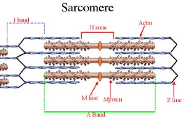
H zone
The part of the sarcomere that refers to the center of the A band, where there are only thick filaments.

M line
The part of the sarcomere that refers to the center of the H zone. It contains stabilizing proteins that are also at the center of the A band. The proteins connect the central portion of each filament to neighboring thick filaments. It stabilizes the position of thick filaments.

I band
The part of the sarcomere that refers to the area with only thin filaments. It extends from A band of one sarcomere to the A band of the next sarcomere.

Zone of Overlap
The region of the sarcomere that is where thin filaments are found between thick filaments.
Myosin
A type of contractile protein that is made of two long skinny proteins that are twisted together, and the ends have little rounded club shapes. About 300 of them bundled together make one thick filament.
Actin
A type of contractile protein that is twisted into a helix to form thin filaments. Each protein has a binding site for a myosin head.
Tropomyosin
A type of regulatory protein that forms a long chain that covers the active sites on actin, preventing actin-myosin interactions. They covering binding sites and prevent them from being used when you don’t want to contract the cell.
Troponin
A type of regulatory protein that holds the tropomyosin strand in place. Before a contraction, these molecules must change position, moving the tropomyosin molecules and exposing active sites. They are used to pull tropomyosin off when you want to contract.
Titin
A type of structural protein that extends from the tips of thick filament to attachment sites at the Z line. Made of elastic protein. One of the largest proteins in the body. It stabilizes the whole sarcomere. When the sarcomere is resting, this protein is relaxed. It is tense only when some external force stretches stretches the sarcomere. When the sarcomere stretches, this strand maintains normal alignment of filaments. When tension is removed, the strand returns sarcomere to normal length.
Nebulin
A type of structural protein that wraps around thin actin filaments. It helps attach actin to the Z line. Extends along f-actin strand in the cleft between the rows of G-actin molecules. Holds the F-actin strand together.
Dystrophin
A type of structural protein that links all thin filaments to the cells of sarcolemma. Stabilizes and connects the myofibrils from one end of the cell to another.
Neuromuscular Junction
Connection between neuron and muscle fiber. Chemical communication between neuron and skeletal muscle fiber occurs here. Made of axon terminal of neuron, motor end plate, synaptic cleft, and membrane of muscle fiber. Each fiber has one of these. Here, the axon terminal of neuron attaches to motor end plate of skeletal muscle fiber.
Somatic Motor Neuron
A part of the NMJ that refers to the neuron in nervous system whose job is to talk to the muscle fibers. Consists of axon terminal, synaptic end bulbs, and synaptic vesicles.
Axon Terminal
A part of the somatic motor neuron within NMJ that refers to the end of the axon. It is the expanded tip of the axon at NMJ. Cytoplasm of it contains mitochondria and synaptic vesicles.
Synaptic End Bulbs
A part of the somatic motor neuron within NMJ that refers to the enlarged part at the end of the axon terminal.
Synaptic Vesicles
A part of the somatic motor neuron within NMJ that refers to the small secretory vesicles. It contains acetylcholine. Nervous nucleus is constantly telling ER and golgi apparatus to synthesize these. It gets sent down the axons to the end bulbs and then wait for a signal from cell body to tell axon to release them into the synaptic cleft.
Acetylcholine (ACh)
A neurotransmitter held in synaptic vesicles. It is a chemical released by neuron and it communicates with other cells. It causes change in membrane potential.
Synaptic Cleft
A part of the NMJ that refers the narrow space that separates axon terminal from the motor end plate of the muscle fiber.
Acetylcholine Esterase
An enzyme that breaks down ACh.
Motor End Plate
A part of the NMJ that refers the area where sarcolemma is forming neuromuscular junction. It is where the axon of motor neuron establishes synaptic contact with skeletal muscle fiber.
Junctional Folds
A part of the motor end plate that is meant to hold more ACh receptors.
Acetylcholine Receptors
A part of the motor end plate that is important so that muscle fibers will know when neuron tells it to contract.
When the action terminal arrives at the axon terminal, ACh is released from synaptic vesicles into the synaptic cleft.
ACH then diffuses across the synaptic cleft and binds to receptor sites on the motor end plate of muscle fiber.
This generates an action potential in the sarcolemma and in each T tubule.
Steps to sending an action potential across the NMJ
Neuron signals the muscle fiber across the NMJ: Nerve impulse goes down axon and reaches axon terminal. Triggers exocytosis of acetylcholine because voltage-gated channels open up which allows calcium to rush in and when that happens, positively charged ions triggers neurons to do exocytosis of synaptic vesicles. ACh released into cleft and binds to ion channels in motor end plate. Initiates action potential along T tubule.
Action potential travels along the T-tubules and stimulates the SR to release calcium ions into the sarcoplasm.
Calcium ions expose myosin binding sites on actin: The calcium ions binds to troponin in myofibrils which pulls tropomyosin off of the actin, allowing for contraction to occur. Also active transport pumps Calcium ions back to SR to stop muscle contraction.
Contraction Cycle: Myosin heads walk along actin to contract the myofibrils. Thick and thin filaments slide across each other. Z-lines get closer together as myosin pulls on actin filaments.
Excitation-contraction coupling: Steps to linking action potential to muscle contraction
ATP Hydrolysis: 600 ATP per sarcomere to contract muscle just a smidge. Cuts off third phosphate group, turns ATP to ADP. ATP bound to myosin head, then ATP hydrolysis (enzyme) cuts ATP, discharges energy, now myosin head holds ADP and a phosphate that has been cut off. Charges up myosin head and straighten out.
Cross-Bridge Formation: When myosin head straightens out and attaches to myosin binding site from actin filament. Myosin heads attach to actin filaments. Thick and thin filaments connected. When this happens, myosin head releases extra phosphate group.
Power Stroke: Myosin rotate downwards and pull actin filaments toward it. ADP molecule is released. Pull actin filaments further along.
Myosin Detachment: Myosin head can only detach from actin filament if ATP is provided. Once ATP binds to myosin heads, the two filaments can be separated and the process can restart.
Sliding Filament Model Contraction Cycle
Rigor mortis
Sets in after death because calcium floods muscle cells causing proteins (actin and myosin) to bind, but there is no ATP to detach them, locking the muscles in contracted, stiff state. Body eventually becomes pliable because the same muscle proteins begin to break down due to the action of enzymes released during decomposition, which destroys the bonds holding the actin and myosin together, leading to a release of stiffness.
Motor Unit
One neuron and all muscle fibers connected to it. Muscles contract when these are stimulated. The number of these involved determines the amount of tension produced. All fibers within this contact at the same time, force exerted depends on how many of these contract. They are activated on a rotating basis. Some of these are resting while some are contracting.
Fine movements
What movements are controlled by small motor units (a neuron attached to 1 or 2 muscle fibers)?
Gross movements
What movements are controlled by large motor units (a neuron that controls 150+ muscle fibers)?
Recruitment
Adding more motor units to the contraction to make it stronger. Increase in muscular tension produced by increasing the number of active motor units.
Muscle Tone
Slight tension held in all muscle fibers even while not moving. Because the brain is constantly sending impulses down your motor nerves and those nerves are sending impulses strong enough to exercise the muscle fibers. Maintains body position and firmness. Stabilizes the position of bones and joints.
Flaccid Fibers
Muscle fibers without tone that is only seen as a result of nerve damage (neuron is cut and the muscle fiber cannot contract).
Flaccid Paralysis
A muscle tone disorder that refers to the loss of muscle tone. An example is a spinal cord injury.
Spastic Paralysis
A muscle tone disorder that refers to too much muscle tone. There is no control with when contractions occur. Examples include cerebral palsy. Overstimulates muscles constantly.
Botulism Toxicity
An infection that occurs when this toxin prevents neuron synaptic vesicles from releasing acetylcholine (muscle fibers cannot contract). It causes paralysis, stops all muscle contractions, and is a lethal substance.
Tetanus Infection
An infection that riggers the body to control all muscles. Causes cramping all over.
Slow Fibers
A type of skeletal muscle fiber.
Slow contraction, ATP generatiion
Fatigue Resistant, takes a long time before they run out of energy.
A lot of myoglobin (dark red color), mitochondria (continuously produce ATP throughout contraction), and capillaries.
Aerobic metabolism and large oxygen reserve.
Smaller diameter
Examples: Aerobic exercises, endurance activities, keeping posture.
Fast Fibers
A type of skeletal muscle fiber.
Strong, fast contractions.
White (not much myoglobin or mitochondria or blood)
Make ATP through fermentation: Glycolysis which converts glycogen to lactic acid. Use large amounts of ATP and rely on anaerobic metabolism.
Fatigue quickly: Because fermentation is not efficient in making ATP.
Large diameter, densely packed myofibrils, large glycogen reserves.
Most muscle fibers are these type.
Examples: Where fast reflexes are needed or where there are repeated motions.
Intermediate Fibers
A type of skeletal muscle fiber.
A lot of myoglobin and mitochondria.
Can switch between aerobic respiration and fermentation. (Do both slow and fast)
Make a lot of ATP, fast contraction but not as fatigue resistant as slow fibers.
Contract faster than slow fibers but slower than fast fibers.
Examples: Walking and sprinting muscles.
Convert fast fibers into intermediate fibers.
Helps make muscles fatigue resistant.
Helps develop new blood vessels and more mitochondria in muscle fibers.
Helps with digestion
Skeletal strength
Cardiovascular health
Aerobic Exercises
Builds muscles by widening muscle fibers.
You cannot make new muscle fibers.
Can increase size and strength of fibers.
Must be consistent (muscle loss begins within 2 weeks of use).
Strength Training
Anabolic Steroids
Mimic testosterone and stimulates myofibril development in muscle fibers. Comes with many side effects including heart attacks. strokes, and impotency (can’t produce sperm). Also testicles of a man get smaller, and in women, voices deepen, hair growth, and clitoris enlarges. The muscles enlarge but tendons and ligaments are weak (injury prone).
After 30, muscle tissue is slowly replaced with connective tissue and adipose tissue.
10% muscle loss by 50, 40% by 80.
Fast and intermediate muscle fibers are the first to go.
Skeletal muscle fibers become smaller: Decrease in number of myofibrils. Fibers have less ATP, glycogen reserves and myoglobin. Reduction in muscle strength and endurance. Fatigue quickly.
Skeletal muscles become less elastic: Develop more fibrous connective tissue within endomysium and perimysium (fibrosis). Makes muscles less flexible, collagen fibers restrict movement and circulation.
Tolerance for exercise decreases: Rapid fatigue and less ability to eliminate heat.
Ability to recover from muscular injuries decreases: Myosatellite cells steadily decrease and the amount of fibrous tissue increases. Repair capabilities are limited, scar tissue usually forms.
Aging (muscle tissue effects)