Harp Pigmentation Disorders - MSK Exam 1
1/67
There's no tags or description
Looks like no tags are added yet.
Name | Mastery | Learn | Test | Matching | Spaced |
|---|
No study sessions yet.
68 Terms
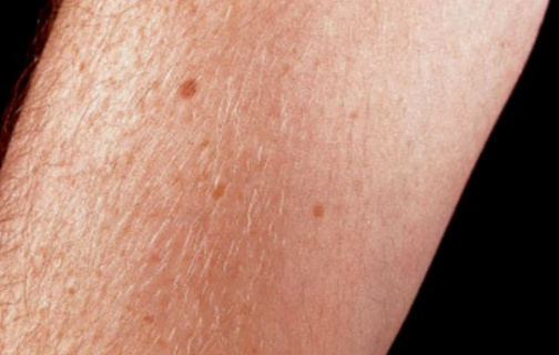
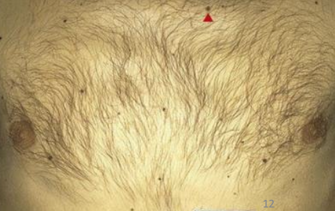
Freckles (ephelides)

Freckles (ephelides)
Freckles (ephelides)
Freckles (ephelides)
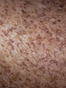
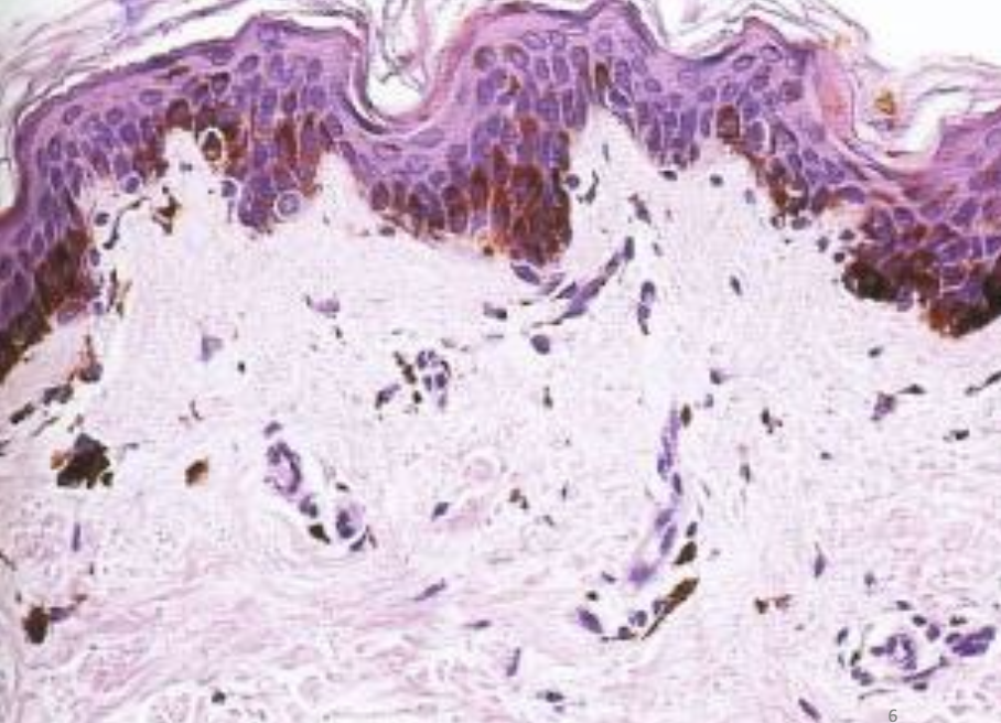
senile lentigo (lentigo senilis) / liver spots / age spots / solar lentigo / actinic lentigo
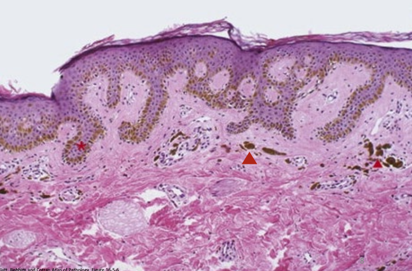
senile lentigo (lentigo senilis) / liver spots / age spots / solar lentigo / actinic lentigo
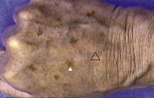
hockey sticks/dirty socks
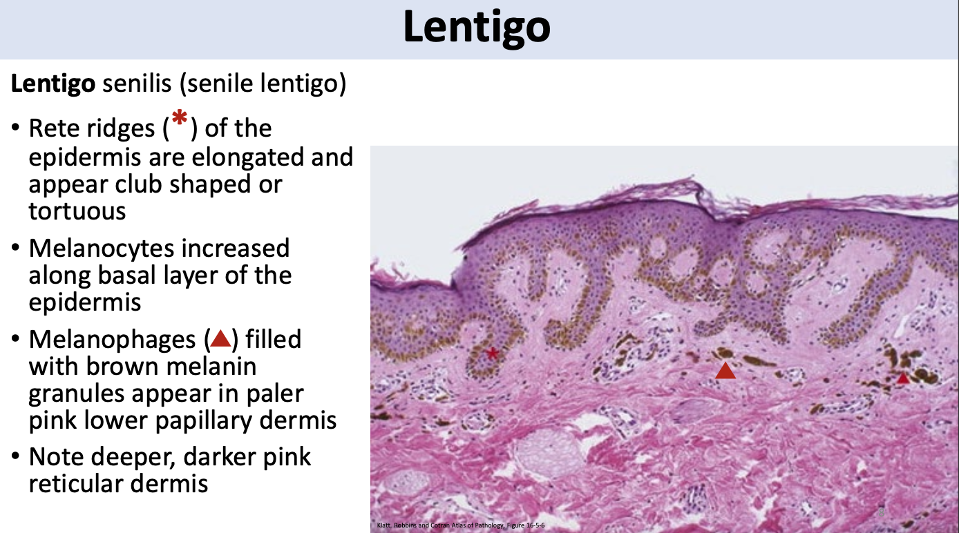
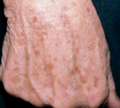
senile lentigo (lentigo senilis) / liver spots / age spots / solar lentigo / actinic lentigo
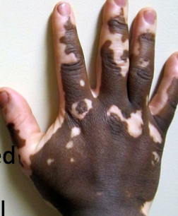
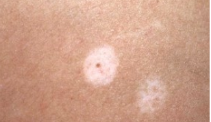
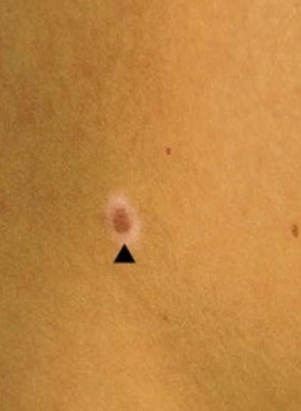
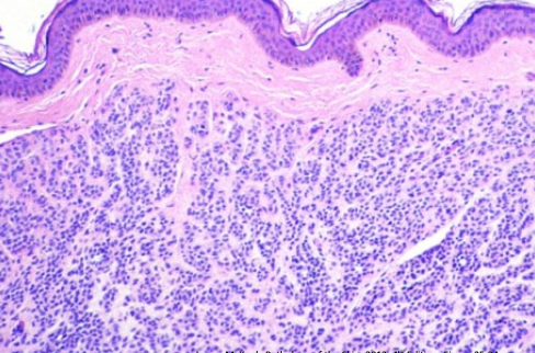
Vitiligo (leukoderma)
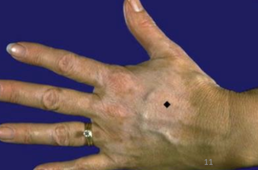
Vitiligo (leukoderma)
Vitiligo (leukoderma)
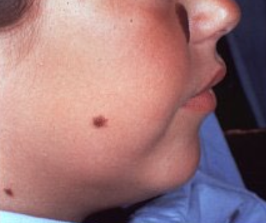
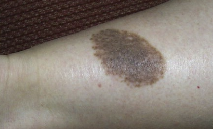
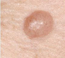
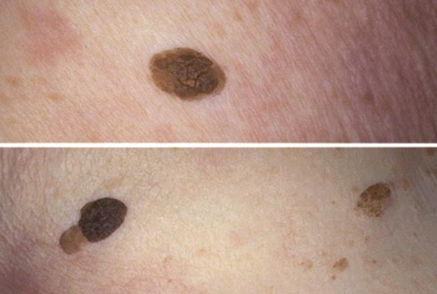
Melanocytic nevi
Melanocytic nevi
Congenital nevus
Congenital nevus
extends into deep dermis
halo nevus
due to immune response to nevus cells
halo nevus
due to immune response to nevus cells
halo nevus
due to immune response to nevus cells
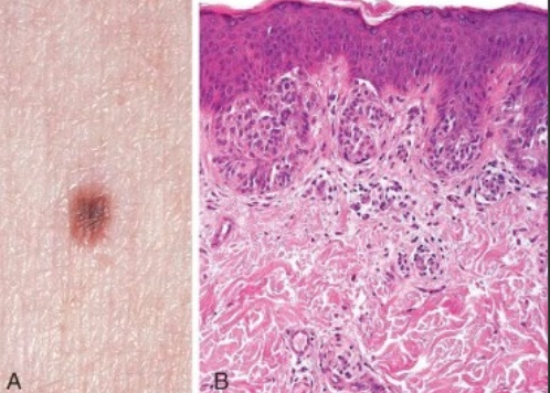
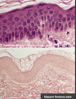
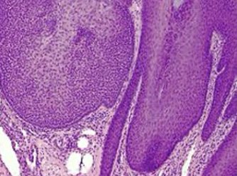
junctional melanocytic nevi
compound melanocytic nevi
junctional melanocytic nevi
nests present at dermal-epidermal junction
begin in early childhood
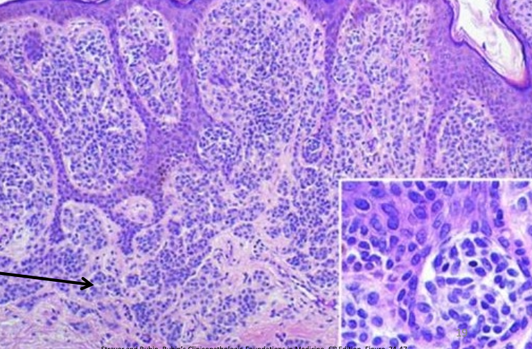
compound melanocytic nevi
nevus cells at junction and within dermis (appear to have fallen off/down into dermis)
intradermal melanocytic nevi
intradermal melanocytic nevi
intradermal melanocytic nevi
pigmentation lost at this point due to loss of junctional component —> nevus cells are only within dermis
MC nevi in adults - develop around puberty
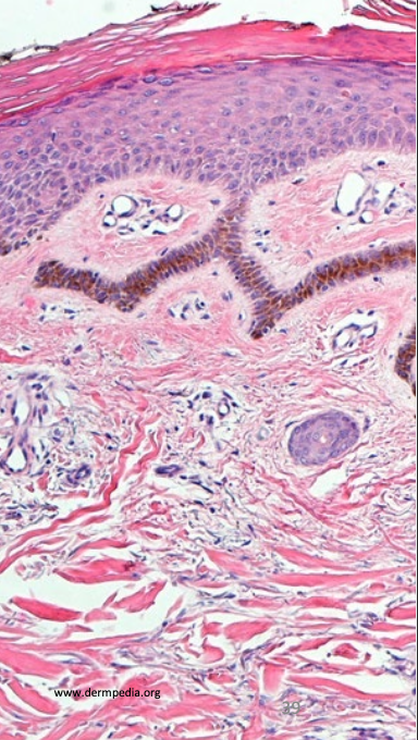
poroma
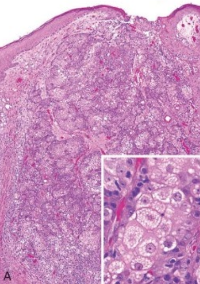
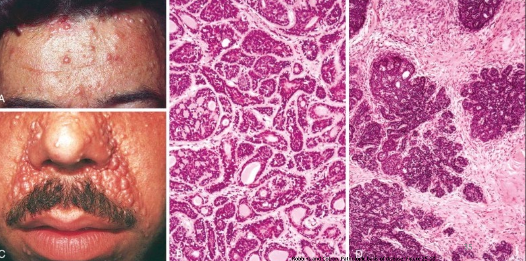
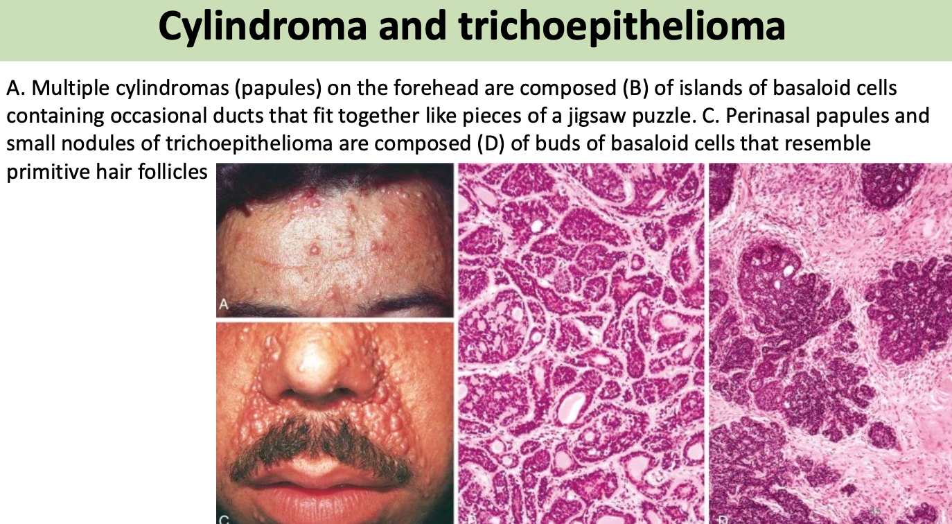
cylindroma
cutaneous horn of actinic keratosis
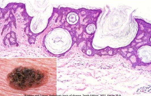
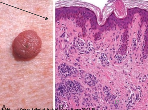
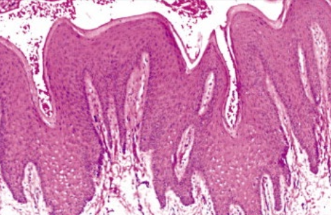
Seborrheic keratosis
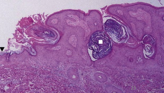
Seborrheic keratosis
white square = horn pseudocysts
Seborrheic keratosis
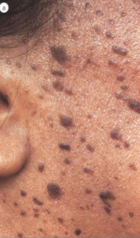
Seborrheic keratosis
Seborrheic keratosis
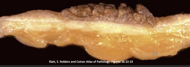
Seborrheic keratosis
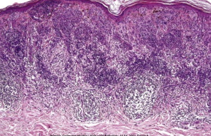
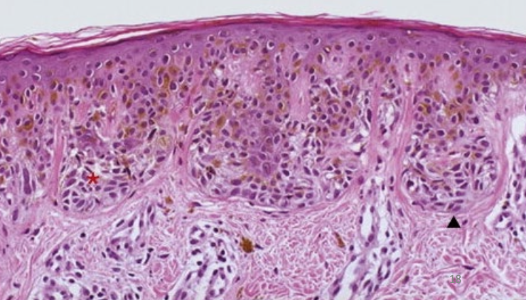
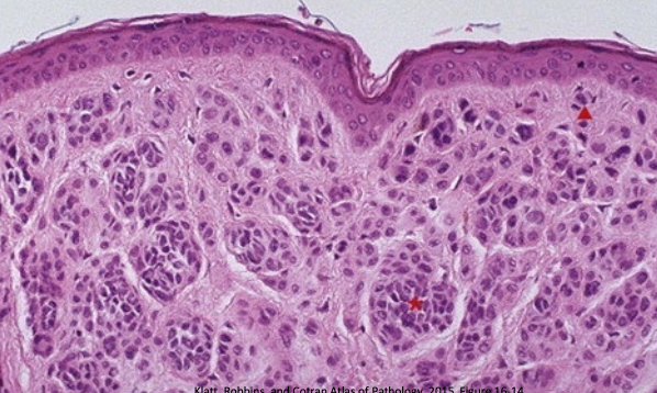
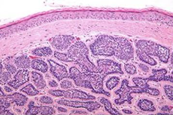
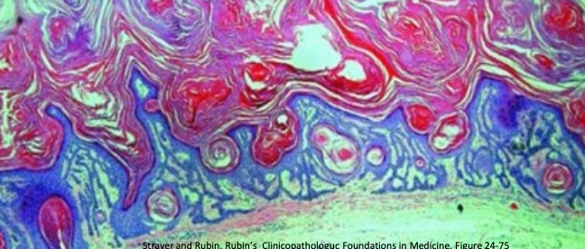
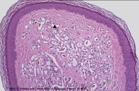
Acanthosis nigricans
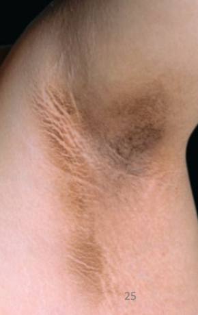
Acanthosis nigricans
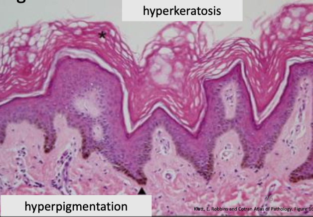
Acanthosis nigricans
brown color at arrow shows increased melanin granules in epidermal basal layer
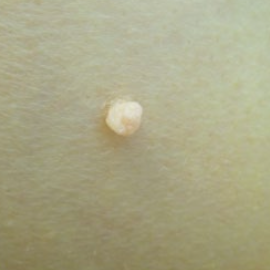
Fibroepithelial polyp
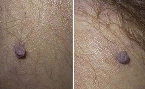
Fibroepithelial polyp
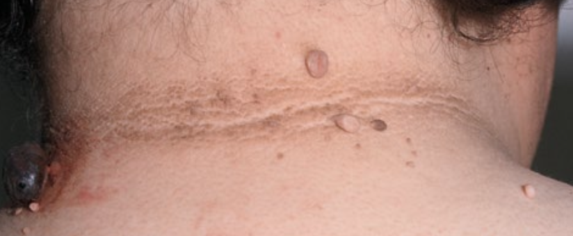
Fibroepithelial polyp
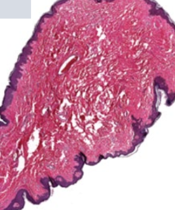
Fibroepithelial polyp
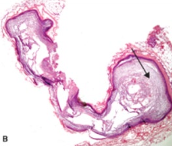
Epidermal Inclusion cyst
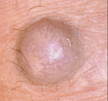
Epidermal Inclusion cyst
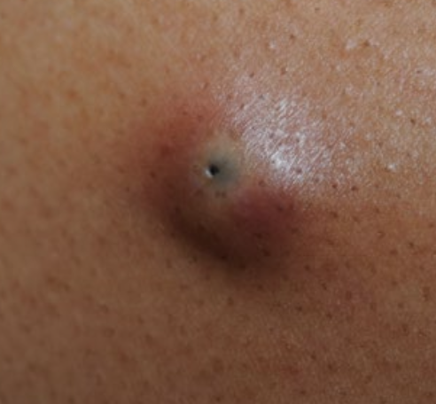
Epidermal Inclusion cyst
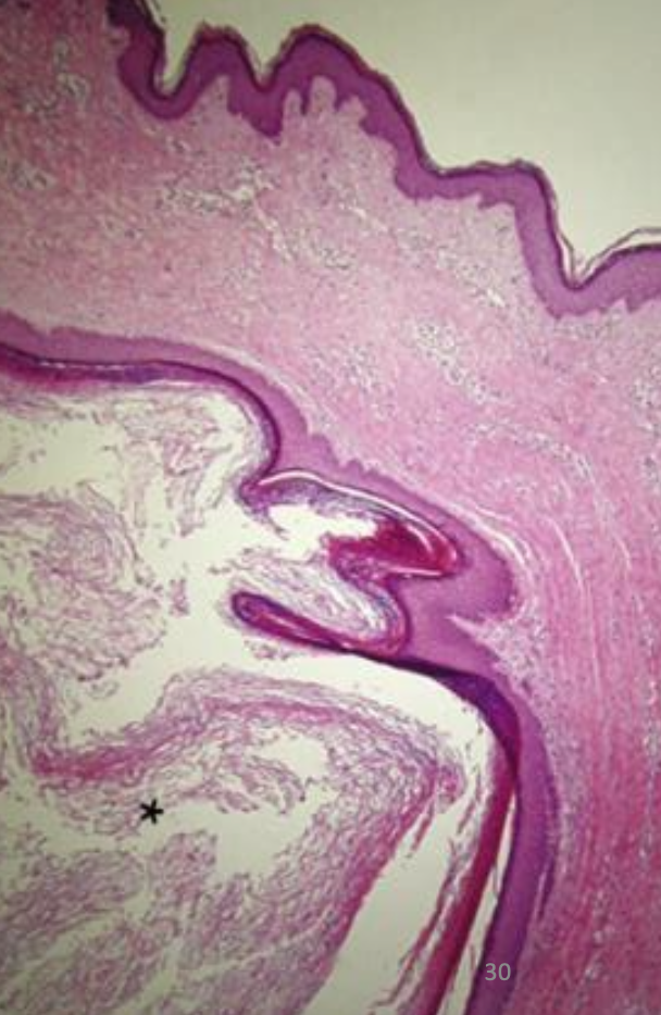
Epidermal Inclusion cyst
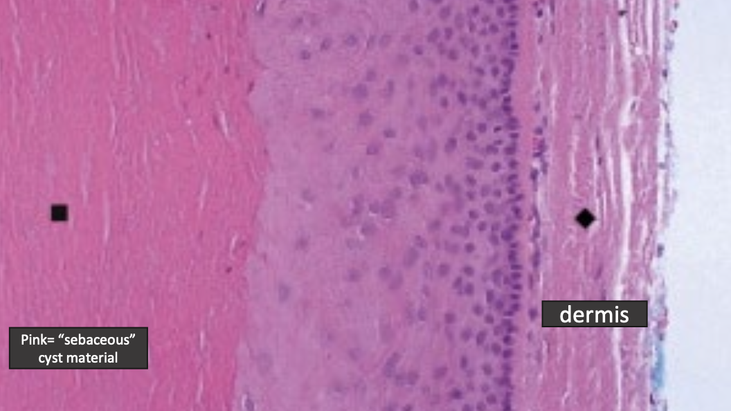
Epidermal Inclusion cyst
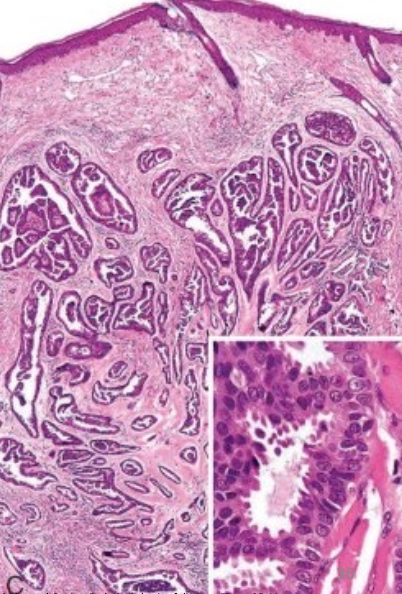
eccrine poroma
adnexal/appendage tumor with sweat gland differentiation
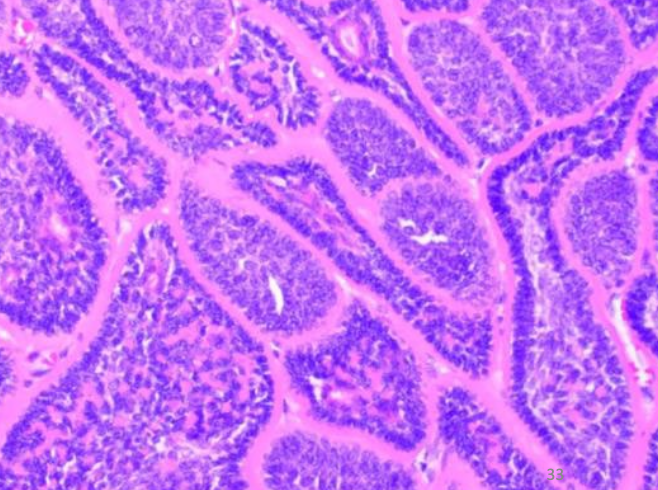
cylindroma
adnexal/appendage tumor of head/neck with apocrine sweat gland differentiation
histology: jigsaw puzzle-like
trichoepithelioma
adnexal/appendage tumor with hair
syringoma
adnexal/appendage tumor of eyelids and upper cheeks with eccrine duct differentiation
dense fibrous stroma
pilomatrixoma
adnexal/appendage tumor of hair matrix (in kids)
sebaceous adenoma
adnexal/appendage tumor of sebaceous gland
apocrine carcinoma
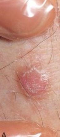
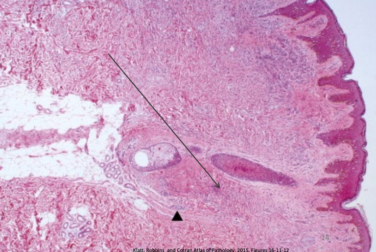
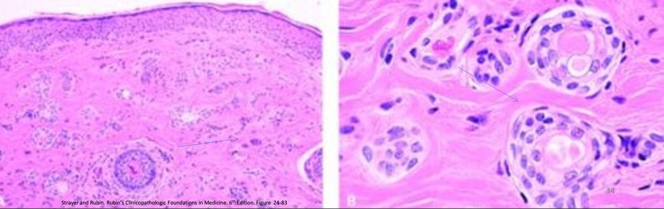
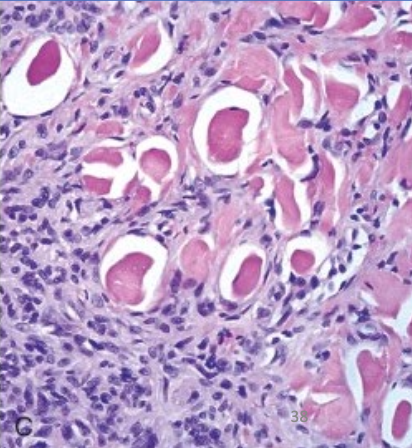
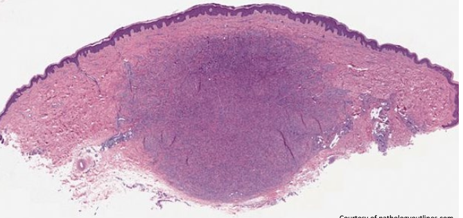
Benign fibrous histiocytoma (Dermatofibroma)
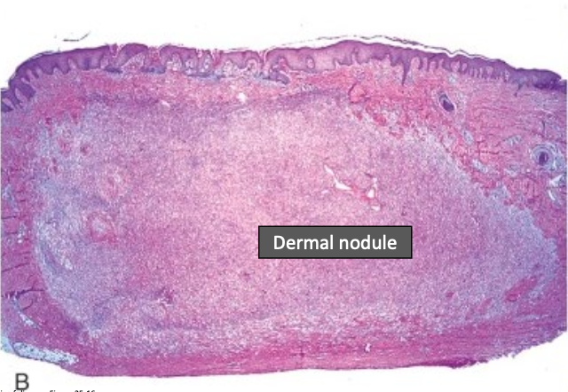
Benign fibrous histiocytoma (Dermatofibroma)
Composed of benign, spindle-shaped cells arranged in a well-defined, nonencapsulated mass within the mid-dermis
Overlying epidermal hyperplasia, characterized by downward elongation of hyperpigmented rete ridges
Benign fibrous histiocytoma (Dermatofibroma)
collagen balls (collagen trapping)
Benign fibrous histiocytoma (Dermatofibroma)
Composed of benign, spindle-shaped cells arranged in a well-defined, nonencapsulated mass within the mid-dermis
Overlying epidermal hyperplasia, characterized by downward elongation of hyperpigmented rete ridges
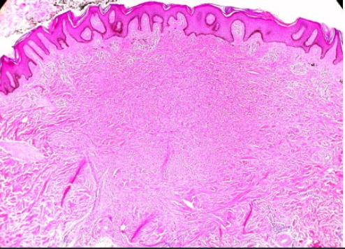
Benign fibrous histiocytoma (Dermatofibroma)
Composed of benign, spindle-shaped cells arranged in a well-defined, nonencapsulated mass within the mid-dermis
Overlying epidermal hyperplasia, characterized by downward elongation of hyperpigmented rete ridges
Benign fibrous histiocytoma (Dermatofibroma)
Composed of benign, spindle-shaped cells arranged in a well-defined, nonencapsulated mass within the mid-dermis
Overlying epidermal hyperplasia, characterized by downward elongation of hyperpigmented rete ridges
capillary hemangioma
capillary hemangioma
Delicate appearing vessels lined by flattened endothelial cells
vascular spaces are small or collapsed; intervening loose connective tissue stroma may contain larger arterioles or venules
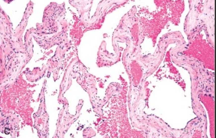
cavernous hemangioma
contains large, dilated vascular spaces that may extend into underlying adipose tissue
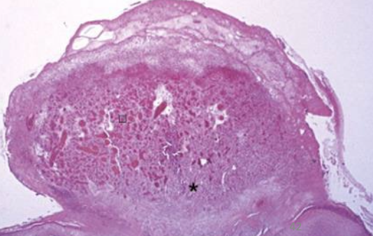
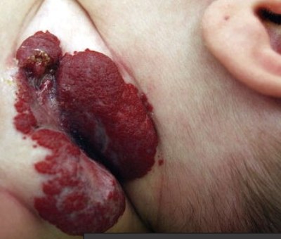
Pyogenic granuloma
Inflammatory cell infiltrate often surrounds capillary-
like vessels
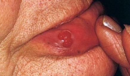
Pyogenic granuloma
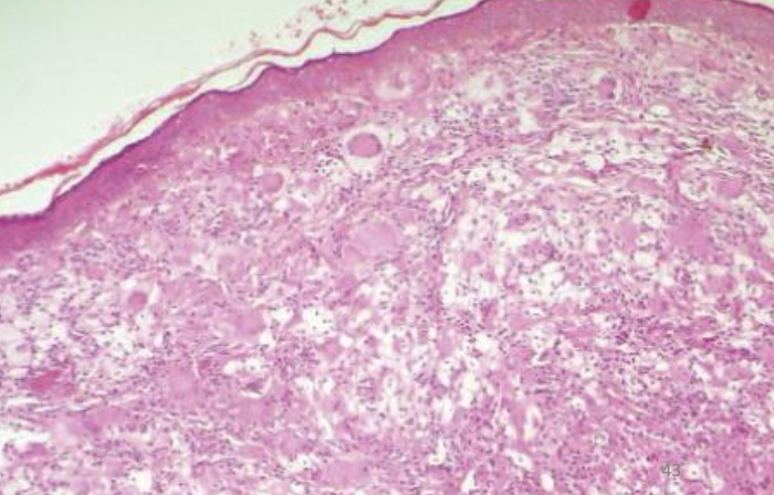
Xanthoma and xanthelasma
Foamy macrophages have accumulated lipid material (phospholipid, triglyceride, cholesterol)
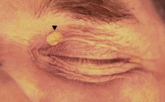
Xanthoma and xanthelasma
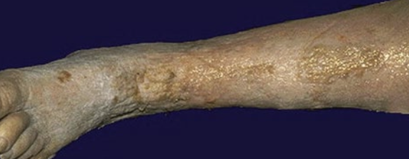
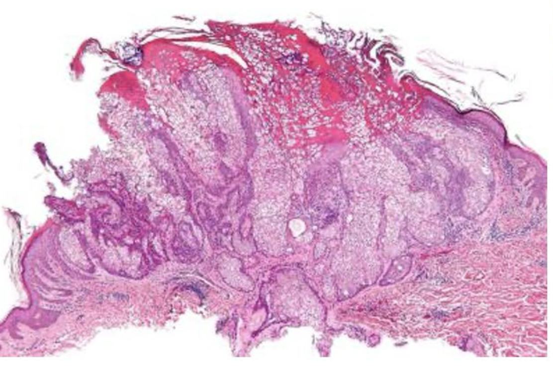
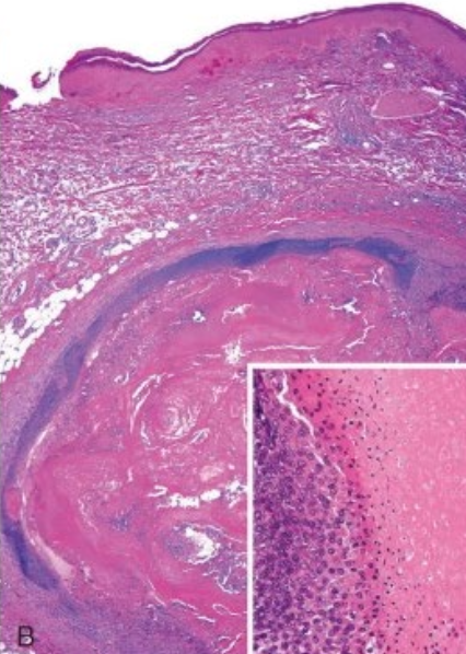
Stasis dermatitis
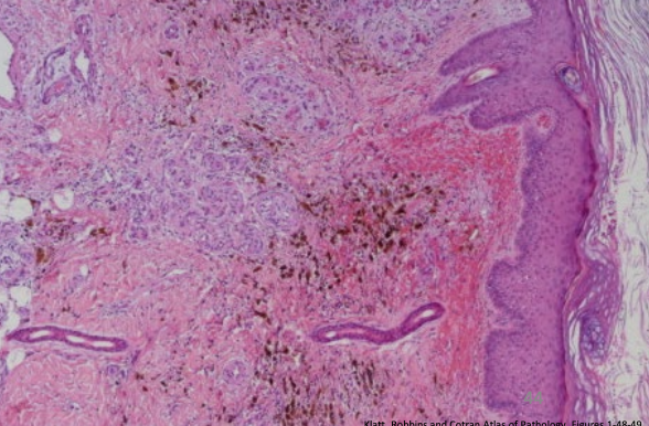
Stasis dermatitis
Hyperkeratotic and acanthotic epidermis at the right
Underneath shows bright red recent hemorrhage admixed with dark-brown hemosiderin granules and proliferating, thickened vascular channels