LOWER LIMBS: FEMUR, PATELLA, TIBIA, & FIBULA
1/60
There's no tags or description
Looks like no tags are added yet.
Name | Mastery | Learn | Test | Matching | Spaced |
|---|
No study sessions yet.
61 Terms
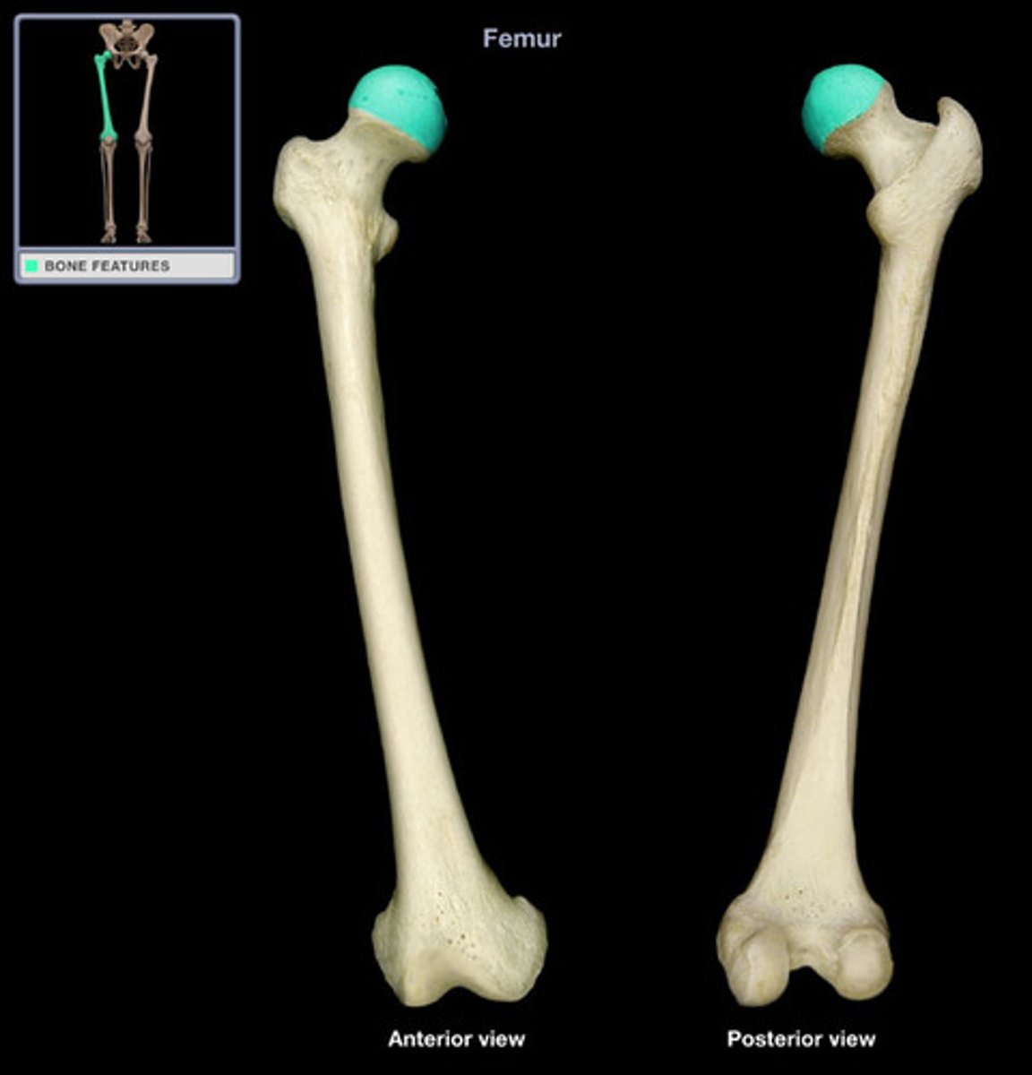
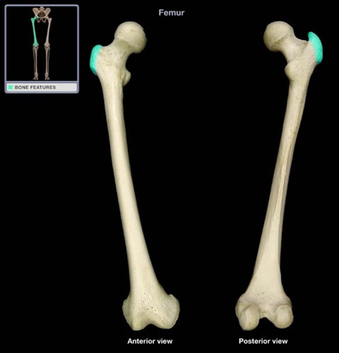
FEMORAL HEAD
rounded proximal part of the FEMUR that fits into ACETABULUM
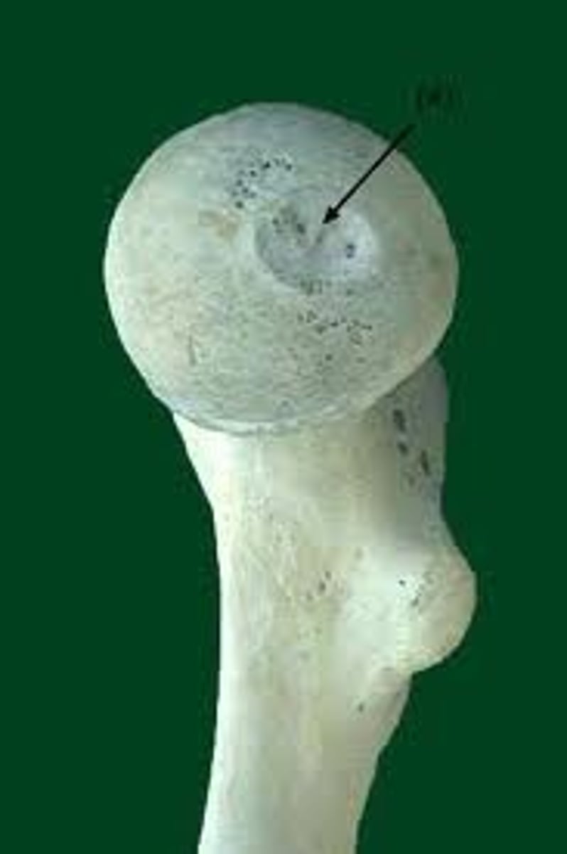
FOVEA CAPITIS (of FEMUR)
small, nonarticular depression near the center of the FEMORAL HEAD
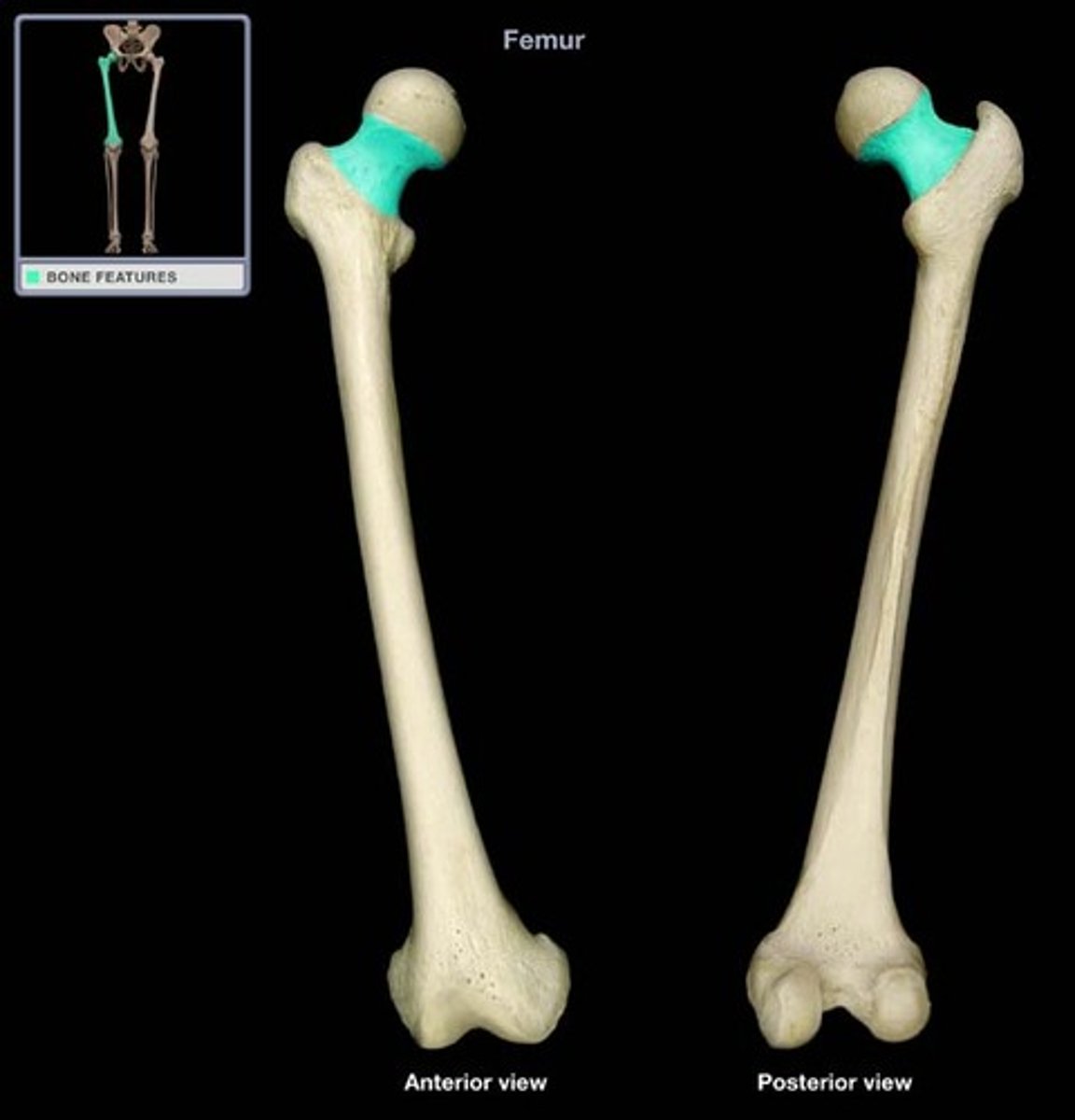
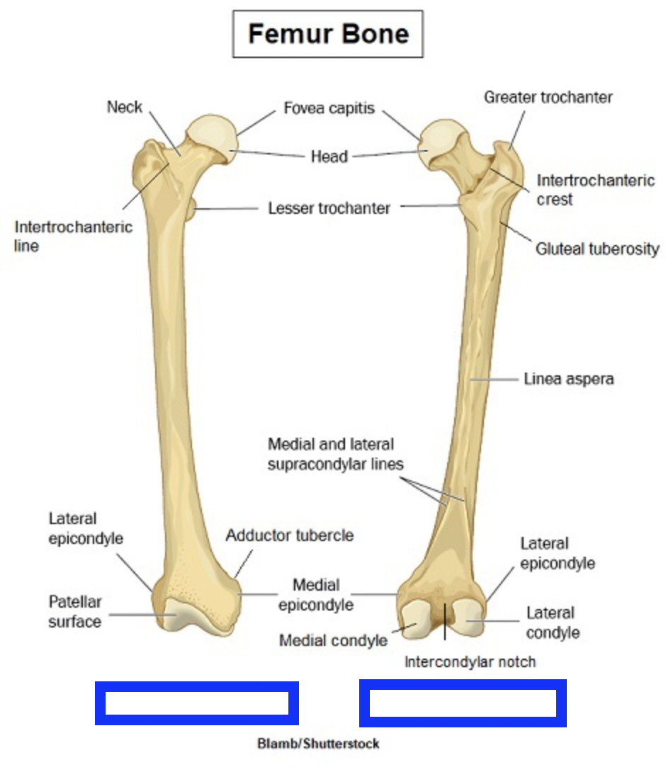
FEMORAL NECK
connects HEAD to SHAFT
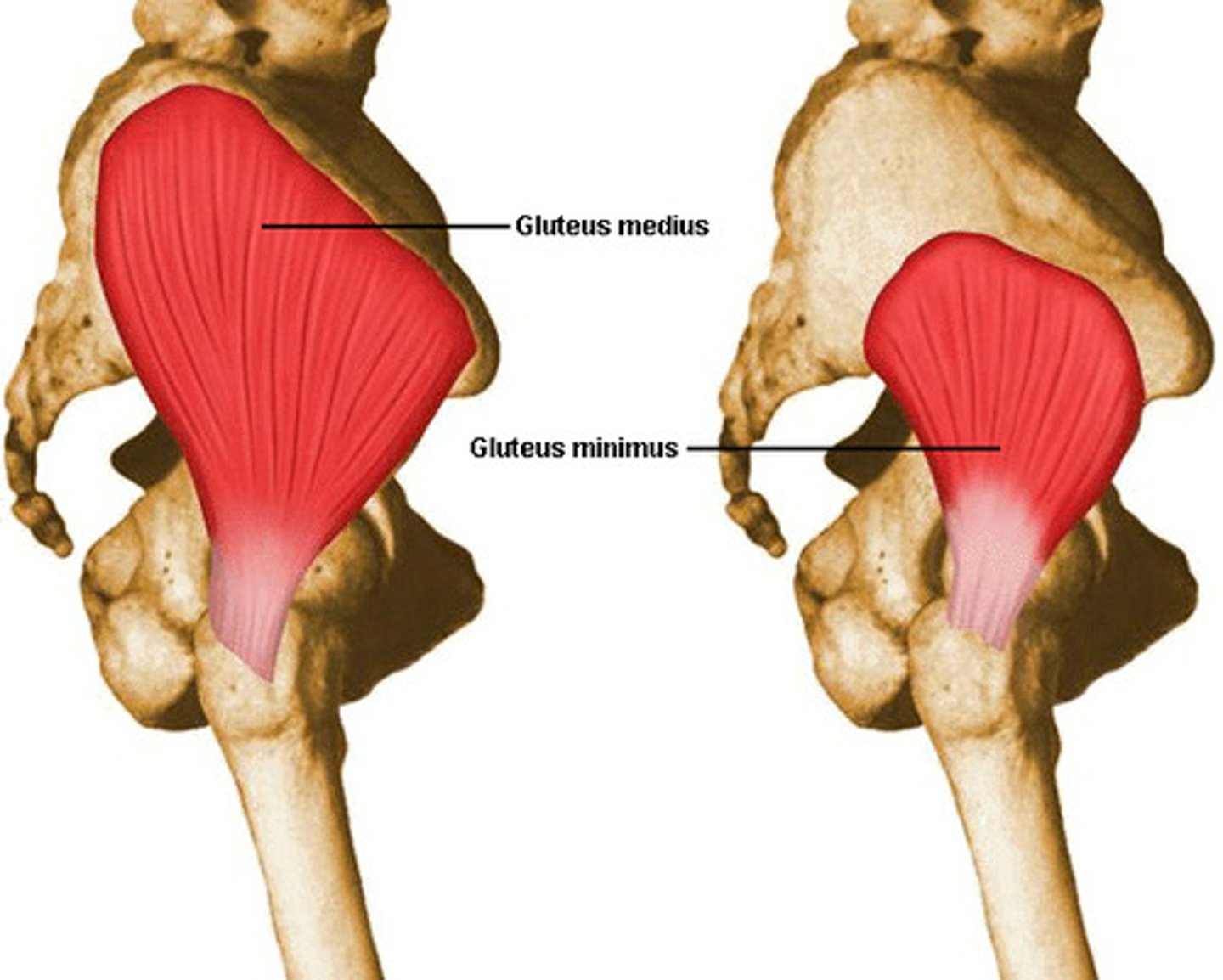
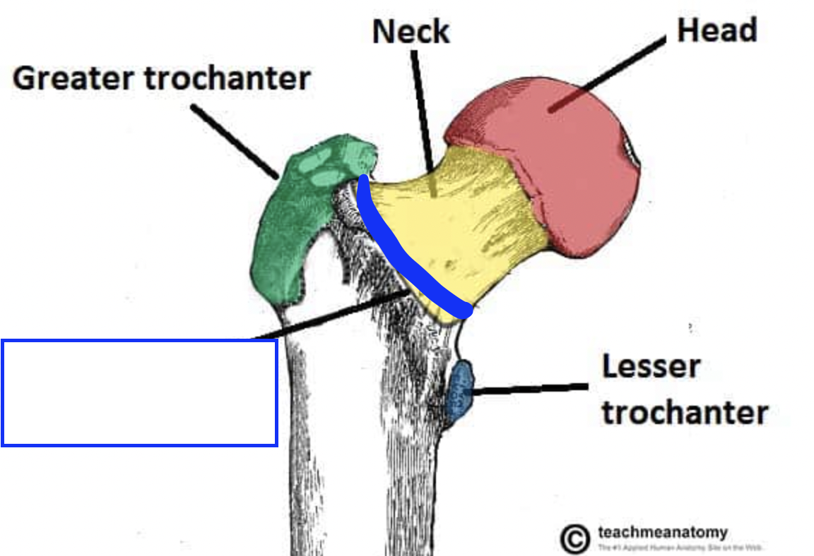
GREATER TROCHANTER (of FEMUR)
large, blunt, nonarticular prominence on the lateral, proximal part of the FEMUR - insertion for GLUTEUS MINIMUS and GLUTEUS MEDIUS
what feature is the insertion for the muscles GLUTEUS MINIMUS and GLUTEUS MEDIUS?
GREATER TROCHANTER (of FEMUR)
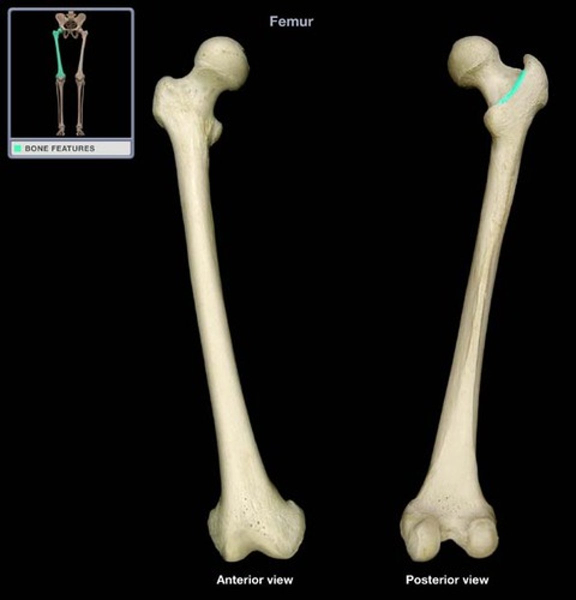
INTERTROCHANTERIC LINE (of FEMUR)
variable, fairly vertical, roughened line that passes between the LESSER and GREATER TROCHANTERS on anterior surface on the FEMORAL NECK
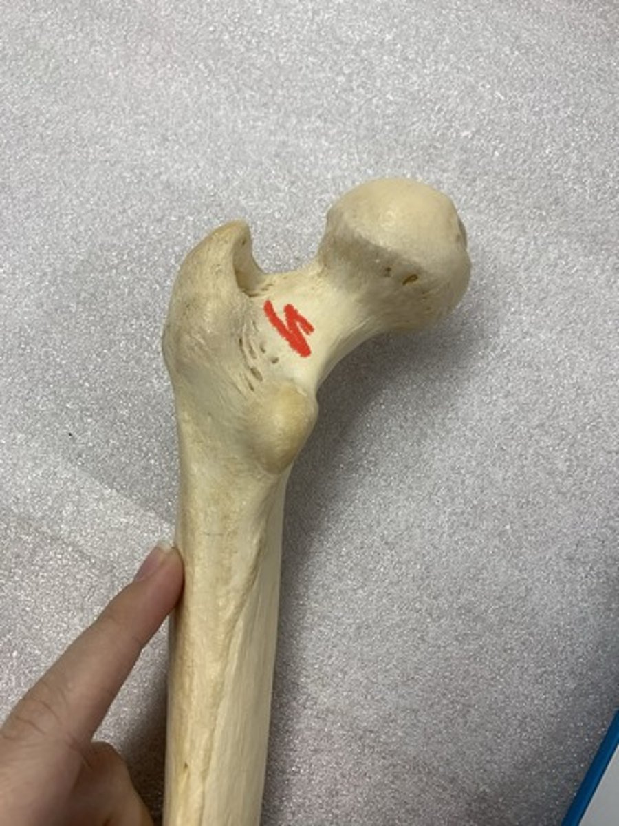
TROCHANTERIC FOSSA (of FEMUR)
pit excavated into posteromedial wall of the GREATER TROCHANTER

OBTURATOR EXTERNUS GROOVE (of FEMUR)
shallow depression aligned laterally and superiorly across the posterior surface of the FEMORAL NECK
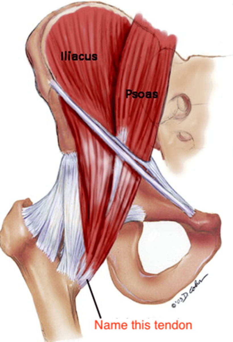
LESSER TROCHANTER (of FEMUR)
blunt, prominent tubercle on the posterior FEMORAL SURFACE just inferior to the point where the NECK joins the SHAFT - insertion of ILIOPSOAS TENDON
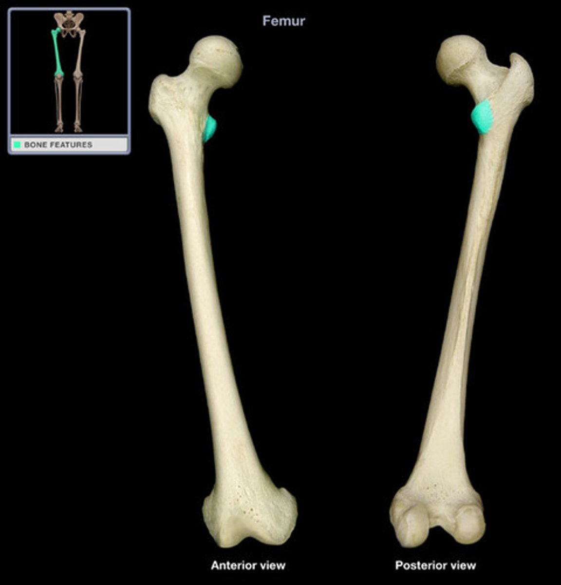
what feature is the insertion for the ILIOPSOAS TENDON?
LESSER TROCHANTER
INTERTROCHANTERIC CREST (of FEMUR)
elevated line on the posterior surface of the proximal femur between the GREATER and LESSER TROCHANTER
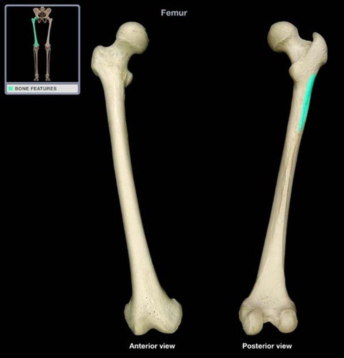
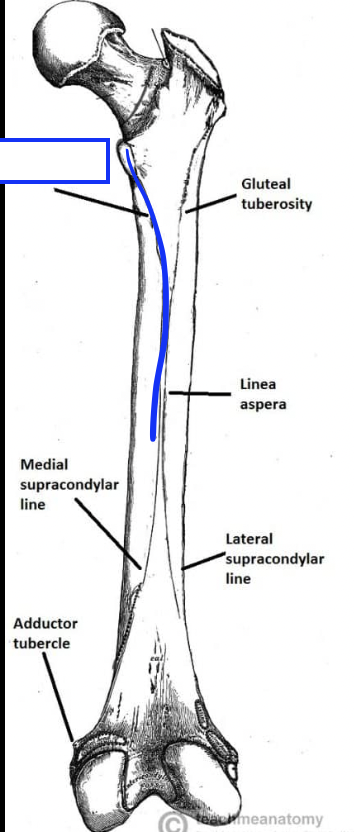
GLUTEAL LINE/TUBEROSITY (of FEMUR)
long, wide, roughened, posterolaterally placed feature that extends from the base of the GREATER TROCHANTER to the lop of the LINEA ASPERA
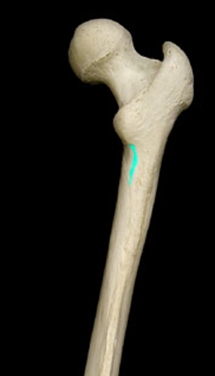
SPIRAL LINE (of FEMUR)
spiraling inferior to LESSER TROCHANTER, connects the inferior end of the INTERTROCHANTERIC LINE with the medial lip of the LINEA ASPERA
PECTINEAL LINE (of FEMUR)
short, curved line that passes inferolaterally from the base of the LESSER TROCHANTER, between the SPIRAL LINE and GLUTEAL TUBEROSITY
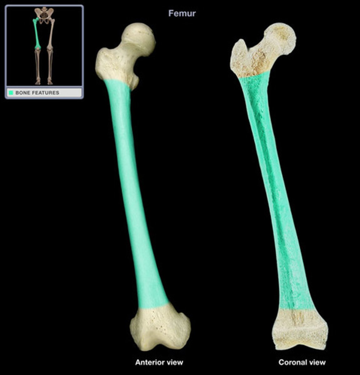
FEMORAL SHAFT
long section between the expanded proximal and distal ends of the bone
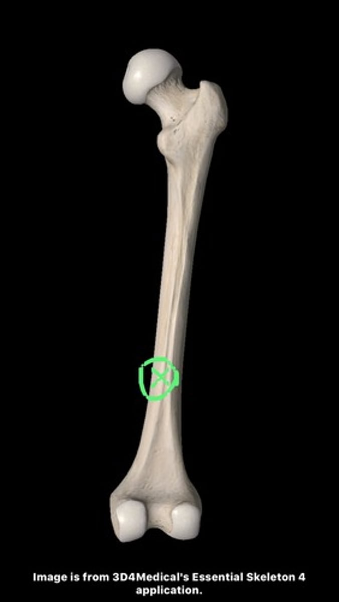
NUTRIENT FORAMEN (of FEMUR)
located about midshaft level on the posterior surface of the bone, adjacent to or on the LINEA ASPERA
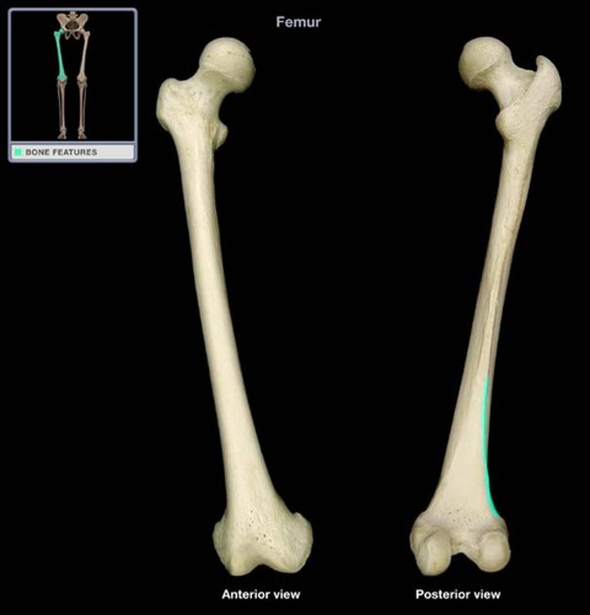
MEDIAL SUPRACONDYLAR LINE/RIDGE (of FEMUR)
inferior, medial extension of the LINEA ASPERA, marking the distal, medial corner of the SHAFT
LATERAL SUPRACONDYLAR LINE/RIDGE (of FEMUR)
inferior (distal), lateral extension of LINEA ASPERA, marking the distal, medial corner of the shaft
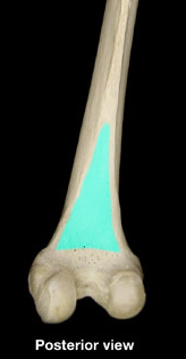
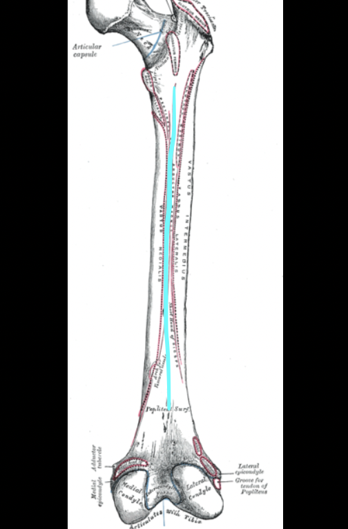
POPLITEAL SURFACE (of FEMUR)
wide, flat, triangular area of the posterior, distal FEMUR
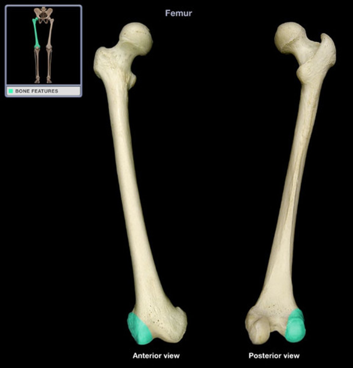
LATERAL CONDYLE (of FEMUR)
large, protruding, articular knob on the lateral side of the distal FEMUR
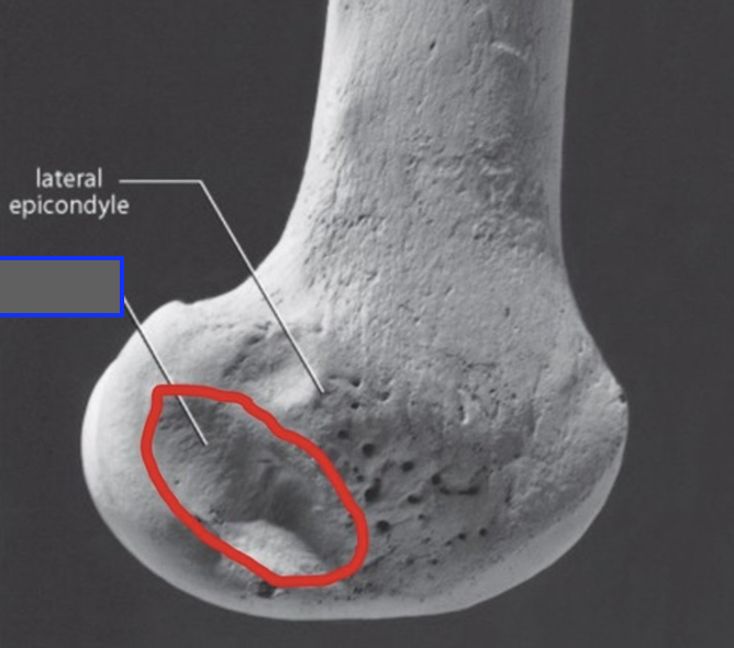
LATERAL EPICONDYLE (of FEMUR)
convexity on the lateral side of the lateral condyle
POPLITEAL GROOVE (of FEMUR)
smooth hollow on the posterolateral side of the lateral condyle of FEMUR
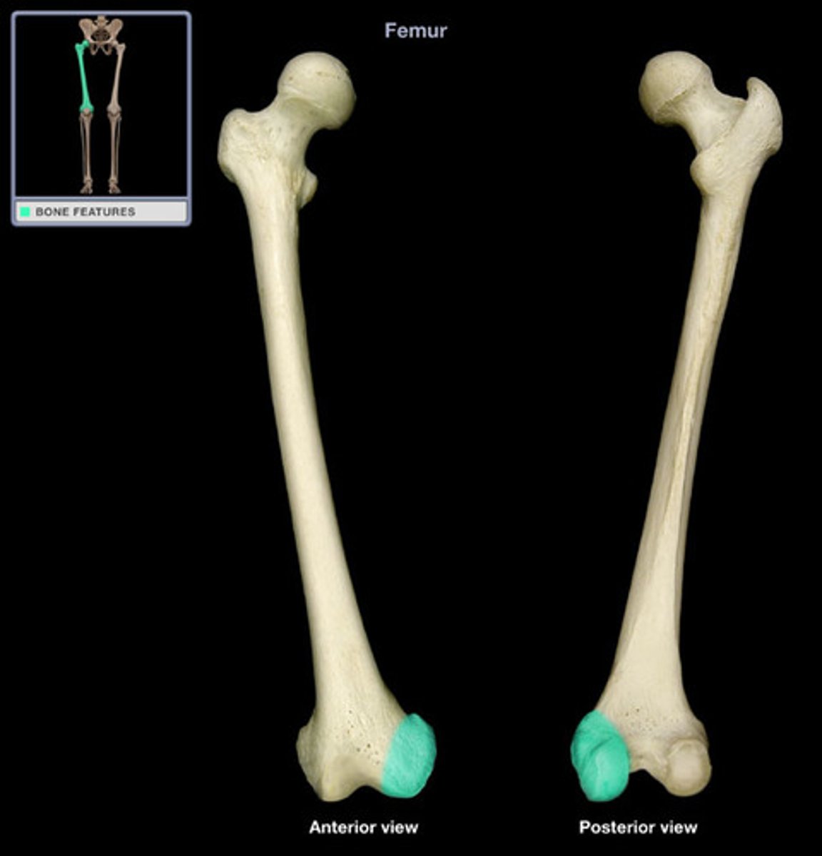
MEDIAL CONDYLE (of FEMUR)
large, articular knob on the medial side of the distal FEMUR
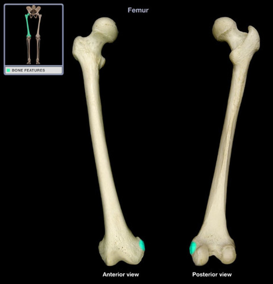
MEDIAL EPICONDYLE (of FEMUR)
convexity on the medial side of the MEDIAL CONDYLE
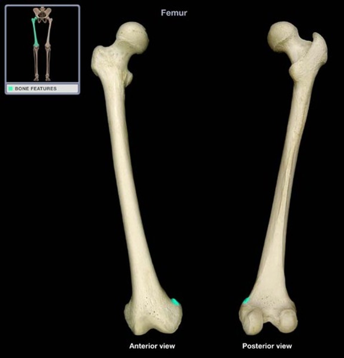
ADDUCTOR TUBERCLE (of FEMUR)
variable, raised tubercle on the medial SUPRACONDYLAR RIDGE just superior to the MEDIAL EPICONDYLE
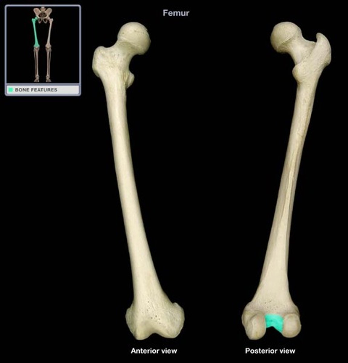
INTERCONDYLAR FOSSA/NOTCH (of FEMUR)
nonarticular, excavated surface between the distal and posterior articular surfaces of the CONDYLES
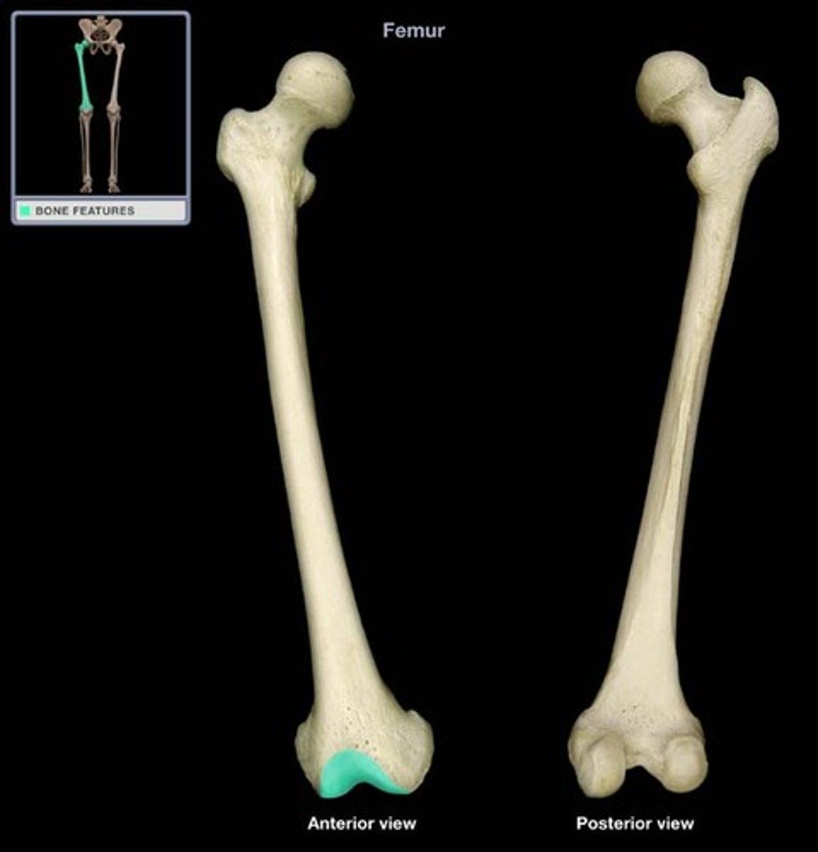
PATELLAR SURFACE (of FEMUR)
notched, articular area on the anterior surface of the distal FEMUR, over which the PATELLA glides during flexion and extension of the knee
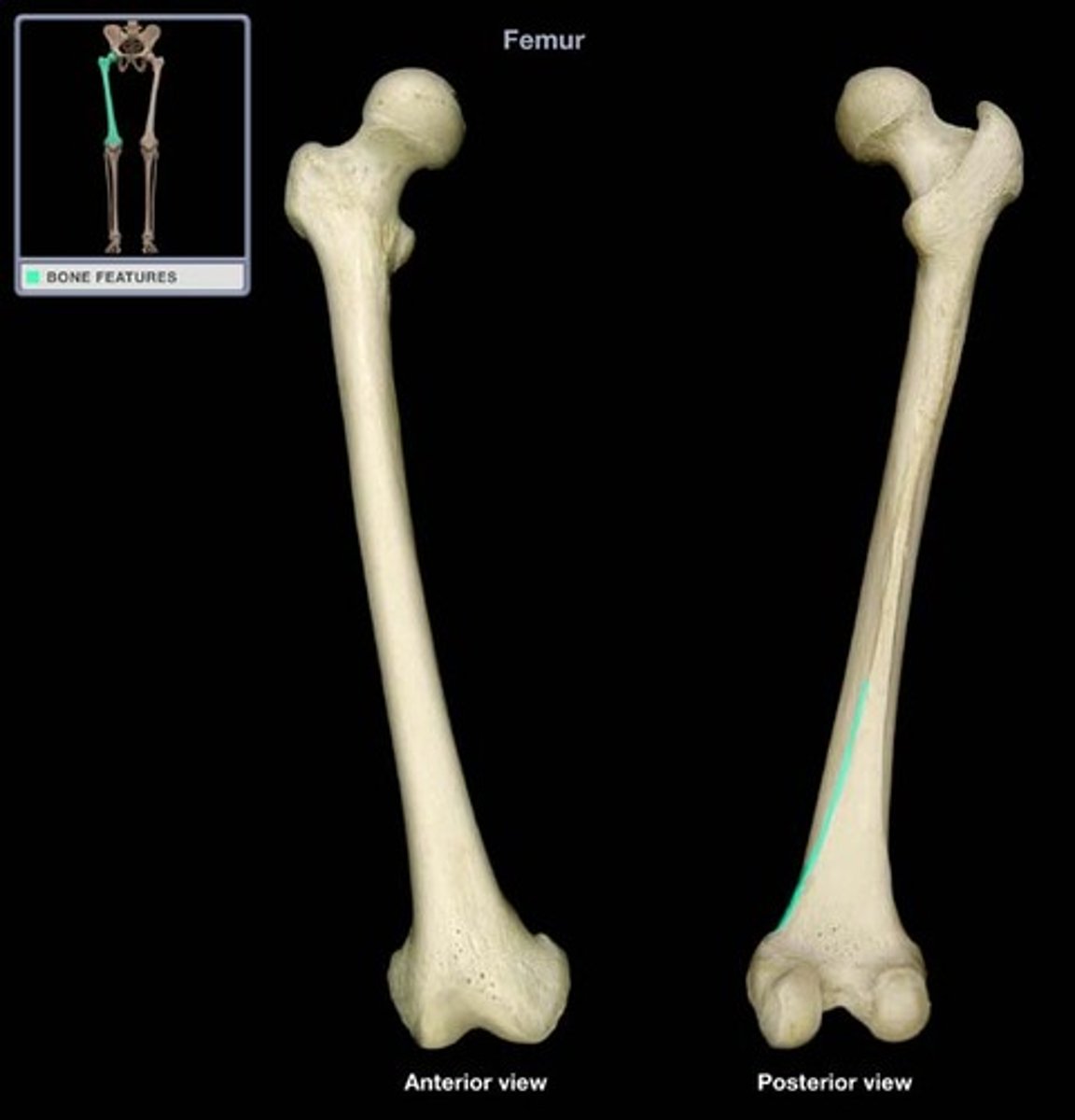
LINEA ASPERA (of FEMUR)
a prominent longitudinal ridge on the posterior surface of the femur, serving as an attachment site for muscles
the intertrochanteric ____ is anterior, and the intertrochanteric ____ is posterior.
line, crest
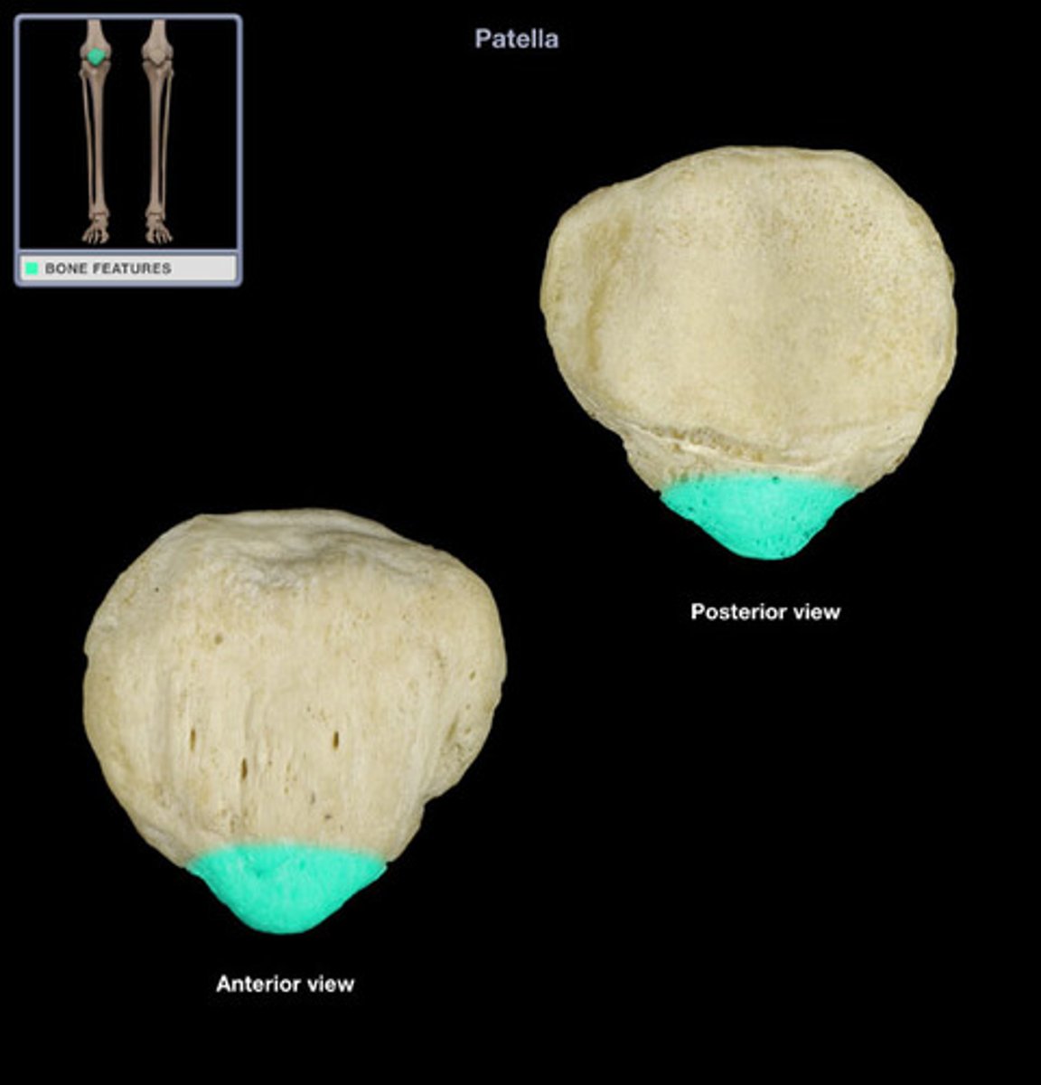
APEX (of PATELLA)
PATELLA is nonarticular point on the bone
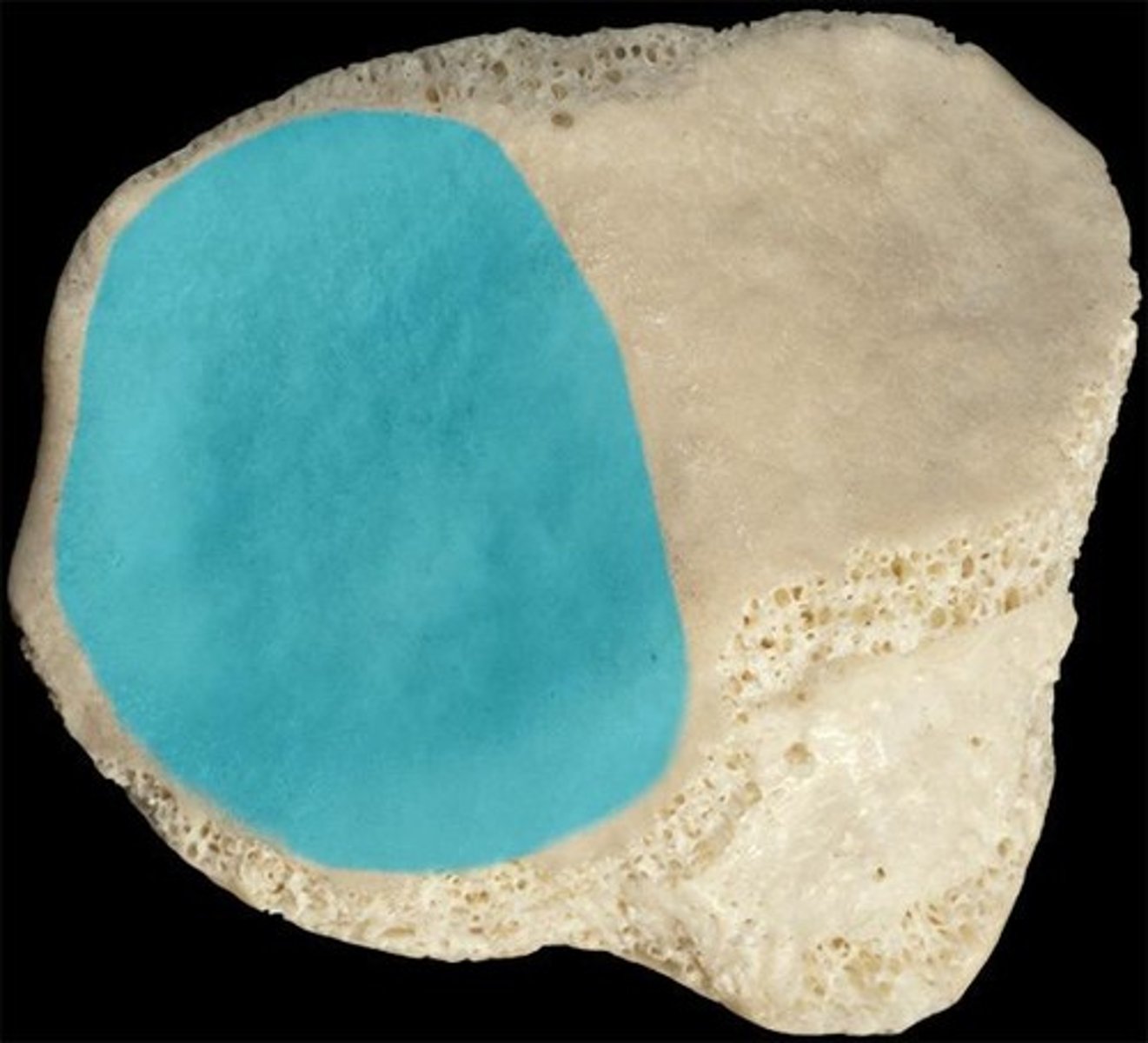
LATERAL ARTICULAR FACET (of PATELLA)
for the distal FEMUR - faces posteriorly and is the largest part of the large articular surface of the PATELLA
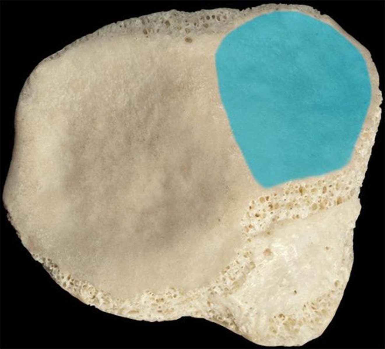
MEDIAL ARTICULAR FACET (of PATELLA)
for the distal FEMUR - faces posteriorly and is smaller than the lateral articular facet
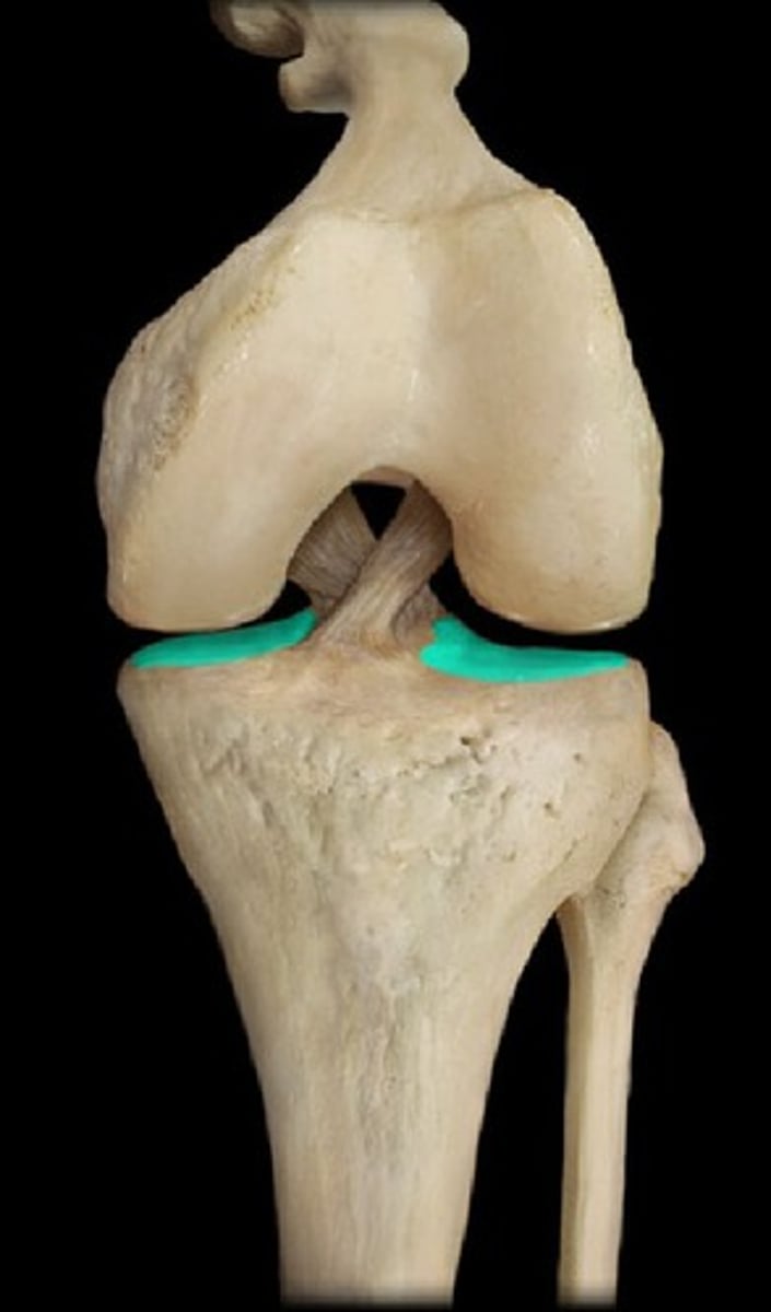
TIBIAL PLATEAU (of TIBIA)
proximal tibial surface on which the FEMUR rests
MEDIAL CONDYLE (of TIBIA)
medial part of the tibial plateau
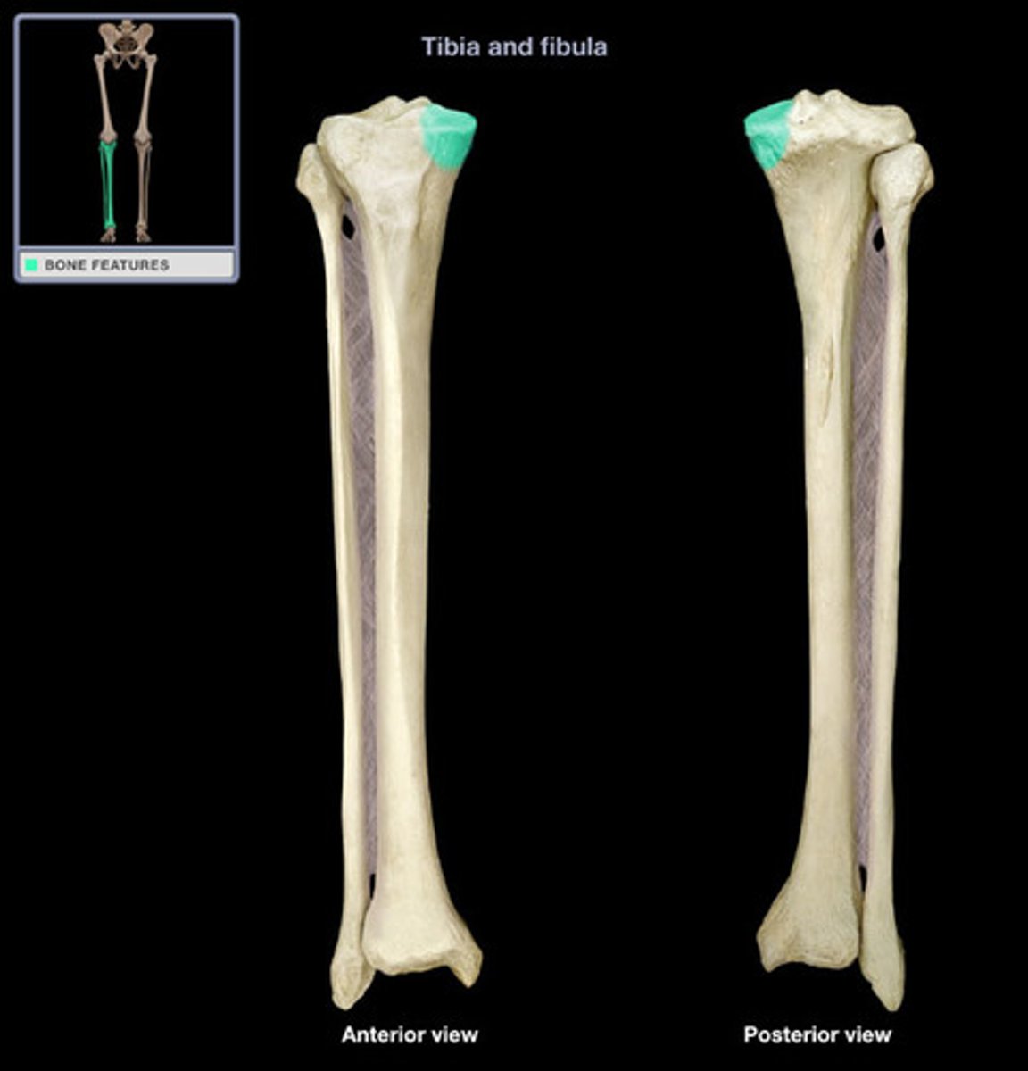
LATERAL CONDYLE (of TIBIA)
lateral part of tibial plateau
INTERCONDYLAR EMINENCE (of TIBIA)
raised area on the proximal tibial surface between articular facets
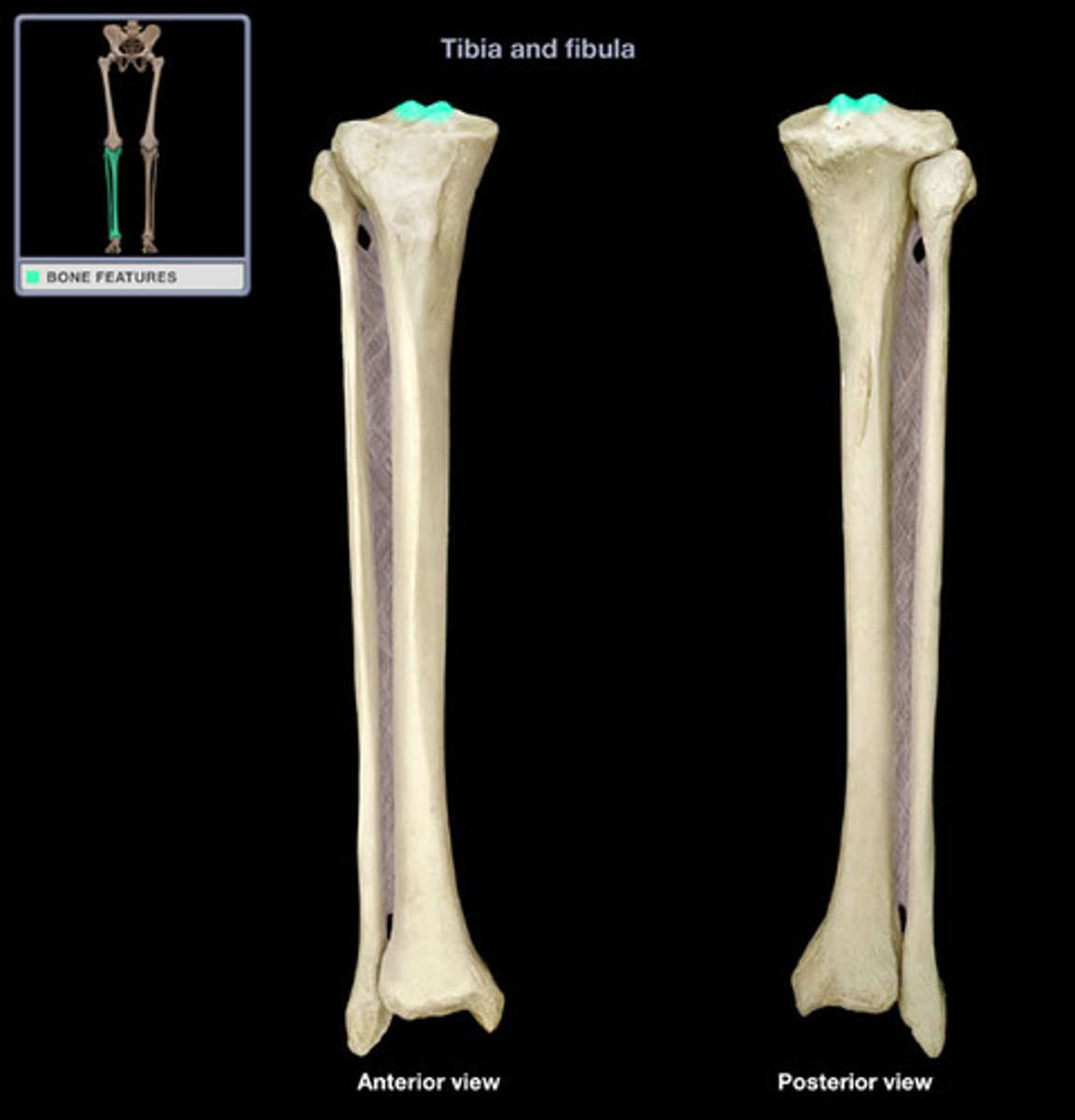
MEDIAL INTERCONDYLAR TUBERCLE (of TIBIA)
forms the medial part of the INTERCONDYLAR EMINENCE
LATERAL INTERCONDYLAR TUBERCLE (of TIBIA)
forms the lateral part of the INTERCONDYLAR EMINENCE
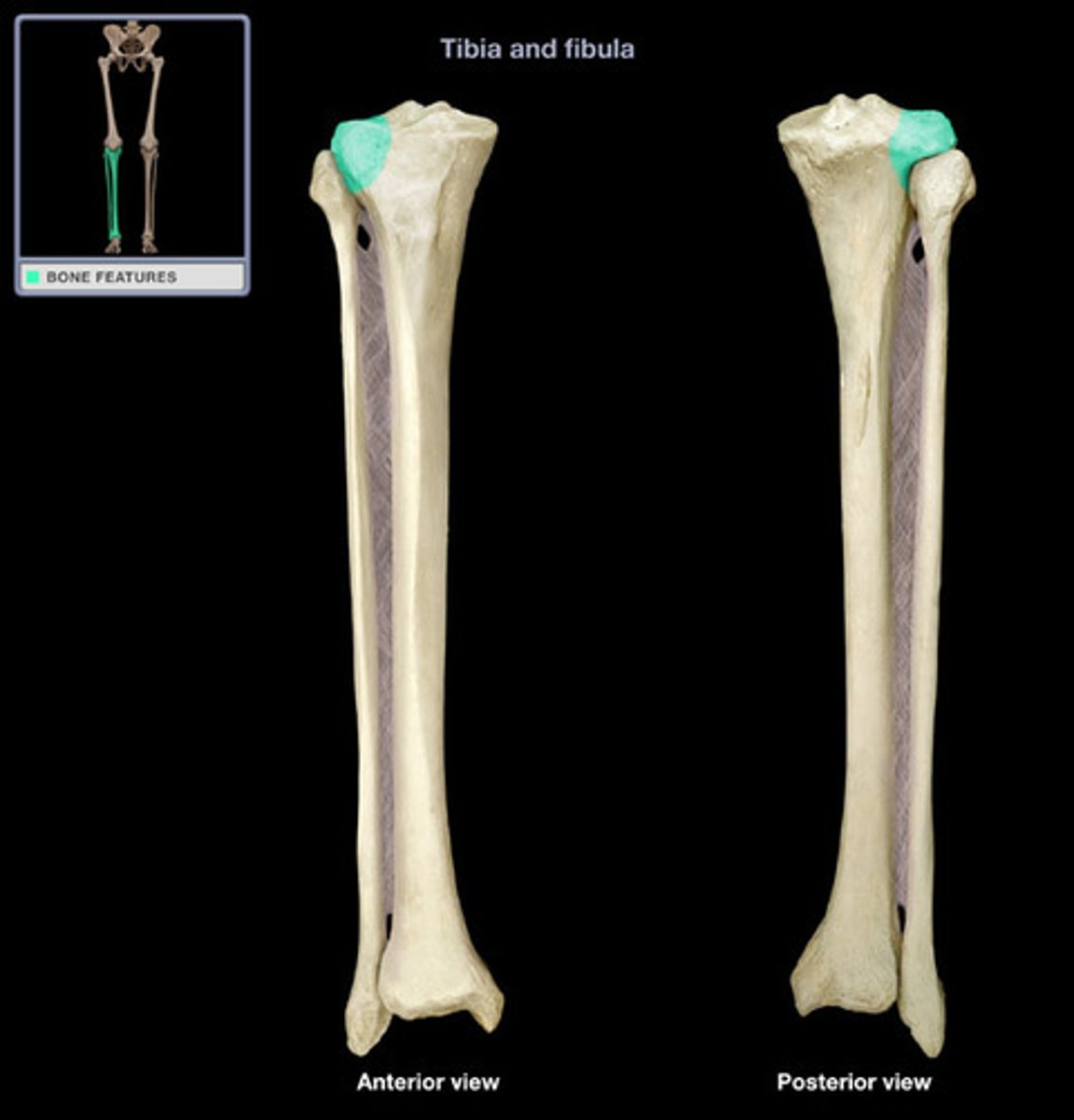
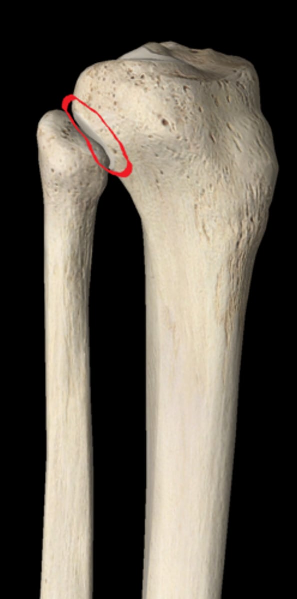
SUPERIOR FIBULAR ARTICULAR FACET (of TIBIA)
located on the posteroinferior edge of the LATERAL CONDYLE
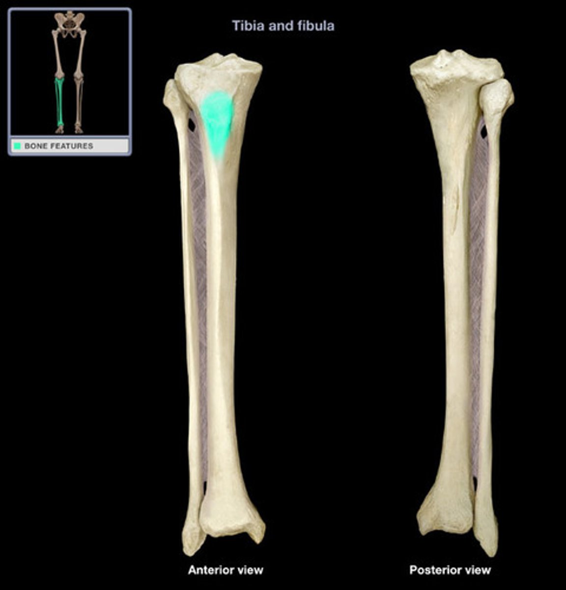
TIBIAL TUBEROSITY
rugose area on the anterior surface of the proximal TIBIA
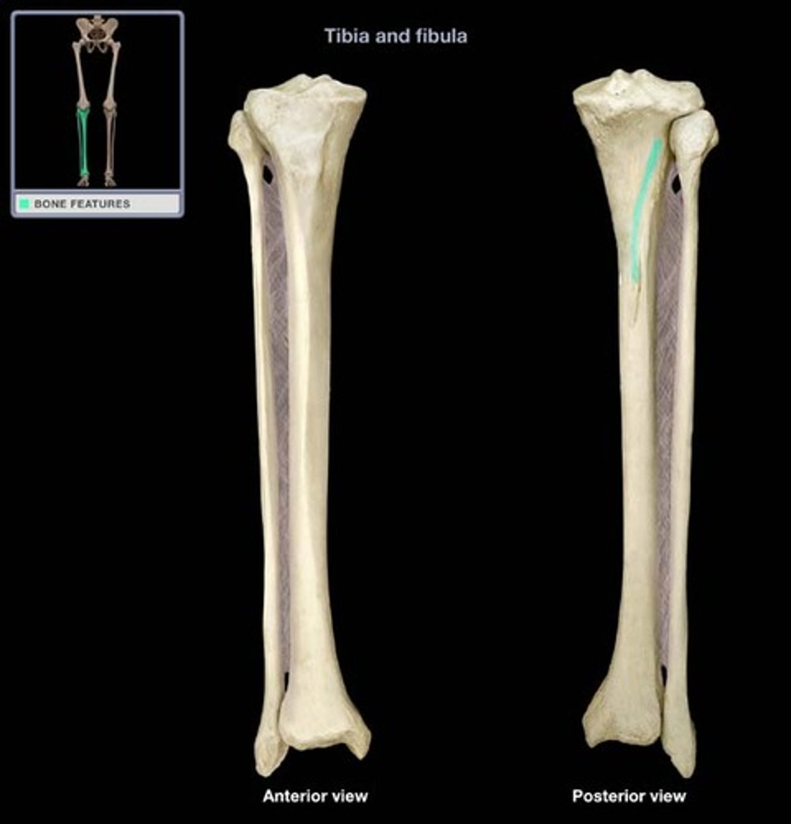
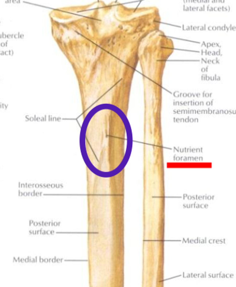
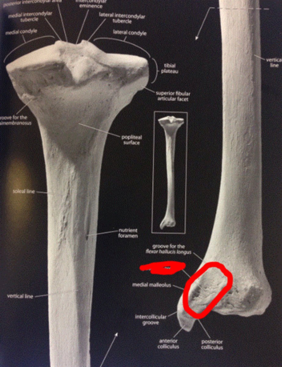
SOLEAL/POPLITEAL LINE (of TIBIA)
crosses the proximal half of the posterior tibial surface from superolateral to inferomedial
NUTRIENT FORAMEN (of TIBIA)
just inferolateral to the POPLITEAL LINE
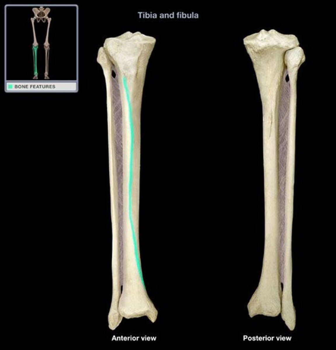
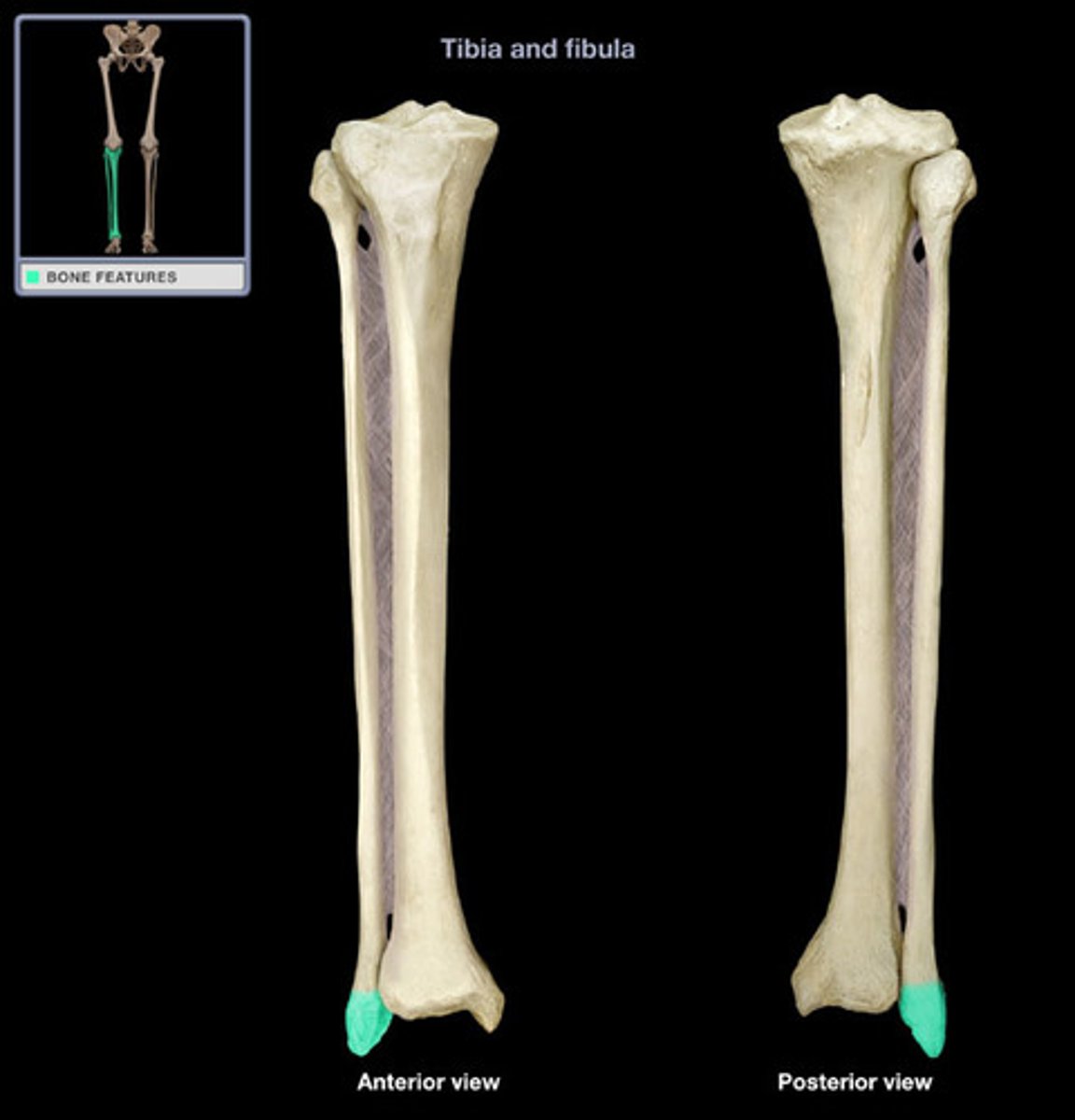
ANTERIOR SURFACE/CREST (of TIBIA)
shaft forms the anterior edge of the "shin"
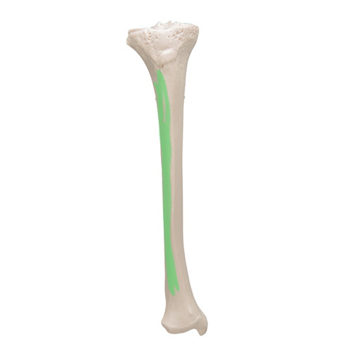
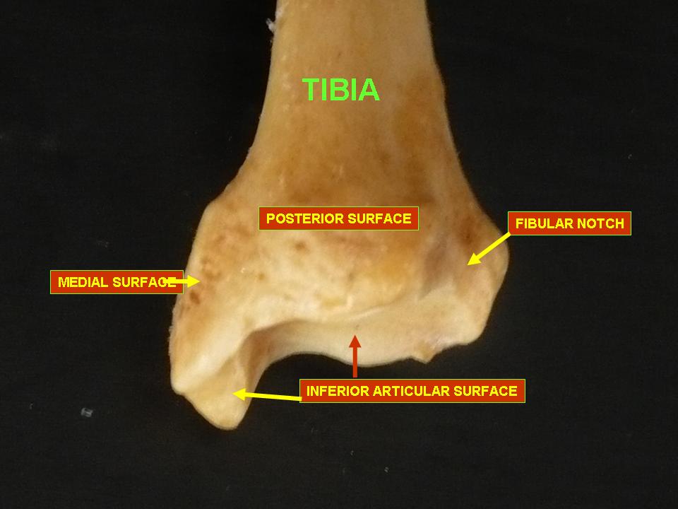
MEDIAL SURFACE (of TIBIA)
shaft forms the medial edge of "shin"
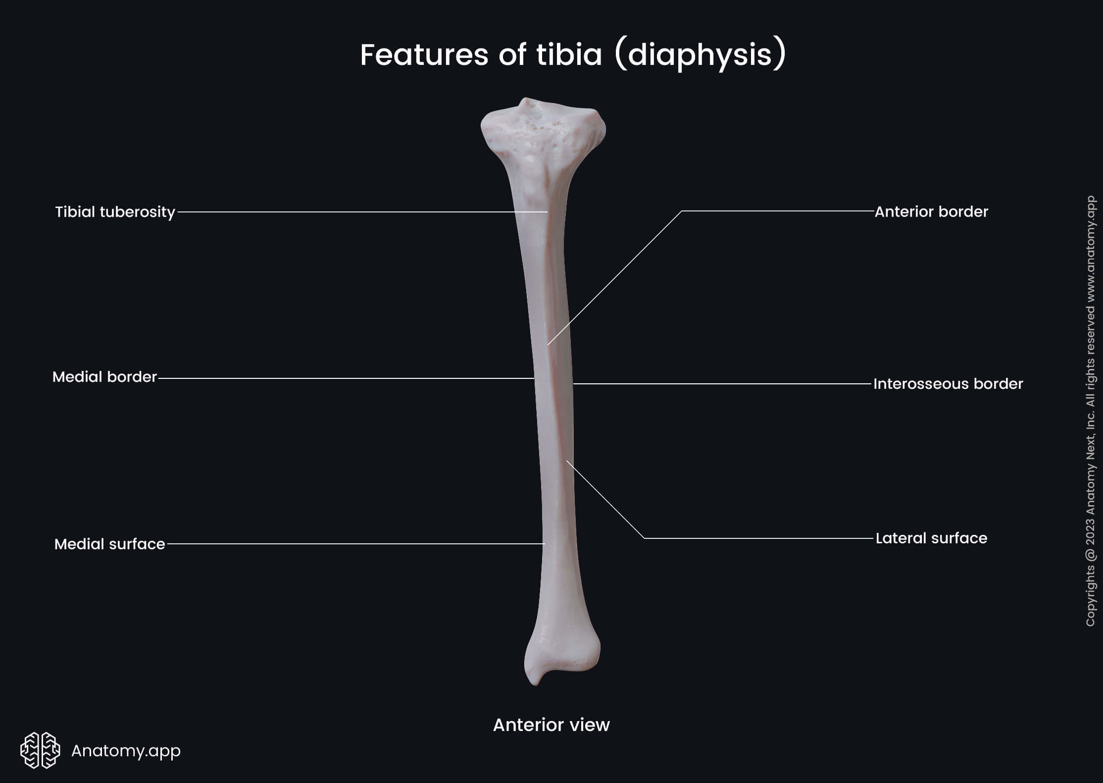
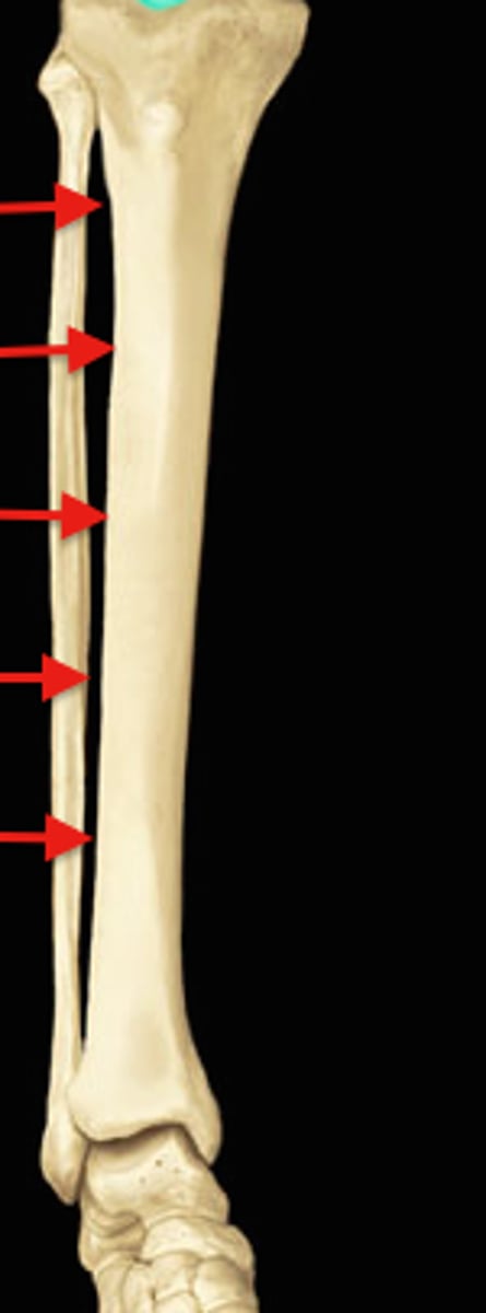
INTEROSSEOUS SURFACE (of TIBIA)
of SHAFT, is lateral, opposite of FIBULA
INTEROSSEOUS CREST (of TIBIA)
lateral crest of the SHAFT, faces FIBULA, attachment of INTEROSSEOUS MEMBRANE
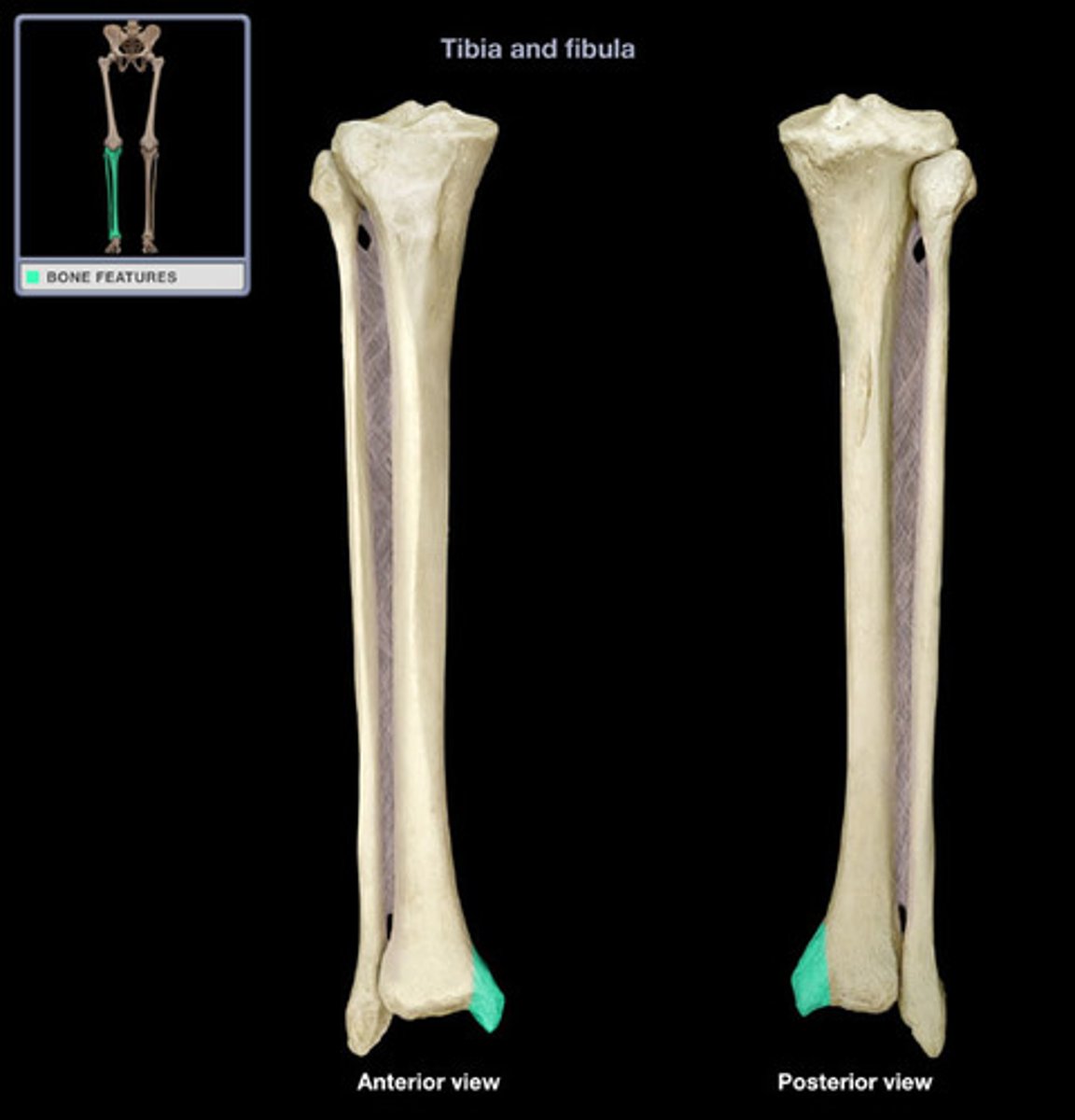
MEDIAL MALLEOLUS (of TIBIA)
projection on the medial side of the distal TIBIA that forms the subcutaneous medial knob at the ankle
FIBULAR NOTCH (of TIBIA)
distolateral corner of the TIBIA

INFERIOR FIBULAR ARTICULAR SURFACE (of TIBIA)
thin articular surface for the FIBULA, which faces laterally at the base of the FIBULAR NOTCH
MALLEOLAR GROOVE (of TIBIA)
posterior aspect of the MEDIAL MALLEOLUS
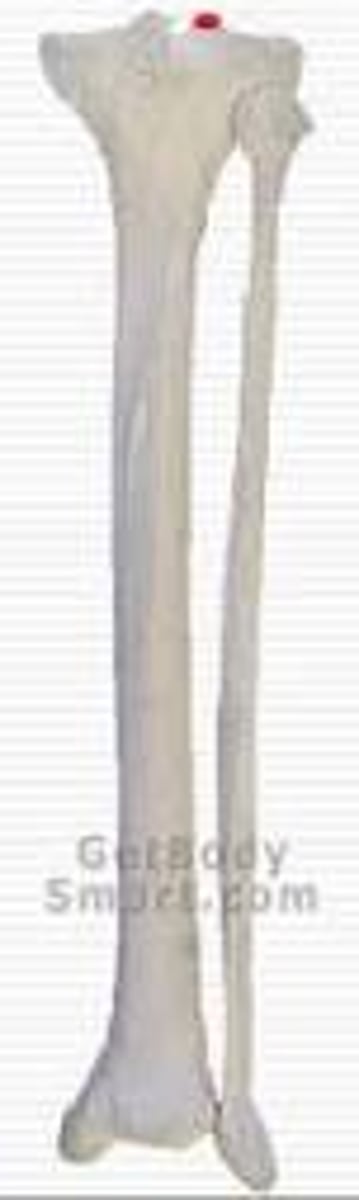
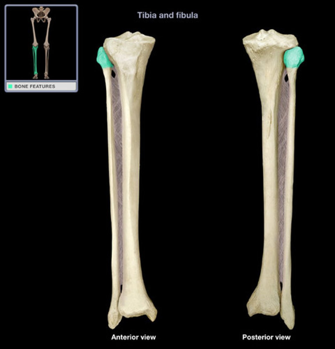
HEAD (of FIBULA)
swollen proximal end of the FIBULA, more massive and less mediolaterally flattened than the distal end
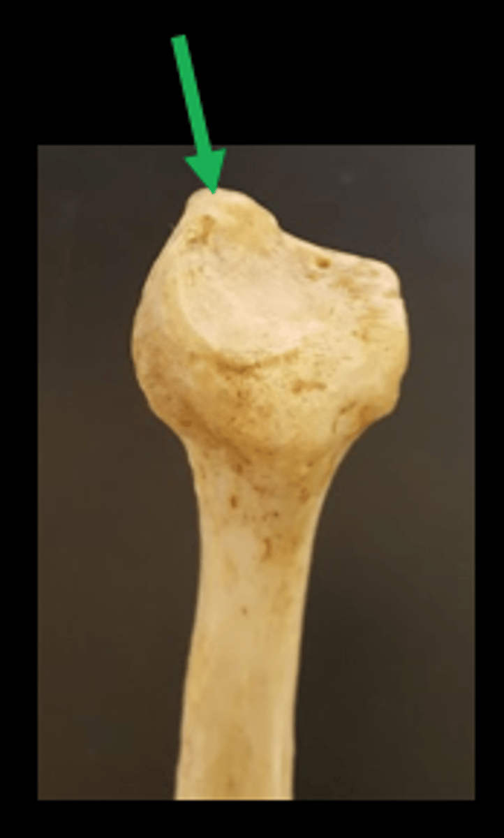
STYLOID PROCESS (of FIBULA)
most proximal projection of the FIBULA, forming the posterior part of the HEAD
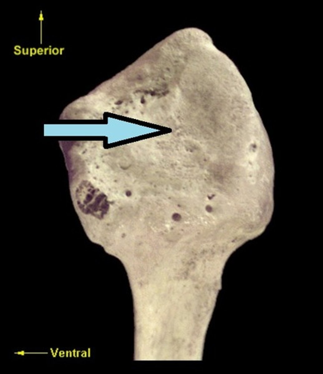
PROXIMAL FIBULAR ARTICULAR SURFACE (of FIBULA)
round, flat, medially oriented surface that corresponds to a similar surface on the lateral proximal TIBIA
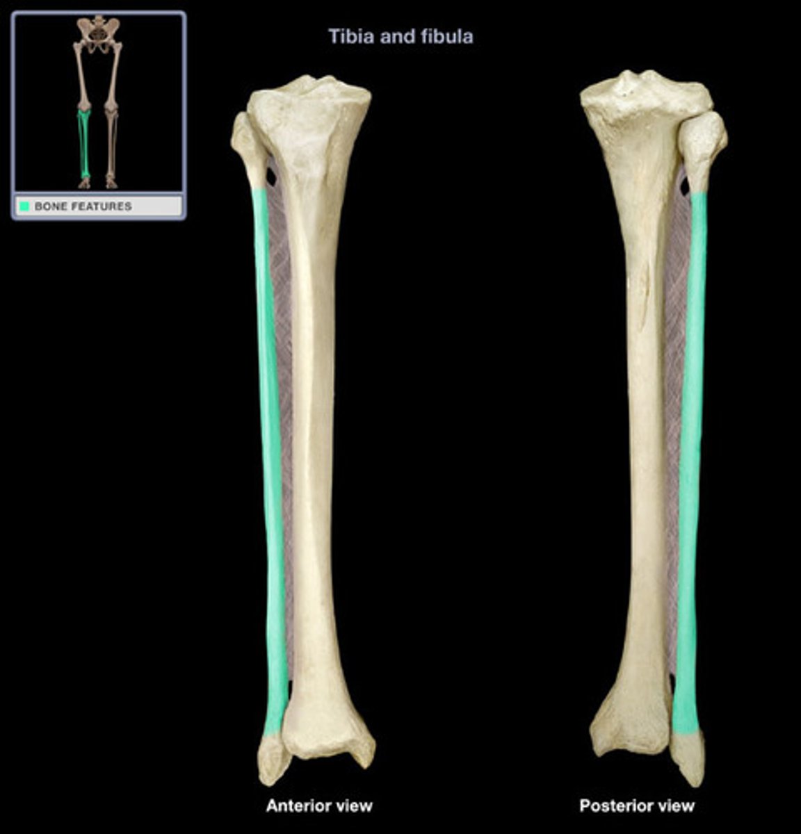
FIBULAR SHAFT
long, thin, fairly straight segment of the bone between the expanded proximal and distal ends
INTEROSSEOUS CREST (of FIBULA)
elevated crest that runs down the medial, slightly anteriorly facing surface of the SHAFT
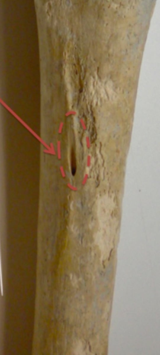
NUTRIENT FORAMEN (of FIBULA)
opens proximally on the posteromedial surface - at about midshaft level
LATERAL MALLEOLUS (of FIBULA)
inferiormost (distalmost) projection of the FIBULA
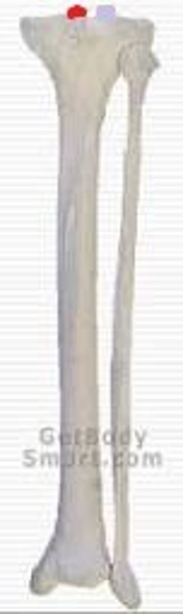
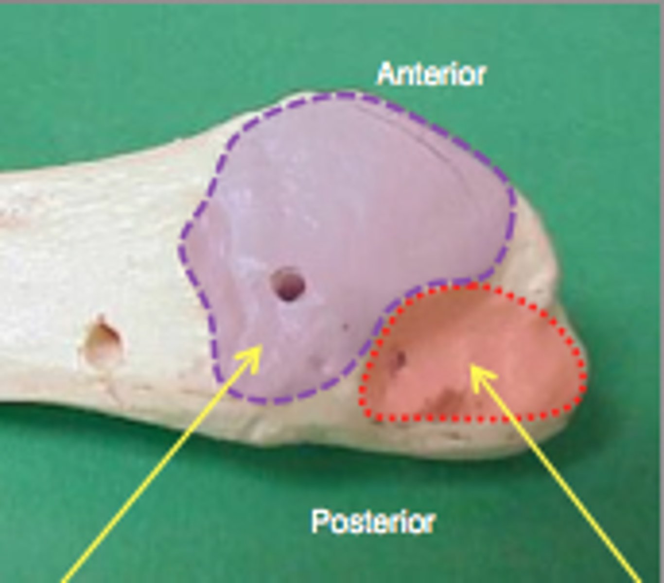
MALLEOLAR (DISTAL) ARTICULAR SURFACE (of FIBULA)
flat, medially facing, triangular surface whose APEX faces inferiorly (purple in image)
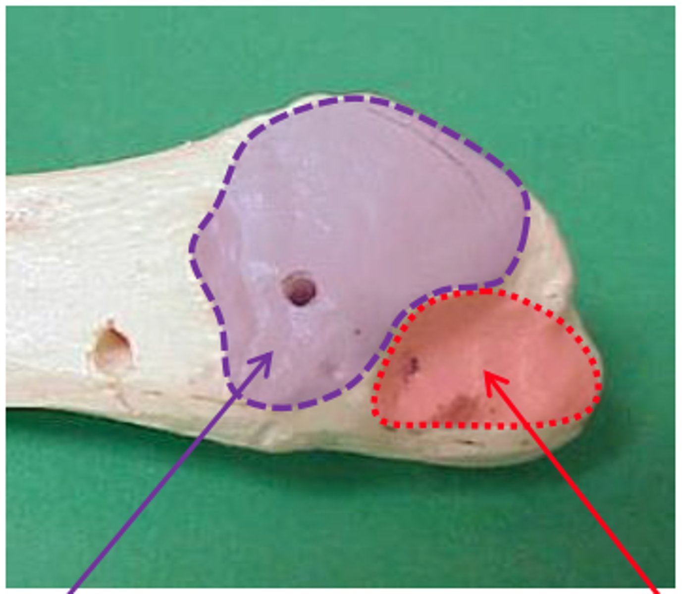
MALLEOLAR FOSSA (of FIBULA)
located just posterior to the distal articular surface (red in image)
FIBULAR (PERONEAL) GROOVE
for tendons of the fibularis longus and fibularis brevis muscles, marks posterior surface of distal FIBULA
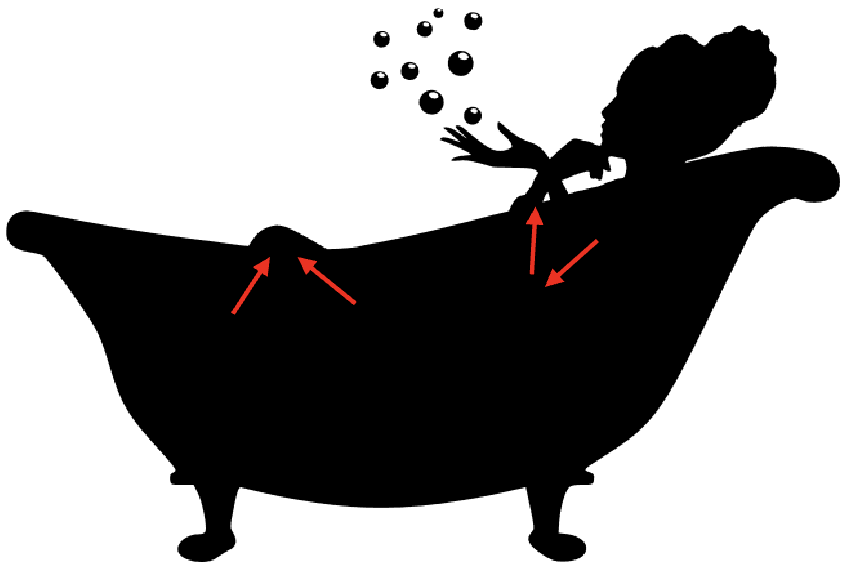
which way do the NUTRIENT FORAMINA face in the FEMUR, TIBIA, and FIBULA?
face like you are sitting in bathtub; FIBULA and TIBIA opens superiorly, FEMORA opens inferiorly