The rest of the objectives
 Call Kai
 Learn
Learn Practice Test
 Spaced Repetition
 Match
Match Flashcards
 Knowt Play
1/75

There's no tags or description
Looks like no tags are added yet.
Name | Mastery | Learn | Test | Matching | Spaced | Call with Kai | Chat |
|---|
No analytics yet
Send a link to your students to track their progress
76 Terms
How does lead poisening alter the heme biosynthesis pathway?
ALA dehydratase → inhibited
normally converts ALA → porphobilinogen
causes ↑ ALA
Ferrochelatase → inhibited
normally inserts Fe2+ into protophorphyrin IX → heme
inhibition causes ↓ heme production and ↑ protoporphyrin IX
What happens when lead poisening inhibits the heme biosynthesis pathway?
ferrochelatase is inhibited → zinc may be inserted into protoporphyrin instead of iron → zinc protoporphyrin
lead inhibits ALA dehydratase + ferrochelatase → ↓ heme + ↑ ALA + ↑ ZPP/protoporphyrin
What are Erythropoietic Protoporphyrins?
porphyrin compounds that accumulate in developing RBC when heme synthesis is impaired
when iron CANNOT be incorporated into protoporphyrin IX
What is the etiology of Erythropoietic Protoporphyrins?
iron deficiency → insufficient iron is available to insert into protoporphyrin IX to form heme
lead poisoning → lead inhibits enzymes in heme synthesis
Sideroblastic anemia → defective heme synthesis → porphyrin accumulation
What do these lab findings indicate?
↓ serum iron, ferritin
↑ TIBC, RDW
microcytic / hypochromic
Erythropoietic Protoporphyrins caused by Iron deficiency
What do these labs indicate?
↑ blood lead level, basophilic stippling
↑ EP / ZPP
Erythropoietic Protoporphyrins caused by Iron deficiency
What disorders exhibit ineffective erythropoiesis?
sideroblastic anemia
Megaloblastic anemia
vitamin B12 deficiency
folate deficiency
Thalassemia
Secondary Hemochromatosis
What is Macrocytosis?
RBC are larger than normal
What happens in megaloblastic anemia?
issue is impaired DNA synthesis due to vitamin B12 or folate deficiency
= DNA problem → cant divide → cell gets bigger
↑ MCV
cytoplasmic growth continues while nuclear division is delayed
created abnormally larger erythroid cells in bone marrow
How is macrocytosis associated with megaloblastic anemia?
B12/folate deficiency → impaired DNA synthesis → nucleus can NOT divide → RBC precursors take longr to maure → cells continue growing → larger RBC precusor → large mature RBC
What is aplastic anemia?
bone marrow failure disorder
destruction of hematopoietic stem cells
ineffective hematopoiesis
destruction of BM microenvironment
BM cellularity < 25%
What can cause Secondary Aplastic Anemia?
drugs
radiation
infection
viruses
chemicals
pregnancy
SLE
transplants
What symptoms are associated with aplastic anemia?
slow, asymptomatic onset
pallow, fatigue
tachycardia
hypotension
cardiac failure
bleeding manifestations
petechiae , bruising
What bone marrow findings indicate aplastic anemia?
marked hypocellular BM aspiration
↓ myeloid progenitors, erythroid progenitors, megakoytic progenitors
fatty replacement of hematopoietic tissue
BM cellularity < 25%
What is used to treat Aplastic Anemia?
Untreated: fatal
BM transplant
Immunosuppressive therapy
What do these labs indicate?
pancytopenia
low Hgb (< 7.0 g/dL)
MCV / MCHC normal
retics decreased < 1%
normal morphology
↑ serum iron / % transferrin
↑ lymphocytes
ANC decreased
Aplastic Anemia
What is the etiology of Fanconis anemia?
inherited form of aplastic anemia
associated with chromosome / DNA repair disorder
breakage of DNA crosslinking agents
faulty DNA repair → chromosome breakage → BM failure
What do these indicate?
pancytopenia
reticulocytopenia
hypocellular BM
macrocytic RBCs
Fanconis anemia
What treatment is used for Fanconis anemia?
stem cell transplant
sucess with sibling donors
What clinical findings can indicate Faconi anemia?
physical malformation at birth in 2/3 of patients
radial hypoplasia, microcephaly, hip dislocation
hyperpigmentation / hypopigmentation
short stature
cross-eyed
low birth weight
symptoms: 5 - 10 yrs old
What can happen with Faconi anemia?
over 90% develop BM failure by 40 yrs old
25% develop sold tumors
1/3 transition to MDS or AML
increased risk of vulvar, esophageal, head / neck cancer
increased telomere shortenings
What is pancytopenia?
decreased number of ALL three major blood cell lines
↓ RBC → anemia < 4.7 - 6.1 × 10^6 ul
↓ WBC → leukopenia: < 4.5 -11.0 × 10³ /ul
↓ PLTs → thrombocytopenia: < 150-450 × 10^9
How is chromosomal breakage associated with Fanconi anemia?
faulty DNA repair → chromosomes become unstable → chromosome breakage
abnormal chromosomes are susceptible to DNA cross linking agents (mitomycin C)
inherited DNA repair defect + chromosome breakage + aplastic anemia
What occurs in a Class I G6PD deficiency?
< 10% enzyme deficiency
chronic hemolytic anemia
What occurs in a Class II G6PD deficiency?
severe enzyme deficiency
intermittent acute hemolysis
What occurs in a Class III G6PD deficiency
10-60% enzymatic activity
intermittent acute hemolysis
What occurs in a Class IV G6PD deficiency
no deficiency or hemolysis
What occurs in a Class V G6PD deficiency
increased enzymatic activity
How are fava beans associated with a G6PD deficiency?
they trigger hemolysis in individuals with the deficiency
fava beans → vicine + convicine → divicine → oxidative stress → RBC damage → hemolysis
What can happen if someone with a G6PD deficiency eats fava beans?
G6PD + oxidative stress from fava beans → hemolytic episode
individuals may be asymptomatic until exposed to trigger
class I would be most sensitive
What are examples of Hereditary Nonspherocytic Hemolytic Anemias?
G6PD deficiency
PK deficiency
What is a G6PD deficiency?
major problem: oxidative damage to RBCs
RBCs can NOT produce enough NADPH to keep glutathione reduced
without reduced glutathione → RBC can NOT detoxify H2O2
leads to: hemoglobin oxidation / RBC destruction

What morphology would this disease indicate?
normocytic / normochromic anemia
reticulocytosis
anisocytosis
poikilocytosis
spherocytosis
schistocytes
Helmet cells
Heinz bodies → due to denatured Hgb
GP6D deficiency
Here are your clinical findings. What is this
hemoglobinuria and jaundice
anemia
hyperbilirubinemia (neonatal)
Kernicterus > 18
brain damage > 25
↑ LDH
dark urine
splenomegaly
fever,chills, headache, nausea, vomiting, back/abdominal pain
asymptomatic till exposed to triggering mechanism
GP6D deficiency
What can trigger the symptoms of a G6PD deficiency?
Drugs
Dapsone (anti-bacterial)
Methylene blue
Nitrofurantoin (anti-bacterial)
Phenazopyridine (urinary tract med)
Primaquine (anti-malarial)
infection
fava beans
What is used to treat G6PD deficiency?
Discontinue implicated drugs
RBC transfusion
What is a Pyruvate Kinase (PK) deficiency
causes an ATP deficiency because PK is needed for glycolysis
↓ PK → ↓ ATP → RBC become rigid → RBC can not change shape properly
autosomal recessive mutation in PKLR gene
incest
What can these clinical findings indicate?
severe neonatal anemia
anemia
jaundice
splenomegaly
gallstones
PK deficiency
Here are your lab findings:
normocytic / normochromic
reticulocytosis
anisocytosis
poikilocytosis
↑ bilirubin / LDH
↓ haptoglobin
normal osmotic fragility
DAT neg
PK deficiency
What is used to treat a PK deficiency?
supportive RBC transfusions
splenectomy
What is Paroxysmal Nocturnal Hemoglobinuria (PNH)?
results from stem cell mutation → lack of GPI proteins
CD55
CD59
these are normally protective → protect RBC from complement
Describe the pathophysiology of PNH.
stem cell mutation → loss of GPI anchor → loss of CD55 / CD59
RBC become susceptible to complement mediated destruction
intravascular hemolysis
hemoglobin released into plasma/urine
abnormal stem cells can affect: RBC, WBC, and PLTs
How is malaria identified in the hematology laboratory?
peripheral blood smears for Plasmodium parasites inside RBC
What are characteristics of a Thick Blood Film?
uses a large drop of blood
determines: whether malaria parasites are present
more sensitive for detecting parasites due to large blood volume
stain: Giemsa or Wright’s Stain
fixed with methyl alcohol
What are characteristics of a Thin Blood Film?
used for: species ID
allow to examine morphology o the parasite and infected RBC
determines: percent parasitemia
count the number of parasitized RBC among approx: 500 - 2,000 RBC → convert to %
What specimen can be used to identify malaria?
blood from venipuncture → EDTA
fingerstick
Describe Plasmodium vivax.
invades: reticulocytes
ring form , trophozoites, infected RBC become large
Describe Plasmodium falciparum
invades: all RBC stages
crescent / banana shaped gametocytes
Describe Plasmodium ovale
invades: reticulocytes
oval shaped RBC / trophozoites
Describe Plasmodium malariae.
invades: mature RBC
older/mature RBC , characteristic band form
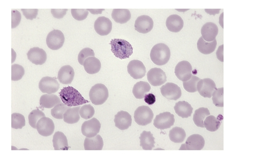
What is this showing?
P . ovale trophozoites in a thin smear

What is this showing?
P. falciparum crescent shaped gametocyte in thin smear

What is this showing?
P. vivax ring form trophozoite

What are these examples of?
P. falciparum
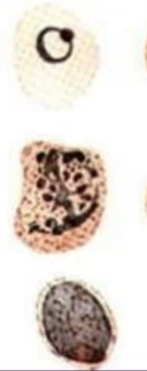
What are these examples of
P. vivax

What are these examples of
P. malariae

What are these examples of?
P. ovale

What is this?
P vivax ring form
What type of parasite is associated with cerebral malaria?
P. falciparum
How is P. falciparum associated with cerebral malaria?
it can infect RBC of all ages → high parasitemia
thats why its so dangerous
infected RBC can sequester in small blood vessels in brain → cerebral malaria
What values indicate Microcytic anemia?
MCV < 80
MCHC < 32
What are examples of Microcytic Anemia?
Thalassemia - normal iron
Iron deficiency - low iron
Chronic disease
Sideroblastic anemia - high iron
Lead poisening
What are examples of Microcytic Hypochromic Anemias?
due to deficiency of iron / abnormal iron utilization
thalassemia
iron deficiency anemia
sideroblastic anemia
What values indicate Normocytic anemia?
MCV: 80 - 100
MCHC: 32 - 36
What is associated with Normocytic aemia?
renal disease
acute blood loss
hemolysis
What indicates Macrocytic anemia?
MCV > 103
What is associated with Macrocytic anmia?
Low B12
pernicious anemia
Malnutrition
GI problems
Low folate
folate malnutrtition
GI problems
liver disease
Normal or High
myeloproliferate disease
liver disease
CDA
What is associated with Iron Deficiency Anemia?
↓ MCV
↓ MCHC
microcytic / hypochromic
What is associated with Megaloblastic Anemia?
↑ MCV
MCHC : usually normal
Macrocytic
What is associated with Aplastic anemia?
MCV normal
MCHC normal
normochromic / normocytic
What causes Babesia?
tick transmitted disease: B. microti
humans are accidental host
also transmitted by transfusion of RBC from asymptomatic donors
What are symptoms of Babesia?
1-9 week incubation
asymptomatic, fever, chills, headache, sweats, nausea, fatigue
jaundice, splenomegaly, hepatomegaly
history of tick infected area

What does this blood smear indicate
Babesia
look for tetrads of merozoites
Here are your lab results, what is this?
low Hgb
↑ retic count
↓ serum haptoglobin
bilirubinemia
leukopenia
thrombocytopenia
hemoglobinuria
proteinuria
Babesia
What is used to treat Babesia?
clindamycin
quinine