Cell membrane & transport
 Call Kai
 Learn
Learn Practice Test
 Spaced Repetition
 Match
Match Flashcards
 Knowt Play
1/28

There's no tags or description
Looks like no tags are added yet.
Name | Mastery | Learn | Test | Matching | Spaced | Call with Kai | Chat |
|---|
No analytics yet
Send a link to your students to track their progress
29 Terms
Functions of biological membranes
an essential feature of every cell is the presence of membranes that define the boundaries of the cell and its various internal compartments
biological membranes protect and organise cells
all cells have an outer plasma membrane, eukaryotic cells also possess internal membranes that encase their granules and control the exchange of essential cell components
Definition of cell’s boundaries
cell surface membrane keeps the interior of the cell physically separated from the surrounding environment
phospholipid bilayer is selectively permeable and allows for desirable substances to be kept within and undesirable substances kept out of the cell
Organisation & localisation of function
molecules or structures with specific functions are embedded in membranes or localised within organelles
for example
many electron carriers are embedded in the thylakoid membranes of chloroplasts and inner mitochondrial membrane
hydrolytic enzymes are found localised within lysosomes
serve to organise and compartmentalise functions within eukaryotic cells
Regulation of cell’s contents
proteins and other components of the membrane help to regulate the transport of substances into and out of the cell and its organelles
for example
to take up and accumulate useful substances such as water, ions and small molecular weight metabolites such as glucose into various compartments
to remove various metabolic waste products
to confine certain chemicals within specific regions of the cell
signal transduction
specific protein receptors on the outer surface of the cell membrane plays a key role in the detection of specific signals (after binding to the receptors) and thus triggering- specific responses within the cell, such as drug or hormone induced responses
cell-to-cell communication
cell membrane has membrane proteins that bind the extra cellular matrix or cell surface constituents to mediate adhesion and communication between adjacent cells
Structure of cell membrane
Fluid Mosaic model
‘mosaic’ —> proteins randomly distributed in or loosely attached to
fluid phospholipid bilaterally is free to move about laterally (due to weak hydrophobic interactions)
Characteristics of the fluid mosaic model
the fluid layer is asymmetrical
the two lipid bilayers may differ in composition and arrangement of proteins and lipids
three major types of membrane lipids are phospholipids, cholesterol and glycolipids
the phospholipid bilayer is fluid or mobile i.e. lateral movement of phospholipids is possible
the unit membrane is a dynamic structure, where the embedded proteins can float, some moving freely while others are fixed in positions by micro filament on the cytoplasmic face
membranes are amphipathic
hydrophilic phosphate head of the phospholipids face outwards into the aqueous environment both inside and outside of the cell
hydrophobic hydrocarbon tails face inwards and create a hydrophobic core

Evidence for the mosaic structure
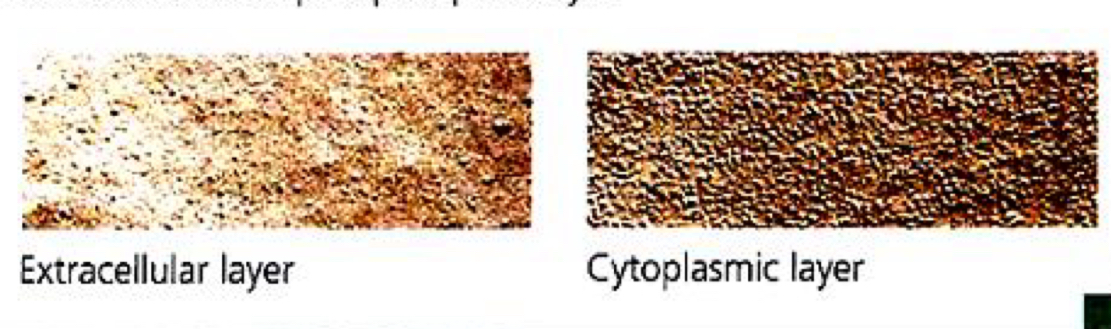
from the use of electron microscopy and freeze fracturing
Freeze-fracture:
a cell membrane can be split into its two layers, revealing the ultra structure of the membrane’s interior
technique: a cell is frozen and fractured with a knife
the fracture plane often follows the hydrophobic interior of a membrane, splitting the phospholipid bilge into two separated layers
membrane proteins go wholly with one of the layers
SEMs show membrane proteins (bumps) in the two layers, demonstrating that proteins are embedded in the phospholipid bilayer
Components of cell membranes
Phospholipids
Cholesterol
Proteins
Carbohydrates
Phospholipids
responsible for the formation of bilayers in an aqueous environment
each phospholipid molecule consists of
glycerol backbone (three-carbon molecule) with 3 hydroxyl (-OH) groups
2 fatty acid chains - hydrophobic hydrocarbon tails
negatively charged phosphate group
additional small, charged molecules which may be linked to the phosphate group
phosphate group and small, charged molecules - contribute to the hydrophilic head
hydrophobic tail + hydrophilic head = phospholipid molecule is termed as amphipathic —> phospholipid bilayer
long hydrocarbon chains of FA form an effective hydrophobic barrier against polar and charged solutes

membrane fluidity
membrane comprises of phospholipid molecules which are held together primarily by hydrophobic interactions between the hydrophobic fatty acid tails
these are weak interactions —> phospholipid molecules are free to move about laterally in the plane of the membrane
rare for molecules to flip transversely across the membrane (hydrophilic part must cross hydrophobic core)

Factors affecting membrane fluidity
Temperature
As temperature increases, membrane fluidity increases
Membranes remain fluid as temperature decreases, until finally the phospholipids settle into a closely packed arrangement and the membrane solidifies —> phase transition (change in state)
temperature at which it occurs —> phase transition temperature
at low temp
kinetic energy of the hydrocarbon chains decreases
hydrocarbon chains are tightly packed
increased hydrophobic interactions between phospholipids molecules and thus their motion is restricted
this bilayer exists in a semisolid state i.e. membrane is less fluid
high temp
kinetic energy and motion of the hydrocarbon chains increases
increased lateral movements of individual molecules, flexing of the chains and transverse flipping
overcoming hydrophobic interactions between phospholipids, resulting in increased space between adjacent phospholipid molecules
bilayer exists in a fluid state (more fluid)
length of fatty acid chains
as length of FA chains increases, membrane fluidity decreases
longer hydrocarbon chains, higher melting point due to increased hydrophobic interactions between hydrocarbon chains
degree of saturation of fatty acid chains
as degree of saturation of FA chains increases, membrane fluidity decreases and vice versa
saturated lipids have long, straight hydrocarbon chains, which allows for close packing and thus enhances membrane solidification
unsaturated lipids have kinks, which prevents the hydrocarbon chains from packing closely together thus enhances membrane fluidity
amount of cholesterol
cholesterol increases the stability and regulates the fluidity of membranes in animal cells
Cholesterol
cholesterols are steroids commonly found wedged between phospholipid molecules in the cell membranes of animal cells

Effect of cholesterol on cell membranes
Membrane stability
cholesterol molecules are usually found in both layers of the cell membrane, intercalated into the lipid monolayers
its rigid steroid ring interferes with the motions of the hydrocarbon chains of phospholipids, thus enhancing the mechanical stability of the membrane
Membrane fluidity
at high temp
cholesterol restrains the movements of phospholipids by interfering with the motions of the hydrocarbon chains
decreased membrane fluidity
at low temp
cholesterol prevents the hydrocarbon chains from packing closely together, thus decreasing the tendency of the membrane to freeze upon
increased membrane fluidity
cholesterol has dual effects on the fluidity of the membrane - resisting changes in membrane fluidity that can be caused by changes in temperature, acting as a temperature buffer for the membrane
membrane permeability
presence of cholesterol molecules decreases the permeability of a lipid bilayer to ions and small polar molecules
fills in spaces between hydrocarbon chains of phospholipids, thereby plugging transient gaps through which ions and small molecules might otherwise pass
membrane stability = fluidity + permeability

Proteins
2 categories of membrane proteins
integral (intrinsic) proteins
peripheral (extrinsic) proteins
classified according to their degree of association with the membrane and their structures

Characteristics of integral and peripheral proteins
Location
Integral proteins
deeply embedded in the hydrophobic interior of the lipid bilayer
2 types exist
unilateral, reaching only a mono layer
transmembrane, spanning the entire bilayer
Peripheral proteins
not embedded but loosely bound to membrane surface, often to exposed parts of integral proteins
found on both sides of the membrane
cytoplasmic side: may be held by network proteins (filaments of cytoskeleton) thus cannot move far
exterior side: may be attached to fibres of extracellular matrix
Structure
Integral/intrinsic proteins
contain both hydrophilic (charged and polar amino acids) and hydrophobic (non polar amino acids) regions i.e. amphipathic
held in place by extensive hydrophobic interactions with the hydrocarbon portions of phospholipids
Peripheral/extrinsic proteins
rich in hydrophilic amino acids so as to allow for interaction with surrounding water and polar surface of the phospholipid bilayer
solubility
integral proteins
usually insoluble in aqueous media
peripheral proteins
usually soluble in aqueous media
how they can be released from membranes
integral proteins
release only through use of detergents or non polar solvents
peripheral proteins
easy release by relatively mild treatment, such as adjustment of ionic strength or pH of the suspending medium
Functions of membrane proteins
enzyme activity
transport
intercellular junctions
cell-to-cell recognition
signal transduction
attachment of the cytoskeleton and extracellular matrix for anchorage

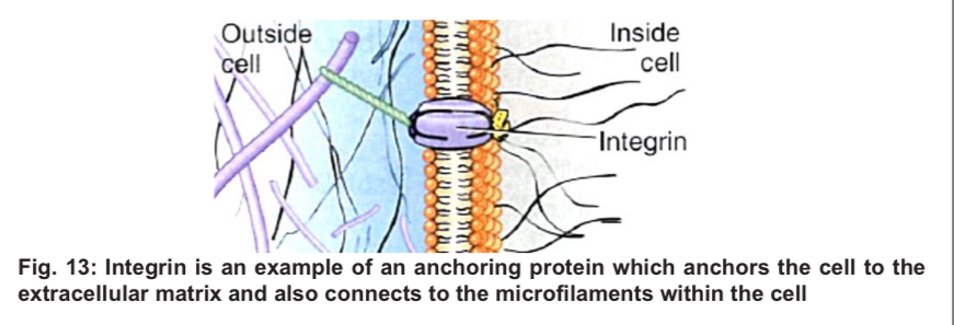
Function of membrane proteins - (1) anchorage
Anchorage
anchoring proteins attach the cell membrane to other substances, stabilise the position of the cell membrane and can help maintain cell shape
anchoring proteins attached to the extracellular matrix can coordinate extracellular and intracellular changes
on the cytoplasmic side, they are bound to microfilaments of the cytoskeleton
on the exterior side, they may attach the cell to fibres of the extracellular matrix
disruptions in cell-cell adhesion can contribute to metastasis stage of cancer
Function of membrane proteins - (2) transport
Transport
Carrier proteins
bind solutes and transport them across the membrane
involves a conformational change of the protein when solute binding occurs, and a return to its original form when the solute is released
energy in the form of ATP may or may not be required
facilitated diffusion (no ATP required): solute moves down concentration gradient
active transport (ATP required): solute moves against a concentration gradient
Channel proteins
some integral proteins contain a water-filled central pore, or hydrophilic channel that forms a passageway to permit the movement (down the conc gradient) of water, ions and small hydrophilic solutes across the cell membrane
2 major kinds of channels
leak channels
permit movement of water at all times e.g. aquaporins
permit movement of ions at all times (though the rate may vary) e.g. Na+ or K+ leak channels
gated channels, which can open or close to regulate ion passage e.g. voltage-gated Na+ or K+ channels

Figure of leak and gated channels

Function of membrane protein - (3) enzymatic activity
enzymatic activity
these enzymes catalyse reactions in the extracellular fluid or within the cytoskeleton, depending on the location of the active site
in some instances, several enzymes can be grouped together to carry out sequential steps in a metabolic pathway
Function of membrane protein - (4) signal transduction
signal transduction
these proteins have very specific 3D conformations, making them ideal as receptor molecules for chemical signalling between cells
chemical signalling works by the binding of a ligand to the receptor protein which triggers changes in the cell
cell membranes differ in the type and number of receptor proteins they contain

Function of membrane protein - (5) cell-to-cell recognition
cell-to-cell recognition
recognition proteins are usually glycoproteins
wide array of possible shapes to the carbohydrate side chains, hence each cell type has its own specific markers
enables cells to recognise other cells, and provides a means for foreign markers to be recognised and attacked by the immune system

Function of membrane protein - (6) Intercellular joining
Intercellular joining
membrane proteins of adjacent cells may adhere together in various kinds of intercellular junctions, such as gap junctions and tight junctions

Carbohydrates
membrane carbohydrates are usually short, branched chains of fewer than 15 units
some of these are covalently bonded to polar ends of phospholipids molecules in the outer lipid layer forming glycolipids
some are covalently bonded to membrane proteins, forming glycoproteins
carbohydrate groups aid in maintaining the orientation of glycoproteins and glycolipids
carbohydrates are highly hydrophilic, glycolipids and glycoproteins are kept in contact with external aqueous environment and are unlikely to rotate towards the interior to diffuse transversely
they are importantly recognition components
sorting of cells into tissues and organs in animal embryos
binding extracellular signal molecules in antibody-antigen reactions
intercellular adhesion to form tissues
cell-to-cell recognition, ability of a cell to distinguish one cell from another e.g. the immune system identifies and acts upon foreign cells

Transport across membrane
Important for reasons
maintain a suitable pH and ionic concentration within the cell for enzyme activity
obtain food supplies for energy and raw materials
excrete toxic substances
secrete useful substances
generate ionic gradients essential for nervous and muscular activity
2 categories of processes for entry into, or exit from cells
Passive processes
Active processes
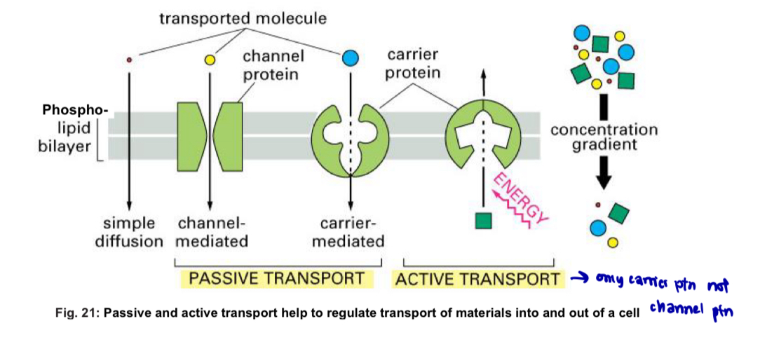
Differences between passive and active transport
Concentration gradient
Passive processes occur down a concentration gradient - substances move from a region of higher concentration to a region of lower concentration
Active processes occur against a concentration gradient - region of lower concentration to a region of higher concentration
Energy requirement
Passive: no cellular energy expenditure (ATP) required
Active: cellular energy expenditure requried usually in the form of ATP
Examples of processes
Passive: simple diffusion, facilitated diffusion, osmosis
Active: active transport, endocytosis, exocytosis
Passive transport
Diffusion: net movement of a a substance from a region of higher concentration to a region of lower concentration down a concentration gradient
molecules have intrinsic kinetic (or thermal motion) energy, which is the tendency for them to spread out evenly into the available space (wrong to assume that passive tpt has completely no energy involved)
diffusion continues until dynamic equilibrium is reached
simple and facilitated diffusion
Simple diffusion
molecules that are able to cross the phospholipid bilayer directly i.e. molecules that have a small molecular weight and/or are readily soluble in the lipid bilayer i.e. hydrophobic molecules
diffusion occurs directly across the plasma membrane without any need for aid of channel or carrier proteins
dynamic equilibrium is reached when concentrations of the diffusing substances are equal n both sides of the membrane = no net movement of substances occurs

Facilitated diffusion
transport of substances down a concentration gradient without the use of ATP (i.e. passive0 until equilibrium is reached
transport protein is used to enhance/increase the rate of transport of the substances across the membrane
transport ptn is specific to the substance being transported i.e. it only transports that substance
is for larger hydrophilic substances e.g. glucose, amino acids and ions
channel or carrier proteins
a. carrier proteins
—> possesses a binding site for solute molecules
—> undergo conformational changes to transport a substance across the membrane
b. channel proteins
—> possesses a central hydrophilic pore that allows free movement of the transported substance across the membrane
—> does not undergo conformational changes to transport a substance across the membrane medium

Factors affecting rate of diffusion
Concentration gradient: steeper conc. gradient = faster diffusion
Distance over which diffusion occurs: shorter distance = faster diffusion (thinner cell membrane = greater no. of molecules diffuse across per unit time)
Area across which diffusion occurs: larger surface area = greater no. of molecules diffuse across per unit time = faster diffusion
Structure through which diffusion occurs: presence of transient gaps in cell membrane may enhance diffusion
type and number of tpt ptn present per unit surface area of the membrane will also affect the diffusion rate
size and type of diffusing molecule
smaller molecules = faster diffusion
temperature
higher temp = faster diffusion
Osmosis
osmosis is the net movement of freely moving water molecules from a region of less negative water potential to a region of more negative water potential through a selectively permeable membrane

Water potential
Water potential: measure of the tendency for water to move from one region to another
pure water has a water potential of 0 (highest possible value)
presence of solutes will make the water potential more negative i.e. any solution will have a negative water potential
water molecules always move from a region of less negative wp to a region of more negative wp
For plant cells:
2 factors affecting wp
solute concentration - solute potential (which is negative), Ψcell = Ψs
pressure exerted by cell wall on its contents, which is generated when water enters the cell = pressure potential Ψp (which is positive
water potential of plant cell = solute potential + pressure potential
For animal cells:
water potential of animal cell is determined primarily by its solute potential cos no cell wall
Ψcell = Ψs

Solute potential
Solute potential Ψs: measure of the ability of a solute to make the water potential more negative
dissolving solute molecules in pure water reduces the number of free