A & P Ch. 12 Nervous Tissue
 Call Kai
 Learn
Learn Practice Test
 Spaced Repetition
 Match
Match Flashcards
 Knowt Play
1/75

There's no tags or description
Looks like no tags are added yet.
Name | Mastery | Learn | Test | Matching | Spaced | Call with Kai | Chat |
|---|
No analytics yet
Send a link to your students to track their progress
76 Terms
organs and cells that make up the nervous system
brain, spinal cord, peripheral nerves, ganglia, neurons
3 basic steps the nervous system uses to coordinate tasks
receives information and transmits to CNS, CNS processes information, signals muscle and gland cells
2 main anatomic divisions of the nervous system
central nervous system CNS, peripheral nervous system PNS
brain, spinal cord; performs most decision-making functionsand integration of sensory information.
central nervous system CNS
nerves, ganglia; connects to CNS, carries signals
peripheral nervous system PNS
sensory/afferent (somatic, visceral) motor/efferent (somatic/visceral)
PNS divisions & subdivisions
carries receptor signals to CNS, informs of stimuli
sensory (afferent) division
carries signals from receptors in skin, muscles, bones, joints
somatic sensory division
carries signals from viscera in thoracic and abdominal cavities, heart, lungs, stomach urinary bladder
visceral sensory division
carries signals from CNS to gland and muscle cells to elicit response/action
motor (efferent) division
carries signals to skeletal muscles producing voluntary movement
somatic motor division
carries signals to glands, cardiac muscle, smooth muscle, operates unconsciously as reflexes
visceral motor division (autonomic nervous system, ANS)
arouse body for action; accelerating hearting heartbeat, increasing respiratory airflow; inhibits digestion, reduces urine production
sympathic division
calming effect, slows heartbeat; stimulates digestion
parasympathetic division
wall of digestive tract, more neurons than spinal cord, enables regions of long digestive tract to communicate, coordinate motility and secretion
enteric plexus
3 physiology properties that allow neurons to communicate with other cells
excitability (respond to stimuli), conductivity (produce electrical signals), secretion (neurotransmitter influencing neighboring cells)
3 functional classes of neurons
sensory/afferent neurons, interneurons, motor/efferent neurons
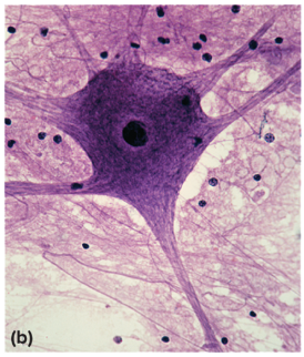
structural classification of neurons
cell body (neurosoma, perikaryon), neurofibrils (actin filament bundles), chromatophilic substance (rough ER in sark-staining regions)
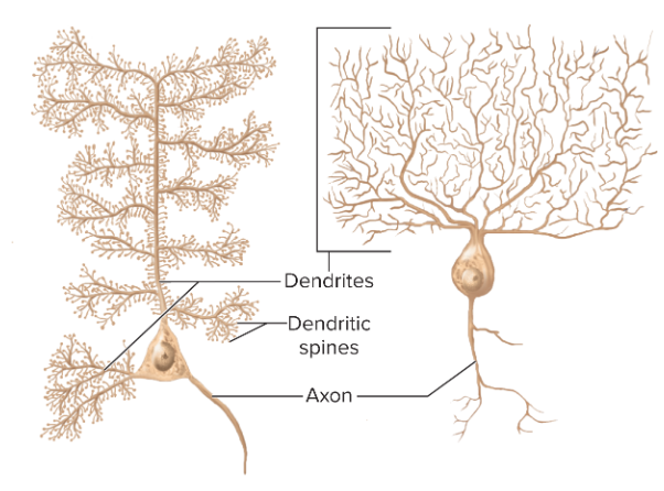
one axon, multiple dendrites; brain, spinal cord
multipolar neurons
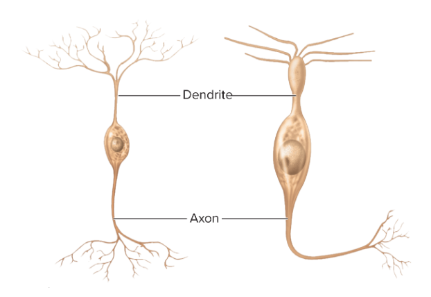
one axon, one dendrite; olfactory nose, certain retina, ear sensory
bipolar neurons
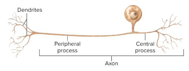
single process leading away from cell body; carry touch and pain signals; spinal cord
unipolar neurons
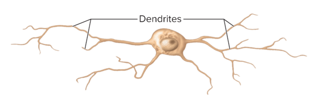
no axon, multiple dendrites, don’t produce action potentials; brain, retina, adrenal medulla; help in visual processes (contrast perception)
anaxonic neurons
process of neurons that usually receive signals from other neurons
dendrites
mound on cell body from which the axon (nerve fiber) originates
axon hillock
solitary neurite
axon (nerve fiber)
cytoplasm of axon
axoplasm
membrane of axon
axolemma
____ send a receive signals while ____ ____ support them and help them function
neurons; glial cells
protect neurons and help them function, bind neurons together, provide supportive framework for the nervous tissue
neuroglia (glial cells)
bulbous body up to 15 arms, each arm reaches to axon and spirals around it forming the myelin sheath to insulate axon from extracellular fluid & speed up conduction in CNS
oligodendrocytes
resemble cuboidal epithelium lining in brain and spinal cord, no basement membrane, rootlike process penetrate underlying tissue; produce cerebrospinal fluid (CSF) which surrounds CNS and fills its cavities
ependymal cells
small macrophages that wander though CNS to probe from cellular debris or issues, phagocytizes dead tissue, etc., become concentrated in areas damaged by infection
microglia
cover entire brain surface and nonsynaptic neuron regions, starlike shape; form blood-brain barrier, nourish neurons by converting blood glucose to lactate, produce growth factors that stimulate neurons, promotes synapse formation, electronically communicate with neurons, removes K⁺ some neurotransmitters from brain & spinal cord ECF, help regulate ECF composition, form scar tissue
astrocytes
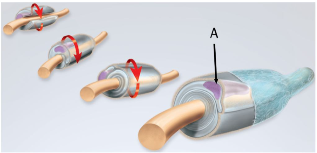
form neurolemma around all PNS axons & myelin around most, aid in damaged nerve regeneration
schwann cells
surround cell bodies of neurons in ganglia, provide electrical insulation & regulate chemical environment of neurons
satellite cells
layers of insulation composed of lipids and proteins around axon formed by Schwann cells in PNS & oligodendrocytes in CNS
myelin sheath
____ axons produce myelin sheaths while ____ axons do not
myelinated; unmyelinated
slow axons sufficient for processes such as secreting stomach acid or dilating pupil in both CNS & PNS
unmyelinated axons
fast axons for motor commands to skeletal muscles and sensory signals for vision & balance in both CNS & PNS
myelinated axons
distal part of axon including Schwann cells degenerate from lack of protein, macrophages clean up tissue debris at and around point of injury
nerve regeneration step 1
some neurons die at this stage; cell body swells, ER breaks up, nucleus moves off center), axon stump sprouts multiple growth processes, distal end continues degeneration (denervation atrophy)
nerve regeneration step 2
Schwann cell neurolemma, endoneurium & basal lamina form regeneration tube, produce cell-adhesion molecules and nerve growth factors
nerve regeneration step 3
regeneration tube guides growing sprout back to original target cells
nerver regeneration step 4
nerve body shrinks and returns to original appearance when synaptic contact is established, reinnervated muscle fibers regrow
neve regeneration step 5
PNS nerves can regenerate if ____ intact and some ____ remains
cell body; neurolemma
which is required for nerve fiber regeneration
endoneurium
CNS nerves can/cannot regenerate
cannot
how long can it take for nerves to regenerate
up to 2 years
charge difference across plasma membrane
resting membrane potential (RMP)
RMP value of unstimulated neuron
-70 mV
to have potential or voltage; separation of electrical charge across cell membrane so that one side is more positive and the other is more negative
polarized
when at rest, high concentrations of K⁺ are located in the ____ fluid
intracellular
when are rest, high concentrations of Na⁺ are located in the ____ fluid
extracellular
what type of transport is responsible for stablishing this gradient
sodium-potassium pumps
what direction would Na⁺ and K⁺ passively diffuse after they are pumped by Na⁺/K⁺ pump if able to
against their concentration gradient
negative membrane potential indicates
interior of cell is more negatively charged than exterior
unmyelinated axons are found
in CNS and PNS
potassium-sodium pumps ____ Na⁺ ion(s) for every ____ K⁺ ion(s)
3; 2
any such case in which the voltage shifts to a less negative value
depolarization
short-range change in voltage where incoming Na⁺ diffuses along inside of plasma membrane creating a wave of depolarization
local potential
graded or vary in magnitude; decremental or get weaker as they spread from point of origin; reversible, if stimulation ceases cation diffuses out of cell; excitatory or inhibitory (less likely to produce action potention)
characteristics of local potentials
a hyperpolarized neuron is
inhibited
a hyperpolarized neuron is caused by
influx of K⁺ ions
rapid voltage change where plasma membrane briefly reverses electrical polarity producing traveling wave of excitation in nerve and muscle cells
action potential
local current arrives at axon hillock; depolarizes membrane
action potential step 1
local potential rises to critical voltage (threshold), about -55mV
action potential step 2
neuron fires; Na⁺ channels open quickly, depolarization spike; K⁺ channels open more slowly
action potential step 3
as voltage peaks K⁺ channels fully open and exit cell repolarizing cell
action potential step 4
more K⁺ leaves cell than amount of Na⁺ entering so membrane voltage drops more negative than original RMP (hyperpolarization)
action potential step 5
if ftimulus depolarizes neuron to threshold, neuron fires at max voltage or doesn’t fire at all
all-or-none law
follow all-or-none law; nondecremental or don’t get weaker with distance;irreversible
characteristics of action potentials
period of resistance to restimulation during action potential and a few seconds after
refractory period
refractory period lasts from start of action potential untel membrane returns to resting potential
absolute refractory period
refractory period that lasts until hyperpolarization ends
relative refractory period