H-BP Microscopes
1/18
There's no tags or description
Looks like no tags are added yet.
Name | Mastery | Learn | Test | Matching | Spaced | Call with Kai |
|---|
No analytics yet
Send a link to your students to track their progress
19 Terms
History
Hans and Zacharias Janssen - “first” compound microscope
Joseph Jackson Lister → Solved spherical aberration - caused by light passing through different parts of the same lens
Ernst Abbe - Numerical Aperture of Condensor
Carl Zeiss & Ernst Abbe - Objective of microscope
Resolution
Between LM, CM, SEM, TEM; TEM is the best.
Resolving power: the smallest distance between two particles at which they can be seen as seperate objects
In a compound microscope wavelength limits resolution.
Due to diffraction, the limit resolution is 0.2 micrometers.
Magnification
Ocular lens x objective lens
Mechanical System
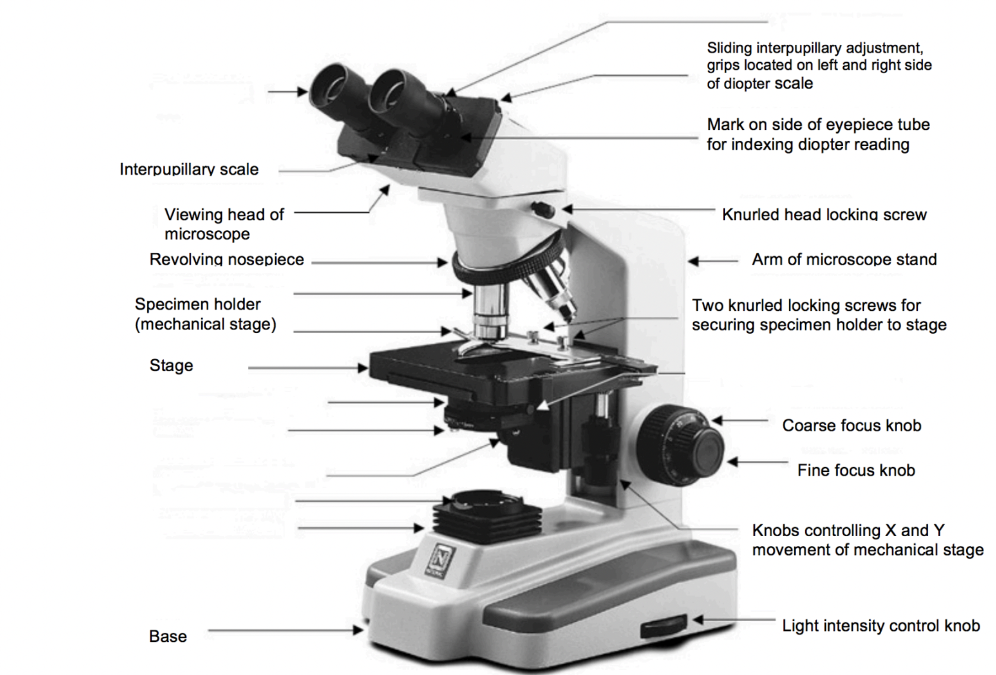
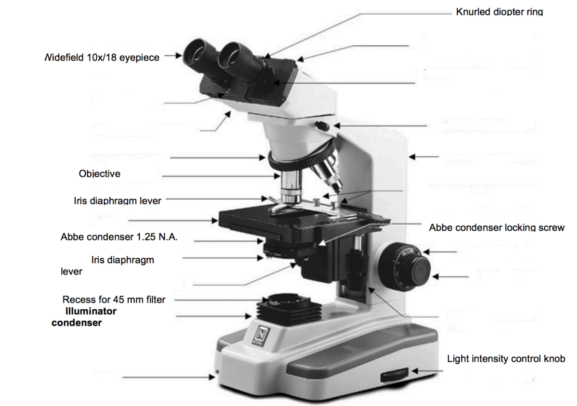
Optical System
Transmitted illumination: light is directed through the specimen from the base
Vertical or Reflected Illumination: Light comes from above and reflects off the specimen
Condenser: Lens system under the microscope stage that focuses light onto the specimen
Optical Principles
The objective lens is a very high powered magnifying glass with a very short focal length → virtual, inverted and enlarged image
Eyepiece is usually a compound lens → focus between the two lenses
The image is viewed with eyes focused at infinity
Bright Field Microscopy
When to use:
Viewing stained or naturally pigmented specimens such as stained prepared slides of tissue sections or living photosynthetic organisms
only dark or strongly refracting objects
Objects absorb light partially or completely
Disadvantages:
Low optical resolution.
Diffraction limits resolution to approximately 0.2 micron.
Out of focus light from point outside the focal plane reduce image clarity
How to enhance:
Oil immersion objective
Staining
Filters on the light source
Dark Field Microscope
An opaque disc is placed under the condenser so that only light scattered by objects on the slide can reach the eye
Used with low magnification (up to x100)
Used in examining:
Urine for crystals (uric acid, oxalate)
Spirochetes (bacteria) (Trepenoma pallidum → syphilis)
suspensions of cells such as mushrooms (yeast), bacteria, small protists
cell and tissue fractions (cheek epithelial cells, blood cells)
determination of motility in cultures (moving independently using metabolic energy)
Live and unstained samples, increase the contrast
Must be strongly illuminated → damaging the sample
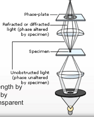
Phase Contrast Microscpy
Preferable to BFM when high magnification is needed (400×, 1000x) but the specimen is colorless or the details are too fine → not enough contrast
Uses:
Cilia and flagella
Amobea
No need to stain
How it works:
The change in phase can be increased to half a wavelength by a transparent phase-plate in the microscope and thereby causing a difference in brightness → object shines out in contrast to the surroundings
Differential Interferance Microscopy
Produces an apparently 3D image of living cells and tissues
Resembles phase-contrast but higher resolution.
Uses polarizing lenses like the polarizing microscope → can be quantitative
Halojen light beam is polarized, split by a beam splitter, and passed through the specimen
Invert Microscope
Modified BFM for special uses
Allow viewing of cells in flasks, welled-plates, or other deep containers
Light source above specimen, objectives beneath stage
Stereo (Dissecting) Microscope
Two compound microscopes which focus on the same point from slightly different angles
Specimen viewed in 3 dimensions
The image upright and laterally correct
Both eyes can see the image
Used for:
Surfaces of solid specimens
Sorting, dissection, microsurgery, watch-making, small circuit board manufacture or inspection
Great working distance and depth of field! (higher the NA, the smaller the depth of field and working distance)
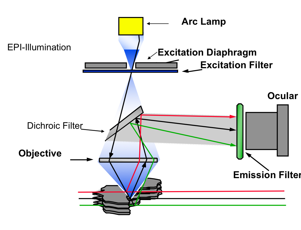
Fluorecence Microscope
The specimen is illuminated with a specific wavelength of light which is absorbed by the fluorophores (fluorescent dyes that absorb excitation light at given wavelength), then emits light with a longer wavelength.
UV → Visible light
Brilliant, shiny particles on dark background
The microscope has a strong UV light source, and special filters that eliminate UV light coming to the eye
Photoluminence: Light energy, or photons, stimulate the emission of a photon
Fluorescence: Type of photoluminescence where light raises an electron to an excited state. The excited state undergoes rapid thermal energy loss to the environment through vibrations, and then a photon is emitted.
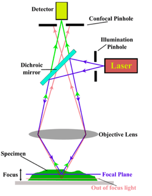
Confocal Microscope
A laser is focused at a plane in the specimen and scans the specimen in a horizontal plane
Only light from the plane of focus reaches the detector
The scanned image is digitally recorded, computer compiles images created from each point to generate a 3D image
Images from consecutive focal planes can be recorded
A very thin section within the tissue
Used for:
Live cells
Fixed cells
Localize/measure enzyme activity
Advantages:
Reduced blurring
Optical sectioning
highly sensitive photomultipliers → improved signal to noise ratio
3D
Magnification can be adjusted digitally
Disadvantages:
Slow scan
Limited use in dynamic tracking
Photobleaching or damage to living cells by laser
Lower resolution than camera detection
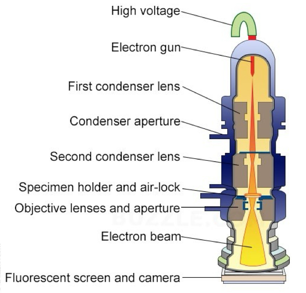
Transmission Electron Microscopy (TEM)
Fixed, dehydrated specimens are embedded in a resin, hardened, sectioned, stained with heavy metals such uranium and lead, and inserted into electron column
The electron beam is absorbed or deflected by the heavy metal stains and shadows are cast onto film or a phosphorescent plate (image is a shadow) at the bottom of the column
Features:
2D image
reveals internal structure
high resolution, high magnification
electron beam is focused by magnetic field
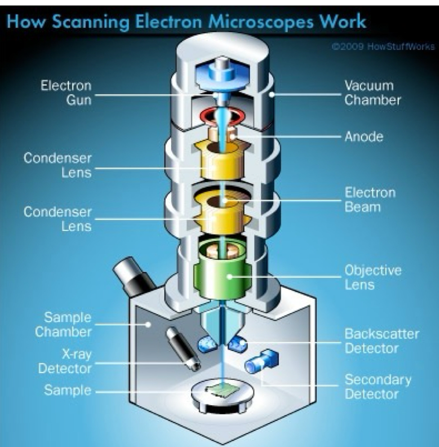
Scanning Electron Microscope
Fixed, dehydrated specimens are mounted stubs and surface-coated with gold, palladium or rhodium.
The specimen is placed in a vacuum and an electron beam scans back and forth over it
Electrons that bounce off the metal-coated specimen surface are collected, converted to a digital image and displayed on a monitor.
3D image
Electron beam is focused using a magnetic field
Gives information about external topography of specimen
Higher resolution and magnification than LM
Can be used to observe individual atoms
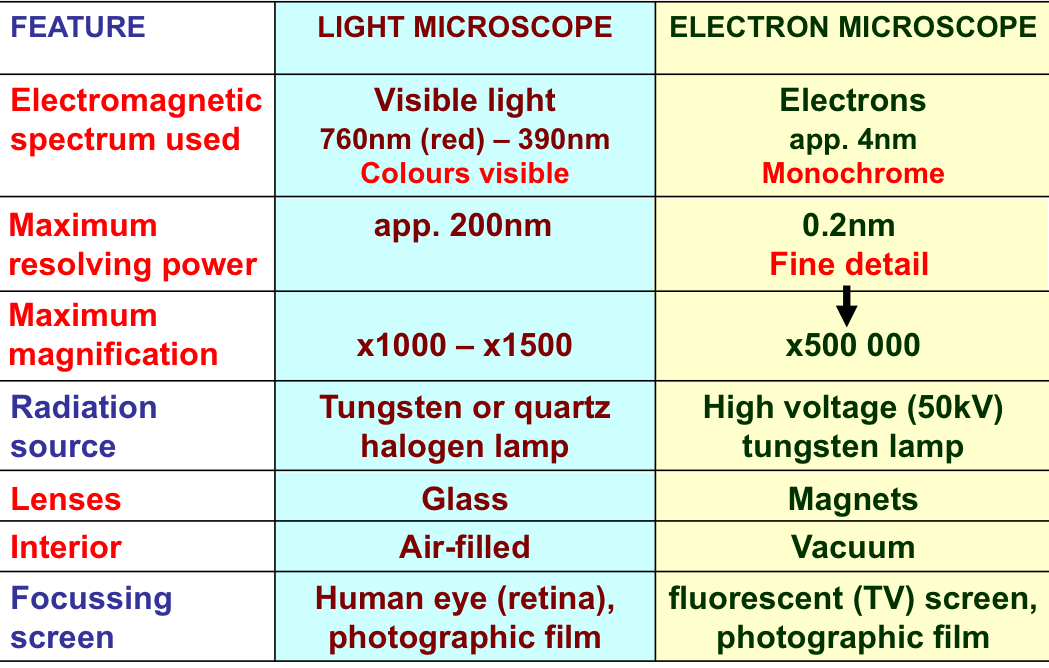
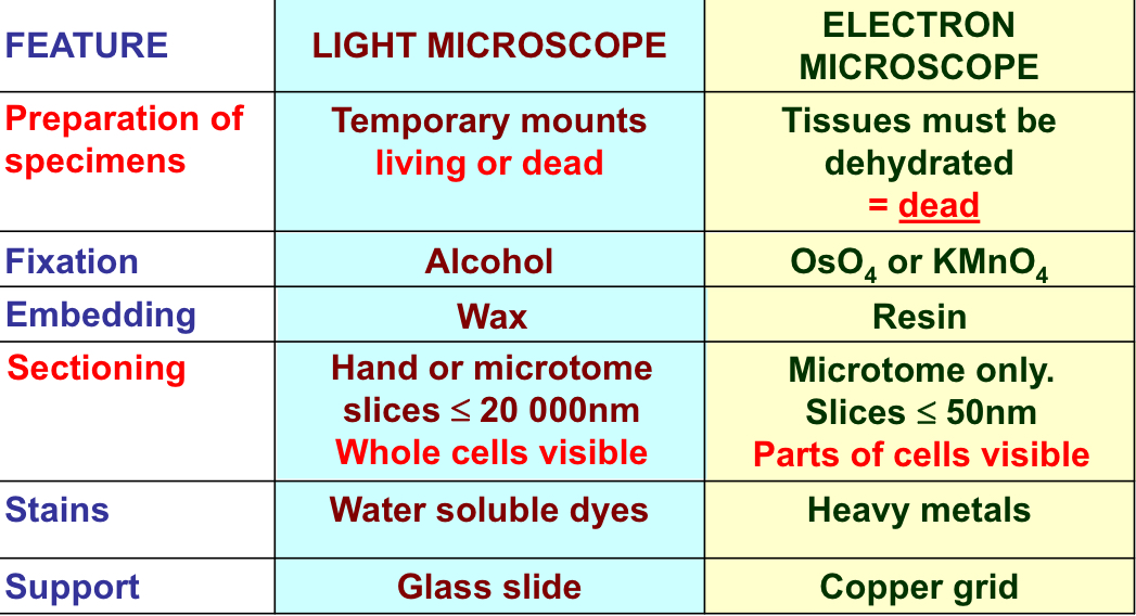
Light VS Electron Microscope
Scanning Tunneling Microscopy
An extremely fine conducting probe is held about an atom’s diameter from the sample
Electrons tunnel between the surface and the tip, producing an electrical signal
While it slowly scans across the surface, the tip is raised and lowered in order to keep the signal constant and maintain the distance
This enables it to follow even the smallest details of the surface it is scanning.
Atomic Force Microsopy
AFM consists of cantilever with a sharp tip at its end that is used to scan the specimen surface
The cantilever is typically silicon or silicon nitride with tip radius of curvature on the order of nanometers
When the tip is brought into proximity of a sample surface, forces between the tip and the sample lead to a deflection of the cantilever