Neurons
 Call Kai
 Learn
Learn Practice Test
 Spaced Repetition
 Match
Match Flashcards
 Knowt Play
1/84

There's no tags or description
Looks like no tags are added yet.
Name | Mastery | Learn | Test | Matching | Spaced | Call with Kai | Chat |
|---|
No analytics yet
Send a link to your students to track their progress
85 Terms
Neurons
basic signaling units
3 main classes: sensory, motor, interneurons
human brain: 86 billion interneurons, averaging 1000 synapses on each
cells in the nervous system that carry information from one place to another by means of a combination of electrical and chemical signals
Glial cells
give structure and support
provide electrical insulation to neurons
modulate neural activity
outnumber neurons by at least 10 to 1
critical to the functioning of the nervous system and maintaining the blood-brain barrier
(glial = glue)
Glial cells DO NOT carry nerve impulses (action potentials)
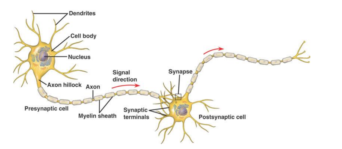
Structure of the Neuron
cell body (like other cells)
also called the Soma
Dendrites (dendritic tree)
Axon
Cytoplasm
Ions: e.g., NA+, K+, CA2+, CL- and
Molecules such as protein
Resting Potential: electrochemical gradient
Electrical Gradient:
Inside of the neuron is negative relative to the outside (-70mV)
Concentration Gradient
more NA+ outside
more K+ inside
Ion channels and Ion pumps
The NA+ and K+ gradients are maintained by the sodium-potassium pump
the ions diffuse back across the plasma membrane through ion channels
since there are more K+ channels than Na+ channels, there is a net outflow of positive ions
The net electrical gradient yields a resting potential of -70mV
Sodium-Potassium Pump
Ion channels pump ions across the membrane

The resting potential of a neuron is
-70mV
There are two opposing forces acting upon potassium ions. Select the correct answer:
A. The electrical force keeps potassium inside the cell, the chemical force drives potassium out of the cell
B. The chemical force keeps potassium inside the cell, the electrical force drives potassium out of the cell
A. The electrical force keeps potassium inside the cell, the chemical force drives potassium out of the cell
True or False: The sodium potassium exchange pump moves 3 potassium ions out of the cell and 2 sodium ions into the cell with each cycle.
False
What does the potassium sodium pump accomplish? Select all that apply:
A. It moves ions from areas of low to areas of high concentration
B. It re-establishes and maintains the resting potential
C. It moves sodium into the cell and potassium out of the cell
D. It lets ions passively flow through the plasma membrane
A. It moves ions from areas of low to areas of high concentration
B. It re-establishes and maintains the resting potential
Action Potential (Nerve Impulse)
if threshold of -55mV is reached, action potential is triggered (all-or-none) — voltage-gated sodium channels open
Absolute vs relative refractory periods
absolute refractory period, Na+ channels temporarily inactive
relative refractory period due to continued outflow of K+ ions
Consequences of Refractory Period
Neuron can only generate ~200 action potentials per second (upper limit on firing rate)
passive current that flows from action potential cannot re-open the voltage-gated channels that generated it
propagation of action potential in one direction
Action Potential Phases

The action potential starts at
the axon hillock
The absolute refractory period is caused by
the temporary inactivation of voltage-gated sodium channels
The voltage across the membrane has to reach a threshold of ___ to initiate an action potential
-55mV
During the course of an action potential, voltage-gated potassium channels start to close …
some time after the membrane potential goes back to its resting state of -70mV
Chemical Synapses
Neurotransmitter release at the synapse, into synaptic cleft
Action potential depolarizes the terminal membrane, which causes Ca2+ to flow into the cell
Ca2+ causes vesicles to bind with cell membrane
Release of neurotransmitter by exocytosis into the synaptic cleft
Transmitter binds with receptor
Ionotropic Receptors
undergo a change in shape when neurotransmitter binds, causing the channel to open. This may have either an excitatory or an inhibitory effect, depending on the ions that can pass through the channel and their concentrations inside and outside the cell.
Metabotropic Receptors
Signaling through these metabotropic receptors depends on the activation of several molecules inside the cell and often involves a second messenger pathway. Because it involves more steps, signaling through metabotropic receptors is much slower than signaling through ligand-activated ion channels
Neurotransmitters
the chemicals that neurons release that allow them to communicate with one another
neurotransmitters have excitatory or inhibitory effects
agonists and antagonists
adrenaline
fight or flight neurotransmitter
Produced in stressful or exciting situations. Increases heart rate & blood flow, leading to a physical boost & heightened awareness.
noradrenaline
concentration neurotransmitter
Affects attention & responding actions in the brain, & involved in fight or flight response. Contracts blood vessels, increasing blood flow.
plays a role in sleep - the only difference between waking and dreaming is noradrenaline
memory processing —> especially memory that has an emotional component, potentially by helping to increase the salience and hence the memorability of such information
dopamine
pleasure neurotransmitter
Feelings of pleasure, and also addiction, movement, and motivation. People repeat behaviors that lead to dopamine release.
serotonin
mood neurotransmitter
Contributes to well-being & happiness; helps sleep cycle & digestive system regulation. Affected by exercise & light exposure.
memory - the function of creating new memories for long-term memory
GABA
calming neurotransmitter
Calms firing nerves in CNS. High levels improve focus; low levels cause anxiety. Also contributes to motor control & vision.
inhibitory
many substances that reduce the activity of the CNS bind to GABA receptors (ex. barbiturates and alcohol)
acetylcholine
learning neurotransmitter
Involved in thought, learning, & memory. Activates muscle action in the body. Also associated with attention and awakening.
depletion associated with Alzheimer’s disease
linked to selective attention (the ability to attend to certain information while tuning out other information) —> acetylcholine sharpens responses of cells to the features of stimuli that are most likely to make them fire, while suppressing responses to less prominent features of a stimulus
glutamate
memory neurotransmitter
Most common brain neurotransmitter. Involved in learning & memory, regulates development & creation of nerve contacts.
excitatory
overactivity —> development of epilepsy (a disease in which an abnormal lowering of a cell’s firing threshold causes it to misfire)
too much produces excitotoxicity
endorphins
euphoria neurotransmitter
Released during exercise, excitement, & sex, producing well-being & euphoria, reducing pain. Biologically active section shown.
Postsynaptic Potentials
IPSP (inhibitory postsynaptic potential)
EPSP (excitatory postsynaptic potential)
Postsynaptic potentials vs. Action potentials
Action Potentials
all or nothing
higher in magnitude
excitatory
Postsynaptic Potentials
graded
smaller in magnitude (0.5 - 5 mV)
excitatory and inhibitory
Information transmission
Within a neuron, the transmission of information usually electrical (action potentials, voltage-gated ion channels).
Between neurons, chemical (neurotransmitters, ligand-gated ion channels).
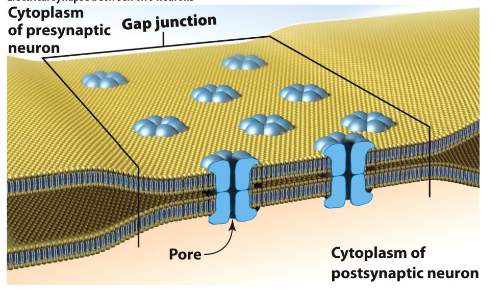
Exception: electrical synapses
Electrical Synapse
How do Neurons Code Information?
Action Potentials = All-or-none
Firing strength does not change
The amplitude of an action potential does not vary
Firing rate can change
Types of Glial Cells in the CNS
Astrocytes
Microglia
Oligodendrocytes
Radial
Astrocyte (Astroglia)
Skeleton of the brain
Homeostasis
Blood-brain barrier (BBB)
Synapse cleanup
influence the communication between neurons by modifying the chemical milieu between them, as well as refining and sculpting the physical connections between neighboring neurons
Microglia
immune defense
recognize foreign substances, engulf and try to destroy them
protects against infection
aid with reorganization after brain damage by removing dead neurons and they serve some of the nutritive needs of neurons and provide structural support
Oligodendrocytes
provide the insulation (myelin) to neurons in the central nervous system
multiple sclerosis: abnormal immune response that causes destruction of myelin
What are the two main classes of cells the human nervous system is composed of?
neurons and glia
Dendritic Tree
the part of the neuron that receives input from other cells
Cell body
the part of the cell containing the nucleus and other cellular apparatus responsible for manufacturing the proteins and enzymes that sustain cell functioning
Axon
the appendage of the cell along which information is carried
varies in length
Sensory Neurons
bring information to the CNS
Interneurons
associate information within the CNS
Motor Neurons
send information from the brain and spinal cord to the muscles
Radial Glia
Guide neurons as they migrate from the site of creation to their final position within the brain
Blood-Brain Barrier
the mechanism by which many harmful substances, such as toxins, are prevented from reaching the brain
consists of tightly packed glial cells between blood vessels and neurons
also blocks certain nutrients, drugs, and immune system cells in the bloodstream from reaching the nervous system directly
Nuclei
distinct groups of neurons whose cell bodies are all situated in the same region in a brain structure called the thalamus
Two broad principles to electrochemical signaling of neurons
information is relayed within a neuron by means of an electrical signal, whereas one neuron influences another via a chemical signal
Resting Potential
at rest, there is a difference in the electrical charge between the inside and outside of a neuron (known as neuron’s resting potential)
typically about -70mV
occurs because the cell membrane of the neuron acts as a barrier separating ions on the inside from those on the outside
Ions
electrically charged particles
ions like sodium and potassium can traverse the cell membrane only through special passageways known as ion channels
Ion channels
in some configurations they allow ions to flow in and out of the cell, and in other configurations the passageway is blocked and ions cannot move from one side of the cell membrane to the other
input from other neurons can affect the opening and closing of ion channels
Action potential
when ion channels open and allow ions to flow in and out of the cell the resulting change in ion concentration on each side of the membrane drives the neuron’s electrical charge away from its resting potential, making it either more negative or more positive
if the cell receives enough stimulation to reduce the voltage across the membrane to -55mV, a threshold is passed and the cell “fires” —> electrical charge rapidly goes to a peak of +40 mV
after reaching the peak (depolarization), the electrical charge then retreats toward the baseline resting potential (repolarization) —> hyperpolarization (voltage briefly becomes more negative than resting potential
then neuron returns to resting potential
Important Properties of Action Potential
self-propagating: once it is set in motion nothing else need be done (like knocking over the first domino)
strength does not dissipate with the distance it travels (peak remains +40mV for its entire trip down the axon)
all-or-nothing (either the cell fires or it doesn’t)
Axon hillock
a specific part of the neuron near the cell body where the action potential is first produced
How does action potential travel through the neuron
first produced at axon hillock
then it is carried along the entire length of the axon to the terminal button
at the terminal bouton action potential ends and the electrical signal gets transformed into a chemical message
Synaptic Vesicles
little balloons in the terminal bouton which are filled with neurotransmitter
some reside in the terminal bouton and others are fused to the outside wall of the neuron
action potential causes synaptic vesicles that are fused to the outside walls of the neuron to burst open, pouring their contents into the area between neurons (synaptic cleft)
Synapse
the region of contact between the neuron
contains the terminal button, the synaptic cleft, and the postsynaptic region
How information is transferred between neurons
at the synapse, neurotransmitter molecules are released from the presynaptic neuron and received by the postsynaptic neuron
when a neurotransmitter reaches the postsynaptic membrane, it fits into a specific region of the receptor (called the binding site), much the way a key fits into a lock
the binding of the neurotransmitter changes the configuration of the receptor, which in turn changes the electrical charge of the postsynaptic neuron in a small local area near the receptor site by altering the flow of ions across the membrane
chemical signal is transformed back into an electrical one
Receptors
the membrane of the dendritic trees of the postsynaptic neuron contains regions known as receptors
these receptors are specially configured proteins that are embedded within the postsynaptic membrane
Excitatory postsynaptic potentials (EPSPs)
make the cell’s electrical charge a bit more positive, bringing it closer to the threshold value of -55mV at which the cell will fire
Inhibitory Postsynaptic Potentials (IPSPs)
make the inside of the cell a bit more negative than the outside and move the cell further away from the threshold at which it will fire
Postsynaptic Potentials
the local changes in the electrical potential that occur near the receptor
either excitatory or inhibitory
whether a particular neurotransmitter has an excitatory or inhibitory effect depends on the receptor to which it binds
combined effect is needed to make neuron fire (two EPSPs/IPSPs have a greater influence if they occur together in time or space)
Postsynaptic Potentials differ from action potentials in three important ways
they are graded: the further they travel from their source, the more they dissipate
postsynaptic potentials are much smaller in magnitude than an action potential, usually in the range of 0.5-5 mV
whereas action potentials are always excitatory, in that they make the cell fire, postsynaptic potentials can be either excitatory or inhibitory
How do neurons code the intensity of a stimulus
via the rate, or pace, of its firing
(not by the size of of the electrical response - because the value of the action potential is always the same)
when there is a strong stimulus, the cell fires many times in succession; when there is a weak input, it fires only occasionally
How postsynaptic potentials can cause an action potential
postsynaptic potentials summate at the axon hillock (where axon meets the body)
if the differential charge across the membrane at the axon hillock reaches -55mV, the cell will fire (otherwise it won’t)
postsynaptic potentials generated close to the axon hillock have a larger influence (because they are graded)
Phases of the action potential
when the threshold of activation is reached, sodium (NA+) begins to enter the cell
Potassium (K+) begins to leave the cell
no more sodium enters the cell, and the voltage reaches its peak positive value
the leakage of potassium drives the voltage in the negative direction, hyperpolarizing the cell; potassium channels then close and the cell returns to its resting potential, at which point it can fire again
Two major classes of neurotransmitters
amino acids
the other main class consists of neurotransmitters that are organized into systems; these are produced by specific sets of neurons whose cell bodies are located subcortically and whose axons project diffusely throughout the cortex
Amino Acids
the smallest and most basic building blocks of proteins
act as the main excitatory and inhibitory neurotransmitters in the brain
two main amino acids
glutamate and gamma-aminobutyric acid (GABA)
excitotoxicity
excessive activity of receptors that can literally excite neurons to death (the neurons get “fried” by too much stimulation)
Barbiturates
reduce seizure activity and induce sedation and sleep
Alcohol and GABA
alcohol produces its anxiolytic (anxiety-reducing) and sedative effects by affecting GABA receptors
Neurotransmitter systems
cholinergic - acetylcholine
serotonergic - serotonin
noradrenergic - noradrenaline
dopaminergic - dopamine
Serotonin-Selective Reuptake Inhibitors (SSRIs)
most popular drug to treat depression
increase the amount of serotonin in the synaptic cleft by inhibiting its presynaptic uptake
ex. Prozac
side effects: interfering with sleep, reducing appetite, and impairing sexual performance
Myelin
the speed at which neurons propagate electrical signals down their axons depends on the degree to which the axon is insulated by a fatty sheath called myelin
the longer the myelin sheath is, the greater the speed with which the electrical signal is propagated down the axon
some have no myelin sheath —> small and generally synapse on nearby neurons
Oligodendrocytes
produces myelin sheath
a portion of oligodendrocyte wraps itself around the axon; such wrapping creates a discrete section of myelin
the more turns there are around the neuron, the greater the insulation and therefore the greater the conduction speed
Nodes of Ranvier
gaps between myelinated sections of an axon
because the electrical signal must jump across these nodes, which have a high concentration of ion channels, they serve to keep the electrical signal constant in size rather than degrading as it travels down the axon
white matter
areas through which myelinated fibers run
because myelin is fatty, it is white
gray matter
concentrations of unmyelinated cell bodies
Fiber tract
when a group of cells sends their axons to the same place, the group of axons is known as a fiber tract
because these axons usually traverse long distances, they tend to be myelinated
ex. corpus callosum
Corpus callosum
the main fiber tract connecting the two halves (hemispheres) of the brain
composed mainly of myelinated fibers
allows a speedy transfer of information from a neuron in one hemisphere to a distant neuron in the other hemisphere
Multiple Sclerosis
causes the myelin surrounding a neuron to be thinned in a patchy or haphazard manner with deleterious effects on motor function, cognitive function, and quality of life
Important elements of neural transmission
action potential reaches terminal
calcium ion channels open, allowing Ca+ ions in
Ca2+ causes synaptic vesicles to release from microtubules
Synaptic vesicles fuse with axon membrane at release sites
vesicles open, releasing neurotransmitters into synaptic cleft
neurotransmitter binds with receptor
vesicle material is recycled
vesicles either return to neuron cell body via retrograde transport or are refilled at axon terminal