Neuro_302
 Note
Note Call Kai
 Learn
Learn Practice Test
 Spaced Repetition
 Match
Match Flashcards
 Knowt Play
1/22

There's no tags or description
Looks like no tags are added yet.
Name | Mastery | Learn | Test | Matching | Spaced | Call with Kai | Chat |
|---|
No analytics yet
Send a link to your students to track their progress
23 Terms
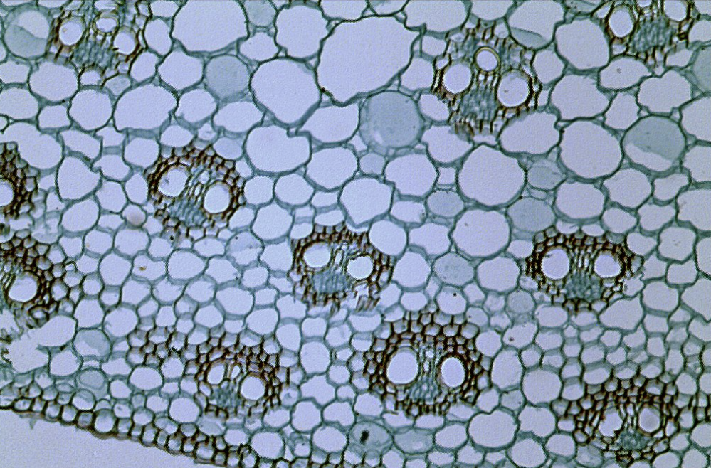
Light Microscopy (Brightfield)
A technique that utilizes light to illuminate the specimen, allowing for visual observation with a limited resolution of 2 micrometers and magnification up to 1500X.
Nissl Stain
A whole-body stain used in microscopy to visualize cell structures, specifically targeting RNA and ribosome-rich areas.
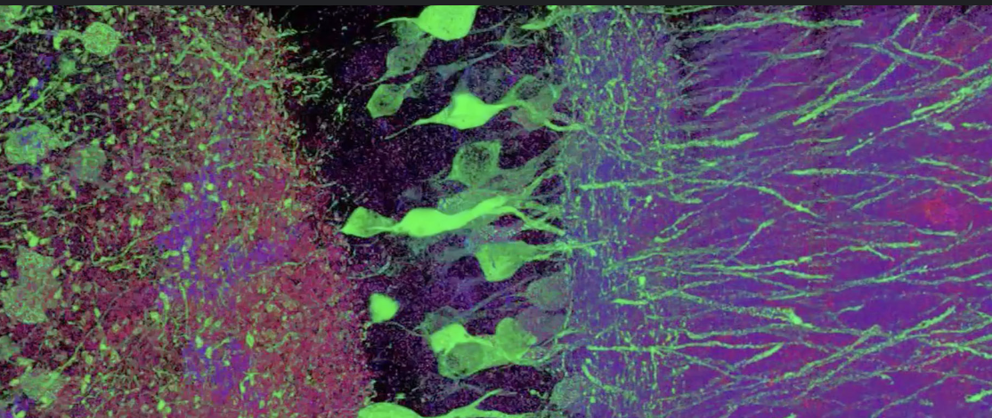
Fluorescence Microscopy (Epi)
A microscopy method that uses specific wavelengths of light to excite fluorophores, resulting in emitted light that is detected to create an image.
Artifacts in Microscopy
Unwanted alterations in an image that may arise from photobleaching, autofluorescence, and phototoxicity.
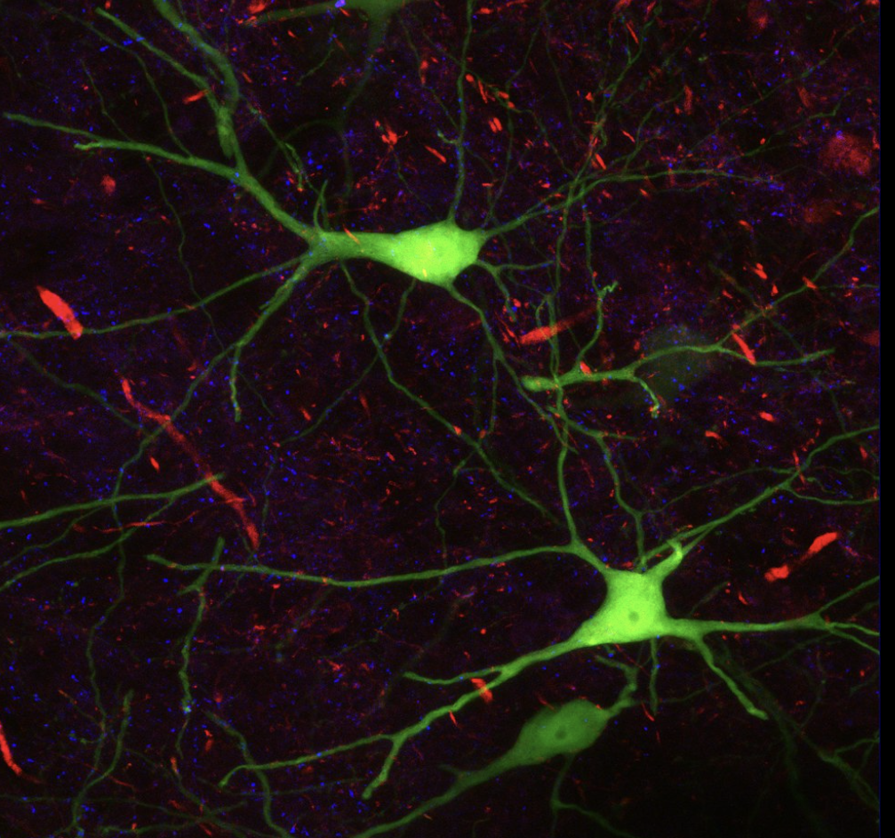
Confocal Microscopy
A type of fluorescence microscopy that reduces scatter and improves resolution, allowing for 3D reconstruction from image stacks with a resolution of 0.2 micrometers.
Golgi Stain
A silver salt stain used to visualize neurons, supporting the reticular theory of no individual cells in the brain.
Neuron Doctrine
The theory proposed by Santiago Ramón y Cajal that states that the nervous system is made up of individual cells called neurons.
Immunohistochemistry
A method using antibodies to detect specific antigens in cells, allowing for the visualization of proteins and cell types.
In-Situ Hybridization
A technique to detect specific RNA or DNA sequences within a cell using complementary probes.
Transgenic Approach
A genetic modification technique where sequences for fluorescent proteins are inserted into the genome to visualize specific cell populations.
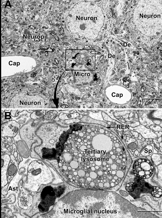
Electron Microscopy
A microscopy method that uses an electron beam for imaging, providing extremely high resolution up to 0.2nm but requiring specimens to be prepared with heavy metal staining.
Propidium Iodine Stain (PI)
A nuclear stain used to identify cell death, as it only penetrates damaged cells, emitting a signal that can indicate cell health.
Phase Contrast Microscopy
A microscopy technique that enhances contrast in transparent specimens by utilizing differences in the refractive index, allowing for visual observation of living cells.
H&E Stain (Hematoxylin and Eosin)
A commonly used stain in histology that highlights cell nuclei (hematoxylin) and cytoplasm (eosin) to differentiate tissue structures.
Transmission Electron Microscopy (TEM)
A type of electron microscopy that allows for visualization of internal structures of cells by transmitting electrons through thin samples.
Scanning Electron Microscopy (SEM)
An electron microscopy method that provides 3D images of the surface of a specimen by scanning it with a focused beam of electrons.
DAPI Stain
A fluorescent stain used to bind to DNA in the nuclei of cells, often used in fluorescence microscopy.
Live-Cell Imaging
A technique for observing living cells over time using various microscopy techniques to study dynamic processes.
Brightfield Staining
A simple staining method used in light microscopy that enhances contrast by coloring specific cell structures.
Fluorescent Dye
A chemical compound that emits light of a specific wavelength upon excitation, often used in fluorescence microscopy to label specific structures.
RNA Stains
Chemical dyes used to visualize RNA within cells, helping in the localization and quantification of RNA.
RNA Scope
A technique that uses in situ hybridization to detect and quantify RNA within tissue sections, providing spatial information about gene expression.
RNA/DNA Scope
A method that combines probes for both RNA and DNA, allowing for the simultaneous detection and localization of these nucleic acids in cells or tissues.