Regional Neuroanatomy I (4)
 Call Kai
 Learn
Learn Practice Test
 Spaced Repetition
 Match
Match Flashcards
 Knowt Play
1/77

There's no tags or description
Looks like no tags are added yet.
Name | Mastery | Learn | Test | Matching | Spaced | Call with Kai | Chat |
|---|
No analytics yet
Send a link to your students to track their progress
78 Terms
6 lobes of brain
frontal, parietal, occipital, temporal, limbic, insular
main functions of frontal lobe
high cognitive & executive functions - planning, decision making, attention
voluntary motor function
borders/landmarks to find frontal lobe
central sulcus, lateral (sylvian) sulcus, singulate sulcus
gyra
folds/ridges of brain
sulci
grooves on the cortical surface

label central sulcus


label lateral sulcus

label parieto-occipital sulcus
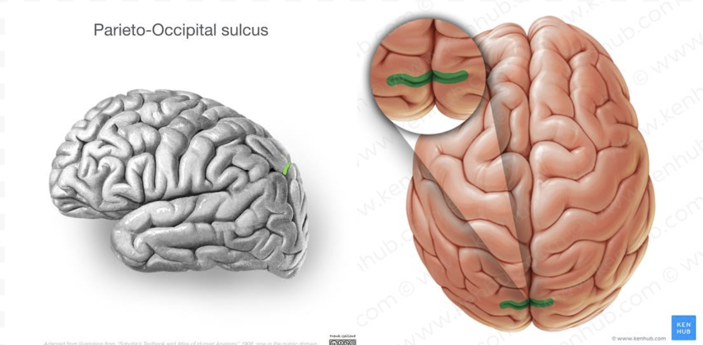

label preoccipital notch

main gyra of frontal cortex & important regions
precentral gyrus → primary motor cortex
superior frontal gyrus
middle frontal gyrus
inferior frontal gyrus → frontal operculum, very involved with language
where is primary motor cortex located
precental gyrus of frontal lobe
important parts of inferior frontal gyrus
pars opercularis
pars triangularus
pars orbitalis
Broca’s area → function, location
required for processing language and producing speech; located in the pars opercularis and pars triangularus of inferior frontal gyrus in frontal lobe

label precentral gyrus
has primary motor cortex

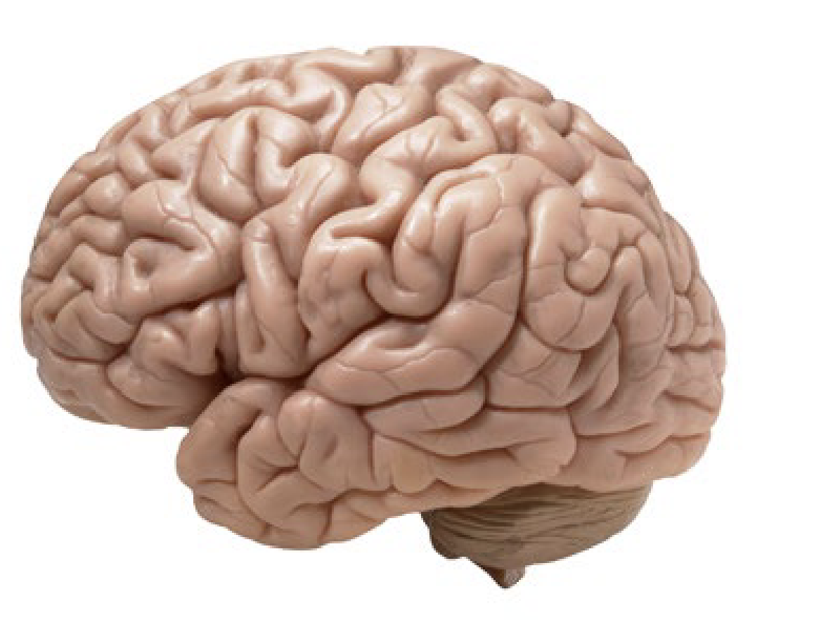
label postcentral gyrus
has primary somatosensory cortex


label superior temporal gyrus
has auditory cortex


label inferior frontal gyrus and relevant pars

parts of the frontal operculum
pars opercularis & pars triangularis (Broca’s area)
important parts of frontal lobe’s medial surface
cingulate sulcus
anterior paracentral gyrus
paracentral gyrus

label cingulate sulcus - why is it important?
separates frontal and limbic lobes

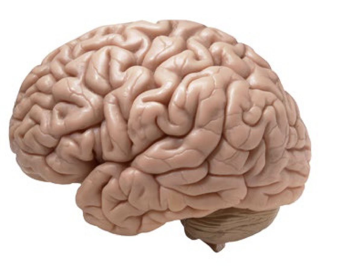
label anterior paracentral gyrus - why is it important?
it contains the medial aspect of the primary motor cortex, which controls the legs


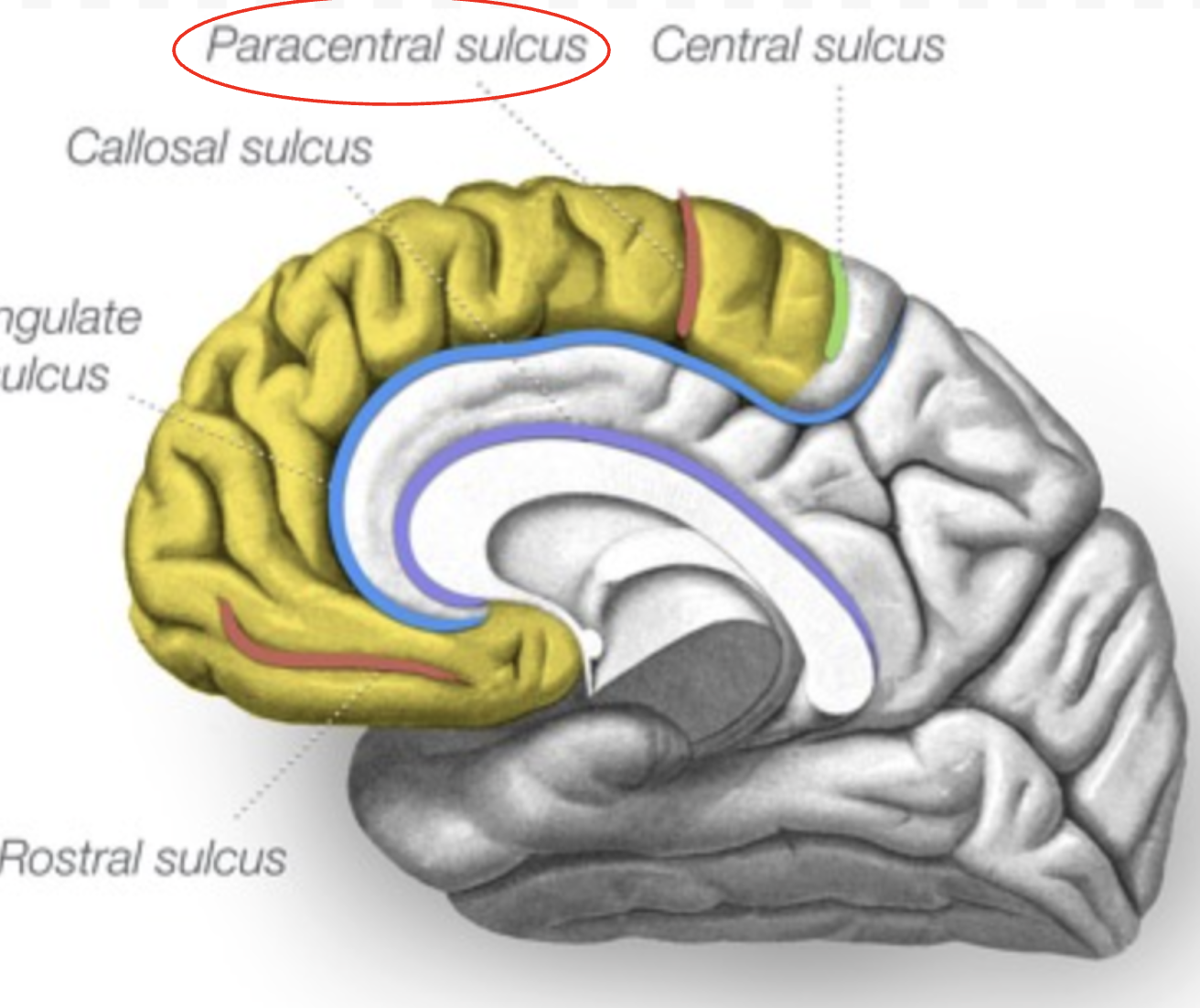
label paracentral sulcus
most important parts of frontal lobe’s ventral surface
olfactory sulcus
gyrus rectus
orbital gyri

label important parts of frontal lobe’s ventral surface

parietal lobe main functions
high cognitive activity
multisensory integration
attention
spatial awareness
parietal lobe location & landmarks
between frontal and occipital lobes - look for central sulcus, lateral sulcus, and parieto-occipital sulcus
main gyra of parietal lobe & important parts
postcentral gyrus → primary somatosensory cortex
superior parietal lobule
supramarginal gyrus & angular gyrus → visuo-spatial awareness, reading, writing
parietal operculum → secondary somatosensory area

label supramarginal gyrus


label angular gyrus
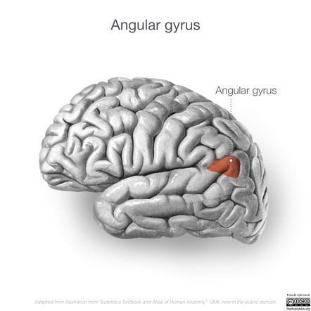
regions of parietal lobe most involved in visuo-spatial awareness and written language (ability to read and write to receive/communicate information)
supramarginal gyrus
angular gyrus
most important regions of parietal lobe medial surface & important parts
posterior paracentral gyrus → medial aspect of primary somatosensory cortex
precuneus → polymodal/multisensory integration
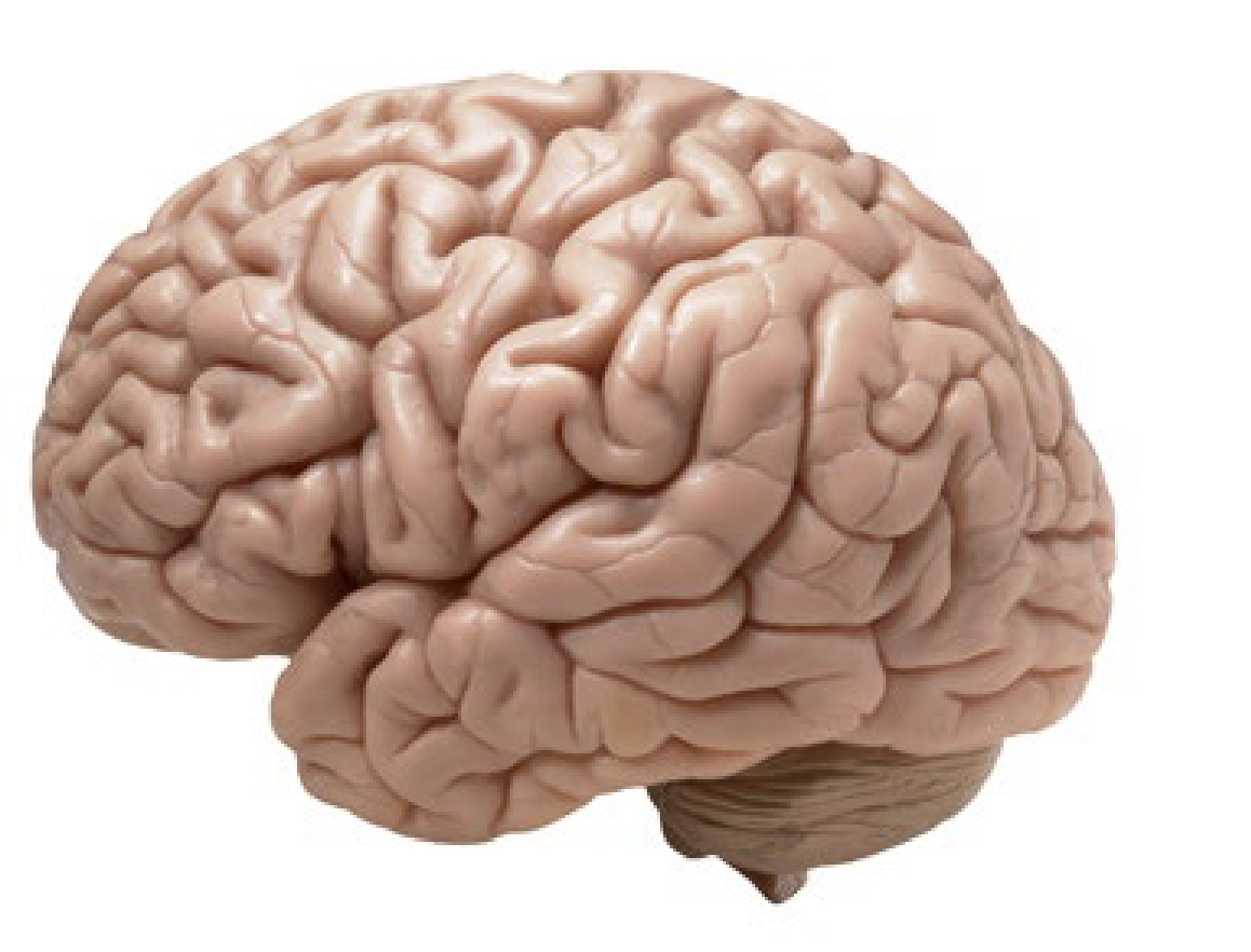
label important parts of parietal lobe’s medial surface

main functions of temporal lobe
audition
multisensory integration
memory
emotion
where is temporal lobe/landmarks
on bottom of brain; look for lateral sulcus, collateral sulcus

label collateral sulcus
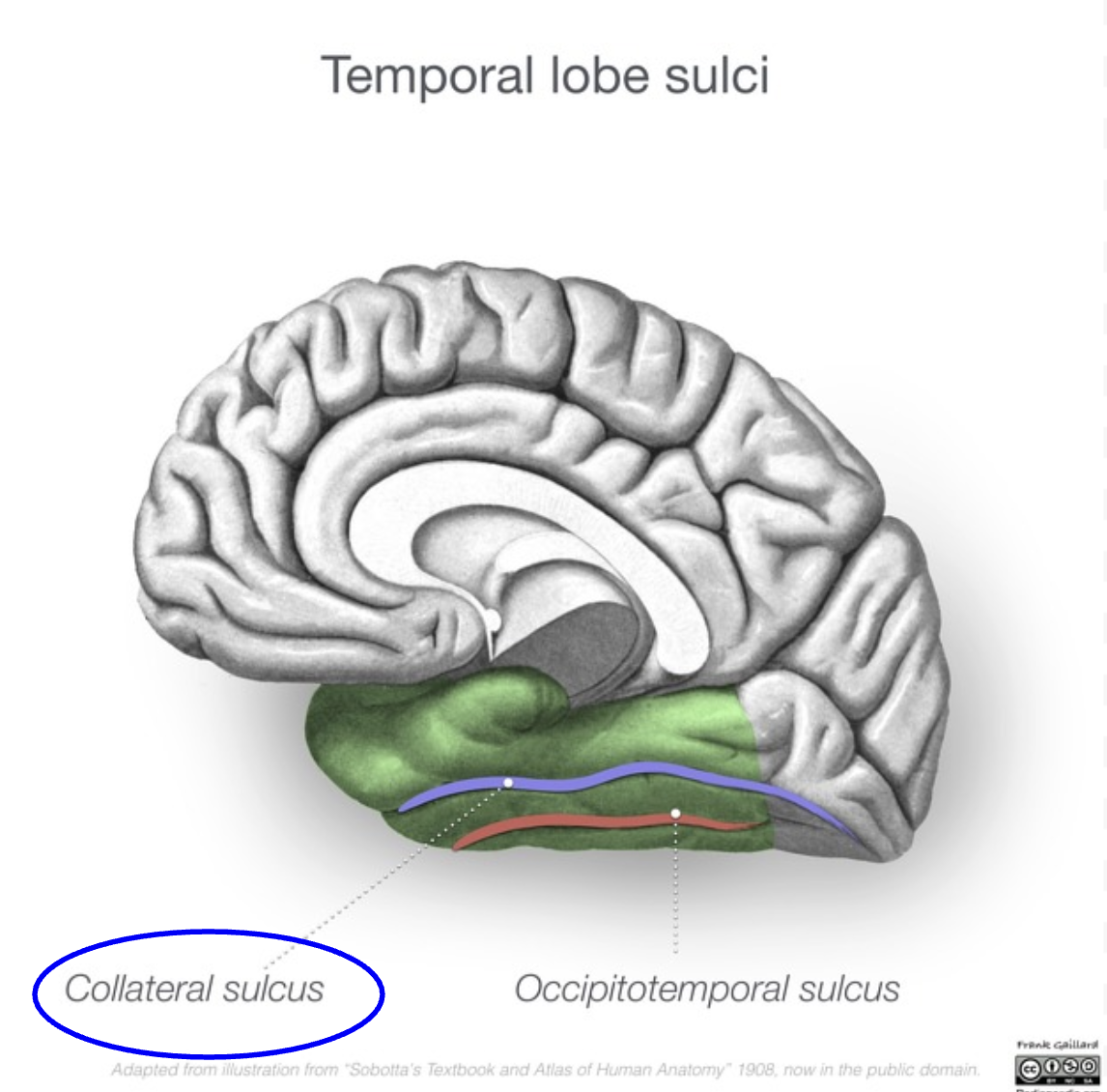
main gyra of temporal lobe, important parts
superior temporal lobe → Wernicke’s area in left hemisphere
middle temporal lobe
inferior temporal lobe
temporal operculum → auditory cortex (includes part of superior temporal gyrus)
what is located in parietal operculum
secondary somatosensory area
what is located in temporal operculum
part of superior temporal gyrus → auditory cortex
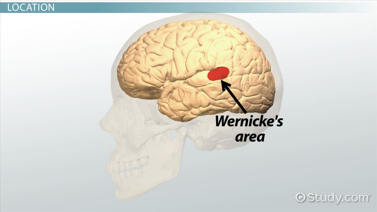
location and importance of Wernicke’s area
in the left superior temporal gyra of temporal lobe
required for language comprehension; damage causes fluent aphasia, where a person can speak fluently/easily, but their words lack meaning or may be made up

label superior temporal gyrus

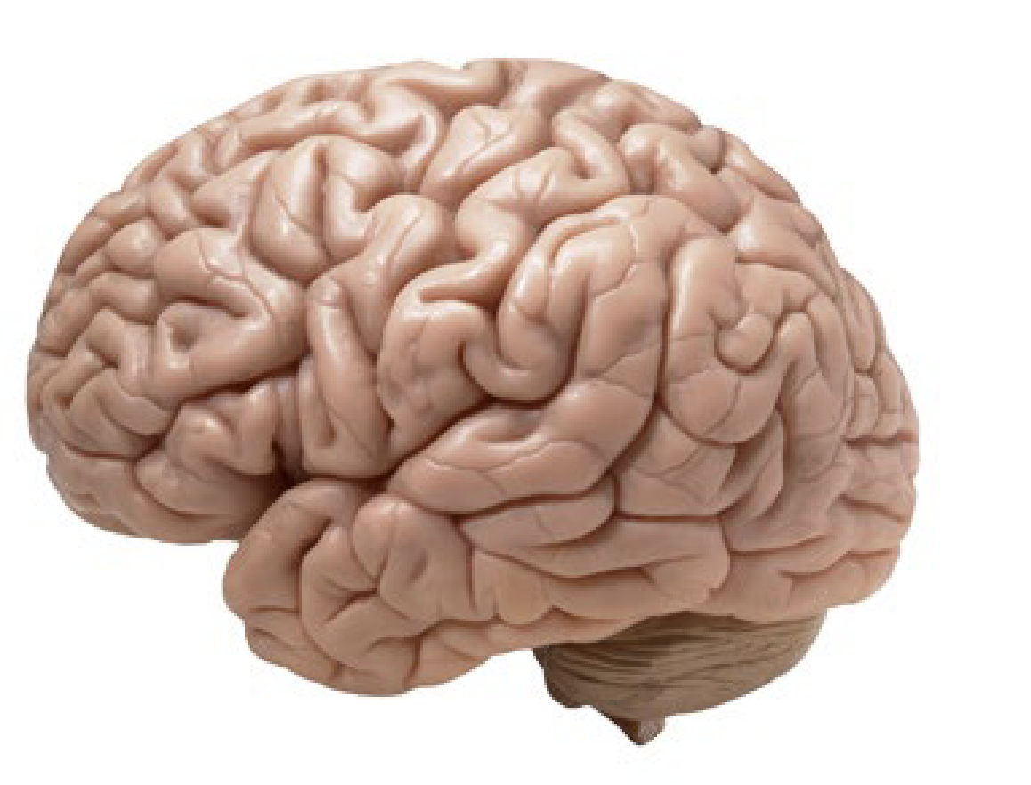
label middle temporal gyrus

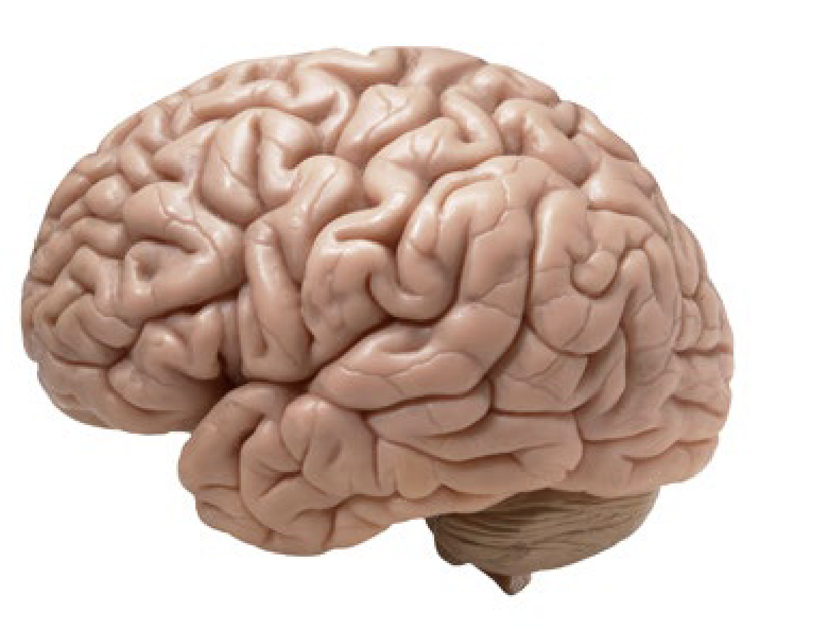
label Wernicke’s area

label operculum and its specific parts

important parts of temporal lobe ventral surface
collateral sulcus
occipitotemporal gyri
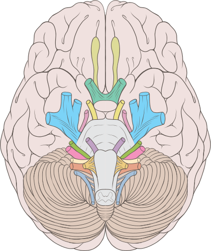
label important parts of temporal lobe ventral surface

occipital lobe main functions
vision
multisensory information
occipital lobe location/landmarks
back of brain, adjacent of temporal and parietal lobes; lateral surface below parieto-occipital sulcus and preoccipital notch
main gyra/structures of occipital lobe
cuneus
lingual gyrus
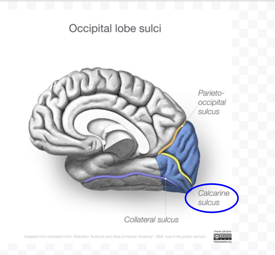
primary visual cortex (borders calcarine sulcus)

label calcarine sulcus

label parieto-occipital sulcus and preoccipital notch

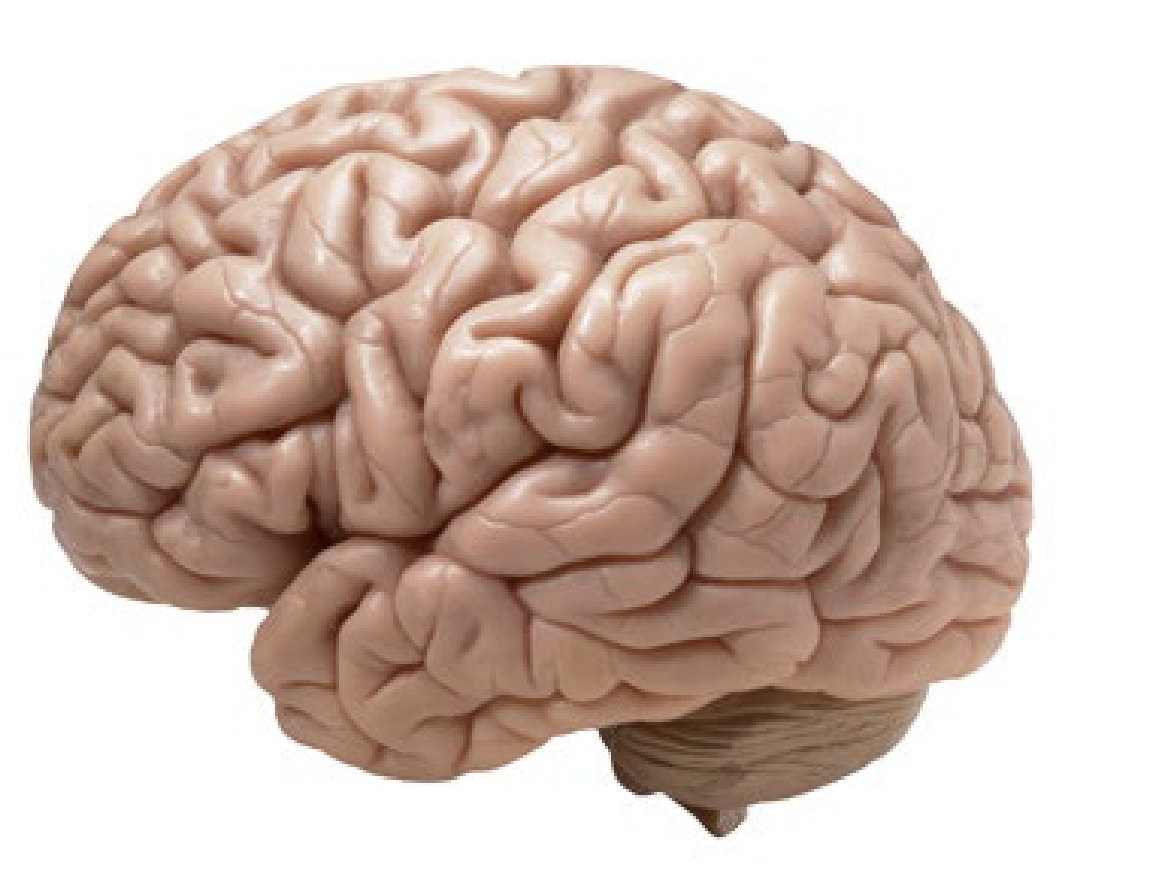
label cuneus, calcarine sulcus, and lingual gyrus

limbic lobe main functions
learning
memory
emotion
limbic lobe location
not visible from surface laterla view; outside of insular lobe but inside. of the four other lobes

main limbic lobe gyra
cingulate gyrus
parahippocampal gyrus
uncus → part of olfactory cortex

label cingulate gyrus
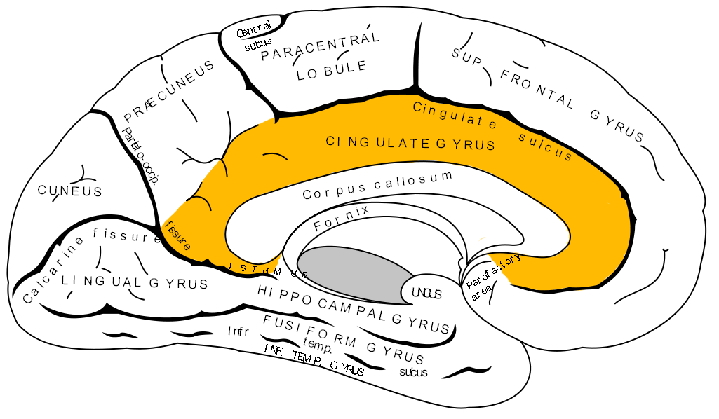
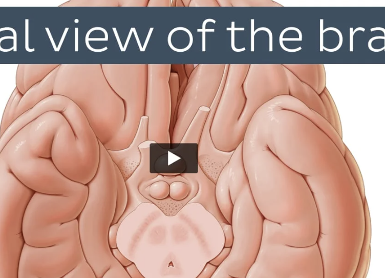
label uncus


label parahippocampus gyrus

main functions of insular lobe
taste
motivation
where is insular lobe
inside limbic lobe

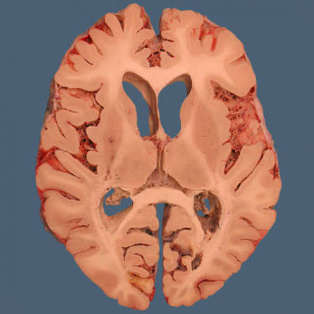
label insular cortex, importance
receives nociceptor and viscerosensory inputs

2 major arterial systems supplying brain with blood
internal carotid artery (ICA)
vertebro-basilar
where does ICA enter brain
go into the dura mater near the optic chiasm
generally where the ICA goes/what it does
internal carotids branch out after entering dura and form the Willis circle system, which distributes blood to the cerebral hemispheres, basal ganglia, and diencephalon
all ICs (branches of ICA)
ophthalmic artery
anterior choroidal
anterior cerebral artery (ACA)
middle cerebral artery (has 4 segments)
posterior communicating arteries (PCom)
ICs NOT in circle of willis
opthalmic artery
anterior choroidal
ICs IN circle of willis
anterior cerebral artery
middle cerebral artery
posterior communicating arteries
opthalmic artery supplies blood to?
cenrtal artery of retina; issues cause vision loss
anterior choroidal artery supplies blood to?
to choroid plexus of lateral ventricles
anterior cerebral artery supplies blood to?
low extremity of motor cortex → optic chiasm, anterior paracentral
low extremity of somatosensory cortex → anterior and posterior gyrus, medial surface of frontal and parietal lobes, posterior paracentral
there are 2 ACAs, one on the right and left
how 2 ACAs connect; importance of this region
with anterior communicating artery; around 30-35% aneurysms are found here
segments of middle cerebral artery
M1, M2, M3, M4
M1 supplies blood to?
basal ganglia → deep in cerebral hemisphere
M2 supplies blood to?
insular cortex
M3 supplies blood to?
this is the opercular segement → blood to frontal, parietal, and temporal opercula, including Broca’s area, motor & somatosensory areas, and the auditory cortex
M4 supplies blood to?
above/below lateral fissure in frontal, parietal, temporal, and occipital lobes
what is the Willis circle, its parts, and where it supplies blood to
ring of vessels connecting anterior and posterior circulation of brain around the optic chiasm
includes ACAs, anterior communicating artery, MCA, PCom, PCA (not IC)
Posterior cerebral artery
PCA → artery that is not an IC branch but is part of the Willis circle. branch off basilar artery that supplies blood to the ventral and medial surface of temporal and occipital lobes. it also has branches supplying the mesencephalon and thalamus.
willis circle looks like/label:
