IB Biology Topic 6
1/147
There's no tags or description
Looks like no tags are added yet.
Name | Mastery | Learn | Test | Matching | Spaced | Call with Kai |
|---|
No analytics yet
Send a link to your students to track their progress
148 Terms
Glucagon
A protein hormone secreted by pancreatic endocrine cells that raises blood glucose levels
Insulin
A hormone that lowers blood glucose levels
Dialysis tubing
Plastic-like cellulose tubing with tiny holes to allow small molecules to pass through, it models absorption of food in the intestine
WIlliam Harvey discovered the blood system
were a combined network, blood flows continusouly, the heart is the pump (arteries from hear) (veins to heart), Blood flow is unidirectional
pulmoary circuit
on the right side of the heart
systemic circulation
on the left side of the heart
Arteries
Blood vessels that carry blood away from the heart
Characteristics of arteries
blood at high pressure, walls are thick, wales stretch or contract with a pulse, walls contain muscles cells and elastic fibres
Capillaries
Microscopic vessel through which exchanges take place between the blood and cells of the body
Characteristics of capllilaries
blood at low pressure, walls made of a single layer, extremly narrow lumen, facilatate material exhange
Cappliaries absorb
cell waste e.g. carbon dioxide and urea
Veins
Blood vessels that carry blood back to the heart
Characterisitc of veins
blood at low pressure, wide lumen, have valves, walls are thin, small amounts of muscles
Role of valves in the blood system
prevent backwards blood flow
heart structure
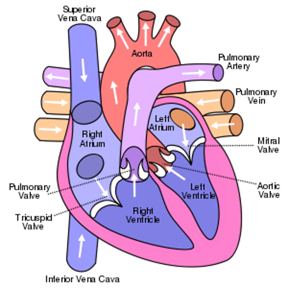
Pathway of deoxygenated blood through the heart
-Vena Cava (veins) from body
-Right Atrium
-Tricuspid valve
-Right Ventricle
-Pulmonary valve
-Pulmonary (artery)
-Lungs
Pathway of oxygenated blood
-from lungs
-Pulmonary veins
-Left Atrium
-bicuspid valve
-Left ventricle
-aortic semilunar valve
-aorta
-body
Sinotorial node
pacemaker on right atrium of heart
Describe the electrical events that trigger the contraction of the heart muscle fibres
- The heart beat is myogenic
- Electrical signals are initiated by the sinoatrial (SA) node
- It stimulates the atria to contract and also relays signals to an atrioventricular node
- The atrioventricular node sends signals via the Bundle of His to Purkinje fibres
- These fibres innervate the ventricles and cause them to contract
how is heart rate increased and decreased?
by impulses brought to the heart through two nerves from the medulla of the brain.
Describe the role of the medulla and epinephrine (adrenaline) in regulating heart rate
- The SA node maintains the heart's normal sinus rhythm (60 - 100 bpm)
- The SA node may be regulated by the medulla, with sympathetic nerves increasing heart rate, by releasing noradrenaline and parasympathetic nerves decreasing the heart rate by releasing acetylcholine
- Heart rate may also be increased by the release of epinephrine (a.k.a. adrenaline) into the bloodstream
Outline the pressure changes in the heart during the cardiac cycle
Blood returning to the heart will flow into the atria and ventricles as the pressure in them is lower
As ventricles fill, atria contract (atrial systole), increasing pressure in atria and forcing blood into ventricles
As ventricles contract, ventricular pressure exceeds atrial pressure and AV valves close to prevent back flow
When ventricular pressure exceeds pressure in aorta, the aortic valve opens to release blood into the aorta
As blood exits the ventricle, ventricular pressure falls below aortic pressure, so the aortic valve closes
When ventricular pressure drops below atrial pressure, the AV valve opens and cycle begins again
Causes of Coronary Occlusion
- Fatty deposits develop in the arteries and reduce the lumen
- The restricted blood flow increases pressure in the artery, = damage to the arterial wall
- The damaged region is repaired with fibrous tissue which significantly reduces the elasticity of the vessel wall
- As the smooth lining of the artery is degraded, lesions form called atherosclerotic plaques
- If the plaque ruptures, blood clotting is triggered, forming a thrombus that restricts blood flow
- If the thrombus is dislodged it becomes an embolus and can cause a blockage in a smaller arteriole
Risk Factors for Coronary Heart Disease
Age
Genetics
Obesity
Diseases
Diet
Exercise
Sex
Smoking
First line of defence
prevent the entry of pathogens into the body
- intact skin
- mucous membranes
Blood clots
1 - Platelets : form a sticky plug at the damaged region (primary haemostasis)
2 - Fibrin strands form an insoluble mesh of fibres that trap blood cells at the site of damage (secondary haemostasis)
Coagulation Cascade
1- Platelets to become sticky and adhere to the damaged region = solid plug
2- Initiate localised vasoconstriction = reduce blood flow. Triggers conversion of the inactive zymogen prothrombin into the activated enzyme thrombin
3 - Thrombin catalyses fibrinogen to fibrin
4 - The fibrin strands = mesh of fibres around the platelet plug and traps blood cells to form a temporary clot
5- When the damaged region is completely repaired, an enzyme (plasmin) is activated to dissolve the clot
Second line of defence
Innate immune system
- non-specific
- non-adaptive
Phagocytosis process
- Phagocytic leukocytes circulate in the blood and move into the body tissue in response to infection
- Histamine released which draw white blood cells to the site of infection via chemotaxis
- Pathogens engulfed when pseudopodia surround the pathogen and then fuse to form an internal vesicle
- The vesicle is then fused to a lysosome forming a phagolysosome and the pathogen is digested
- Antigens may be presented on the surface of the phagocyte in order to stimulate the third line of defence
Third line of defence
Adaptive immune system, which is specific in its response
B lymphocytes
are antibody producing cells that recognize and target particular antigens
T lymphocytes
are regulator cells that reslease chemicals to activate specfic B cells
Antibodies
Proteins produced by B cells that attach to antigens, keeping them from harming the body
Antigen
A protein that, when introduced in the blood, triggers the production of an antibody
Antigens are specific
to a antibody
Antibiotics can only work on
prokaryotic cells
Antibiotics targets
prokaryotic metabolism
Viruses lack a metabolsim and hence
can not be treated with antibiotics
Antibiotic resistance happens by ..
Genes degrade the antibiotic, block its entry, increase its removal or alter the target
Resistance increases due to....
Antibiotics are :
- Over-prescribed
- Misused
- Freely available
Penicillin
the first discovered antibiotic
Florey and Chain experiment
mice made sick with bacteria, half were given penicillin and teh other hald nothing, those who ingestied penicillin lived
Effects of HIV on the immune system
- HIV targets t cells
- the virus is inactive during when the t cells reproduce
- eventually, the virus is active and has spread
- antibodies can't be produced
- one is extremely susceptible to infection
ventialtion maintains
concentration gradients of oxygen and carbon dioxide between air in the alveoli and blood flowing in adjacent capllicaires
Ventilation
movement of air in and out of the lungs
gas exchange
the process of obtaining oxygen from the environment and releasing carbon dioxide
Gas exchange by pressure in lungs:
Occurs via diffusion
O2 concentration is higher in the lungs than in the blood, so O2 diffuses into blood.
CO2 concentration in the blood is higher than in the lungs, so CO2 diffuses out of blood.
cell respiration
the process in cells in which oxygen is used to release stored energy by breaking down sugar molecules
Lung structure
trachea, bronchi, bronchioles, alveoli
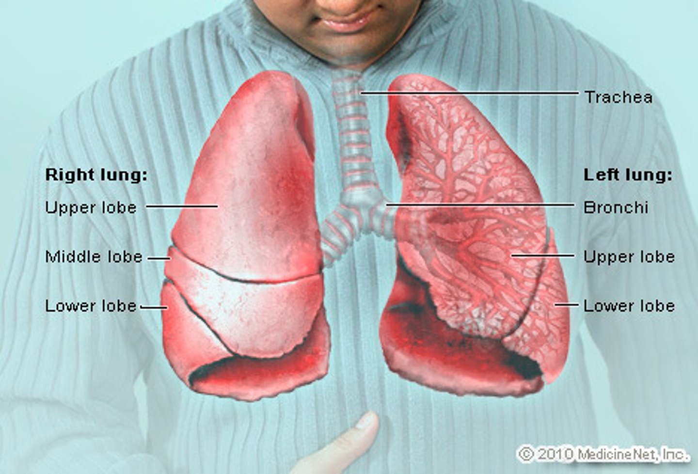
Alveoli structure
- Thin epithelial layer
- Rich capillary network
- Spherical
- Internal surface is covered with a layer of fluid
Type I pneumocytes
extremely thin alveolar cells that are adapted to carry out gas exchange
Type II pneumocytes
cuboidal cells that's produce surfactant which reduces surface tension in the alveoli
What does surfactant do?
It reduces surface tension inside the alveolar or respiratory membrane.
Muscle contrations in the lungs cause
pressure changes in the thorax that force air in and out of the lungs
When the volume of the thoracic cavity increases
pressure in the thorax decreases
When the volume of the thoracic cavity decreases
pressure in the thorax increases
When pressure in the chest is less than atmospheric pressure
inspiration will occur
When pressure in the chest is greater than atmospheric pressure
expiration will occur
Muscular process of inhalation:
1. diaphragm contracts
2. external intercostal muscles pull ribs up
3. this increases pressure in the thoracic cavity
4. pressure in the lungs decreases below atmospheric pressure
5. Air flows in the equalise
Muscular process of exhalation:
1. diaphragm muscles relax
2. internal intercostal muscles pull ribs down
3. abdominal muscles contract
4. decreases volume of thoratic cavity
5. pressure in lungs increases above atmospheric pressure
6. air flows out
Causes of lung cancer:
smoking, passive smoking, air pollution, radon gas, asbestos and silica
Consequence of lung cancer
death, metastasis, blood, wheezing etc.
Emphysema
a condition in which the air sacs of the lungs are damaged and enlarged, causing breathlessness.
causes of emphysema
smoking or second hand smoking
consequences of emphysema
reduced surface area in alveoli, difficulty breathing, volume of alveoli increases
Spirometery
measuring the volume and / or flow at which air can be inhaled or exhaled
Neuron
a specialized cell transmitting nerve electrical impulses; a nerve cell.
Dendrites
Short-branched fibres that convert chemical information from other neurons or receptor cells into electrical signals
Axon
An elongated fibre that transmits electrical signals to terminal regions for communication with other neurons or effectors
Soma
A cell body containing the nucleus and organelles, where essential metabolic processes occur to maintain cell survival
Myelin sheath
Improves the conduction speed of electrical impulses along the axon
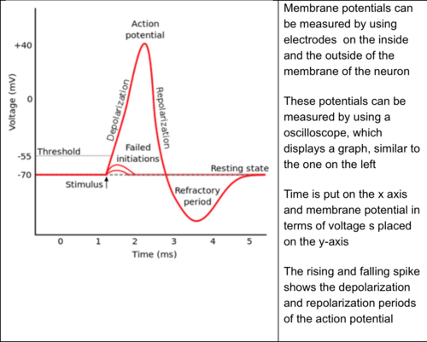
resting potential
the state of the neuron when not firing a neural impulse
How do neurons reach a resting potential
through pumping sodium and potassium ions across their membrame
sodium-potassium pump in neurons
is important for maintaining the resting membrane potential
The resting potential is maintaned by
the Na+/K+ pump
How does the NaK pump work? in neurons
it exchanges 3 sodium ions (3 out) and 2 potassium ion (2 in) so that the membrane mecomes slightly negative
the unequal distribution of ions on different sides of the membrane is called
a membrane potential
Depolirization of a neuron
- The sudden change in membrane potential from a negative to the positive internal charge
- In response to a signal at a dendrite the sodium channels in the axon's membrane open
- As there are more sodium concentrated outside of the neuron the opening of sodium channels causes a passive influx of sodium
- The sodium causes the neuron to be more positive
SODIUM CHANNEL OPENS
Repolarization of a neuron
- The restoration of membrane potential following depolarisation
- Following the sodium influx, potassium channels open
- As K ions are more concentrated in the neuron, a passive removal of potassium is caused
- This means we return to resting potential with a negative interior
POTASSIUM CHANNEL OPENS
refractory period
- the time following an action potential during which a new action potential cannot be initiated
- after re and de polarisation the ionic distribution is reversed using NA K pumps and active transport
- this readies the neuron for the next action potential
NA K PUMP OPENS
Nerve impulses are
action potentials propagated along the axons of neurons
Action potentials are generated within the axon according to the
all or none principal
all-or-none principle
The principle that when a neuron fires, it fires with the same potency each time; a neuron either fires or not—it cannot partially fire.
- An action potential of the same magnitude will always occur provided a minimum electrical stimulus is generated
- This minimum stimulus - known as the threshold potential (-55 mV) - is the level required to open voltage-gated ion channels
- If the threshold potential is not reached, an action potential cannot be generated and hence the neuron will not fire
Local currents in an axon cause
each successive part of the axon to reach the threshold potential
Oscilloscope traces
Measure the changes in membrane potential in axons during action potential
Myelination of the nerve fibres allows
for saltatory conduction
saltatory conduction
Rapid transmission of a nerve impulse along an axon, resulting from the action potential jumping from one node of Ranvier to another, skipping the myelin-sheathed regions of membrane.
Synapses
tiny gaps between neurons and receptor/effector cells
Chemical Transfer Across Synapses
- When an action potential reaches the axon terminal, it triggers the opening of voltage-gated calcium channels
- Calcium ions diffuse into the cell and promote the fusion of vesicles with the cell membrane
- The neurotransmitters are released from the axon terminal (exocytosis) and cross the synaptic cleft
- Neurotransmitters bind to specific receptors on the post-synaptic membrane and open ligand-gated ion channels
- The opening of ion channels generates an electrical impulse in the post-synaptic neuron, propagating the pre-synaptic signal
- The neurotransmitters released into the synapse are either recycled (by reuptake pumps) or degraded (by enzymatic activity)
How are neurotransmitters released?
calcium opens voltage gated channels and allows synaptic vesicle to bind to receptors and exocytosis occurs
When presynaptic neurons are depolarized they release
a neurotransmitter into the synapse
Depolarization in axon terminals causes
Calcium channels to open
centeral nervous system
brain and spinal cord
peripheral nervous system
the sensory and motor neurons that connect the central nervous system to the rest of the body
Acetylcholine
A neurotransmitter that enables learning and memory and also triggers muscle contraction
Acetylcholine must be removed from the synapse as
overstimulation can leas to fatal stuff and paralysis
Acetylcholine is broken down by
acetylcholinesterase
Acetylcholine is made from
choline and acetyl CoA
When Acetycholine is broken down by acetycholinesterase...
choline returns to the axon terminal
Neonicotinoid pesticides
Bind to ACh receptors in the post-synaptic membranes of cholinergic synapses in insects. Cholinesterase does not break down these pesticides, so they remain bound to the receptors, preventing ACh from binding. Thus, they block synaptic transmission, killing the insect.