Unit 1- Biology - [B3] Tissue structure and function
1/143
Earn XP
Description and Tags
This is taken from the applied science foundation diploma BTEC textbook
Name | Mastery | Learn | Test | Matching | Spaced | Call with Kai |
|---|
No analytics yet
Send a link to your students to track their progress
144 Terms
What is epithelial tissue?
a sheet of cells that covers a body surface or lines a body cavity
What is the structure of epithelial tissue?
- layers/sheets of cells
- very little matrix
What are the types of epithelial tissue?
- squamous epithelial tissue
- columnar epithelial tissue
- endothelium tissue
What is endothelium tissue?
The endothelium is a layer of cells that lines the blood vessels and lymph vessels of the body.
How thick is the simple squamous epithelium?
1 cell thick
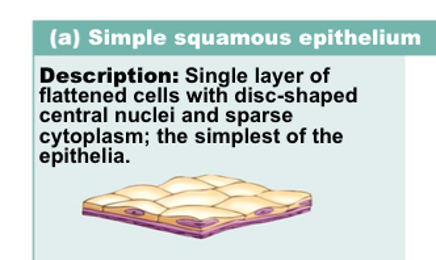
Why are specialised squamous epithelial cells ideal for rapid diffusion?
they form a thin, smooth flat layer, which provides a short diffusion pathway
What is an example of a structure that benefits of this adaptations?
alveoli
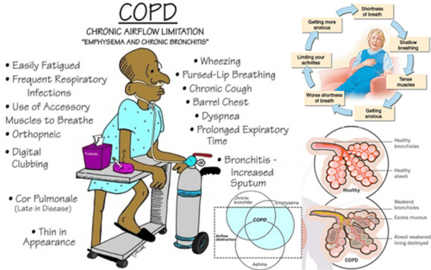
How is COPD caused?
It is a lung disease commonly associated with cigarette smoking irritates the epithelium and the alveoli walls become thicker due to scar tissue. This results in inflammation that limit the airflow in and out of the lungs
What are symptoms of COPD?
chronic bronchitis and emphysema
What are ciliated cells?
Cells with tiny hair like structures
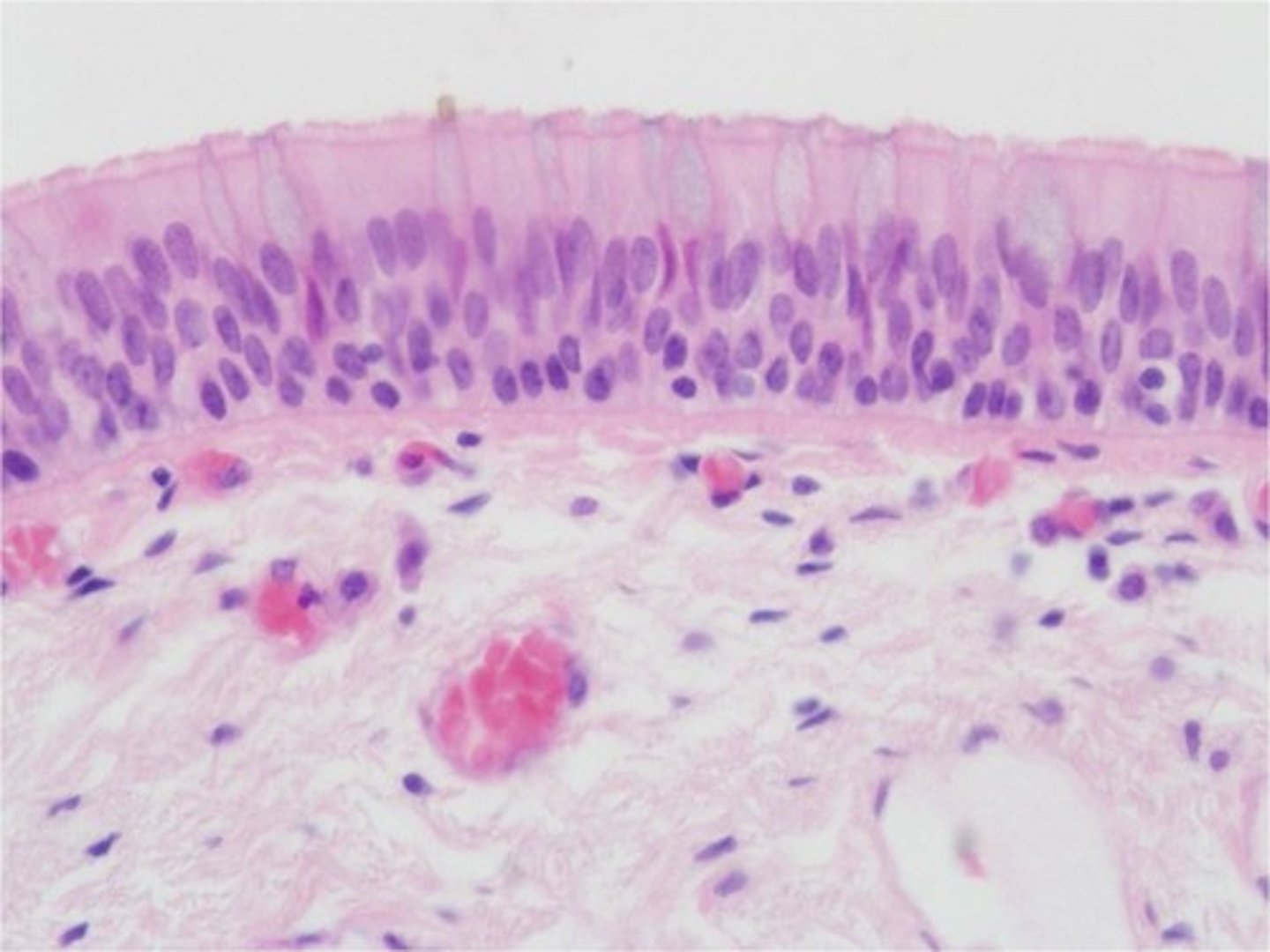
What does ciliated epithelium do in the trachea?
Goblet cells secrete mucus on the lining of the trachea to trap dust and microorganisms, cilia waft the mucus away from the lungs towards the throat
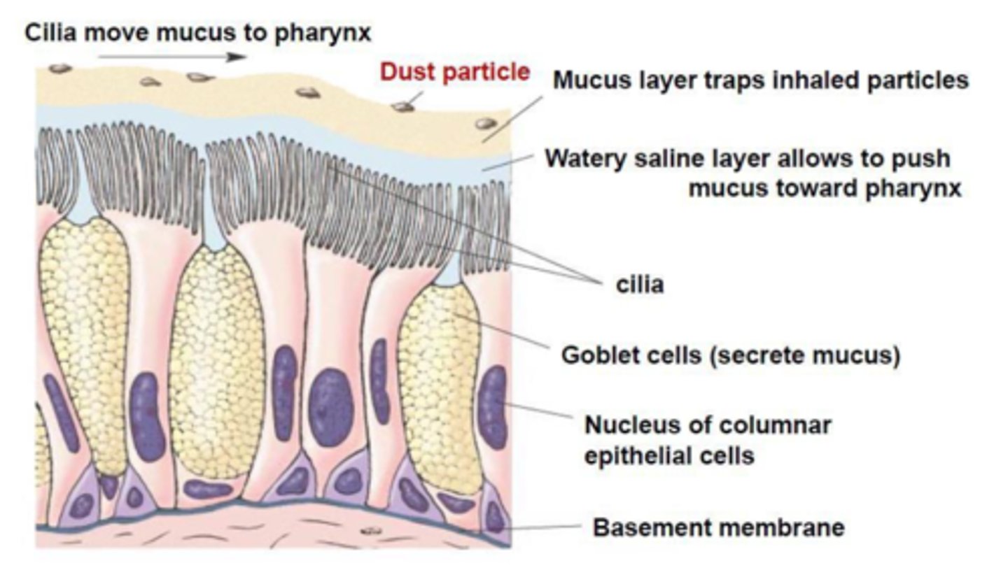
Why do goblet cells secrete mucus?
to protect the lungs and prevent bacteria getting to the alveoli
What is the structure of endothelial tissue?
flattened cells 1 cell thick
Where does the endothelium line?
Heart, blood vessels, lymphatic vessels
What does the endothelium provide?
A short diffusion pathway for the movement of various substances
What can cause damage to the endothelium?
Carbon monoxide and high blood pressure
What is the first step that causes an atheroma to form?
the lining of the artery is damaged
What does the damaged artery trigger?
an immune response by the white blood cells to repair the damaged artery
What does the white blood cells encourage the growth of?
smooth muscle and the deposition of fatty substances such as cholesterol
Where is the location of the fatty substance ?
under the endothelium lining of the arteries
What is the name of the process of deposition?
atherosclerosis
What can these atheromas do?
they can build up enough to break through the inner endothelial lining of the artery
What can this build up of atheromas form?
plaque in the lumen of the artery
If there is plaque in the lumen, what can this lead to?
stroke due to restricted blood flow and CVD
What does CVD stand for?
cardiovascular disease
What is the meaning of lumen?
the space inside a structure
What is the meaning of artery?
blood vessel that carries blood away from the heart
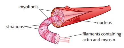
What are muscles composed of?
cells that elongated and form fibres
What do muscle cells contain?
protein filaments
What are the names of these protein filaments?
actin and myosin
What do these protein filaments enable muscles to do?
contract and cause movement
What are the three types of muscles?
skeletal, cardiac and smooth
Where can you find skeletal muscle?
attached to bones
Can you control skeletal muscle?
yes, it is under voluntary control
Where is cardiac muscle found?
only the heart
Is cardiac muscle under voluntary control?
no
Where is smooth muscle found?
in the walls of hollow organs
Is smooth muscle under voluntary control?
No
What is the sarcoplasm?
long strands of cells sharing nuclei and cytoplasm
What is the sarcoplasmic reticulum?
many mitochondria, specialised endoplasmic reticulum
What is the sarcolemma?
a cell membrane of a stratified muscle cell
What is a myofibril?
what each muscle fibre is made up of
What is each myofibril fibre made of?
proteins called myofilaments
What do myofilaments enable?
contractions to take place because of the contractile nature of the proteins
How do myofilaments appear?
different coloured bands
What are the different coloured bands?
A and I
What colour is the A band?
dark
What colour is the I band?
light
What is the sarcomere?
the span from one z-line to the next
Why and when does the length of the sarcomere reduce?
when the muscle contracts because the I-band and the H-zone lengths are reduced
Does the A-band length change during contraction?
no
What is A on the diagram?
Z disc

What is B on the diagram?
H zone

What is C on the diagram?
Z disc

What is D on the diagram?
thin actin filament

What is E on the diagram?
Thick myosin filament
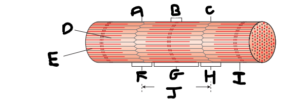
What is F on the diagram?
I band

What is G on the diagram?
A band

What is H on the diagram?
I band

What is I on the diagram?
M line

What is J on the diagram?
sarcomere
What happens during muscle contractions?
actin filaments move and overlap the thick myosin filaments. the sarcomere shortens, decreasing the size of the overall muscle
What are the two types of muscle fibres?
slow twitch and fast twitch
what do fast and slow twitch fibres influence?
how muscle responds during physical activity
What are slow twitch fibres more effective at using?
oxygen to generate energy in the form of ATP
What does this make slow twitch muscles good at?
maintaining continuous and extended muscle contractions over a long period of time
What are properties of slow twitch fibres?
less sarcoplasmic reticulum
more mitochondria for sustained contraction
more myoglobin
a dense capillary network
What is ATP?
an enzyme that transports chemical energy within cells for metabolism
What does ATP stand for?
adenosine triphosphate
What can fast twitch fibres be separated into?
two different kinds
What type of muscle fibres are able to hydrolyse ATP much more quickly and contract quickly and are relatively resistant to fatigue?
fast twitch oxidative muscle fibres
What type of fibre contain a large concentration of glycogen that provides fuel for anaerobic respiration but contract rapidly and fatigue quickly?
fast twitch glycolytic muscle fibres
What are the properties of fast twitch oxidative muscle fibres?
many mitochondria
myoglobin
blood capillaries
What are properties of fast twitch glycolytic muscle fibres?
less myoglobin
few mitochondria
few capillaries
What is the meaning of hydrolyse?
a chemical reaction involving the breaking down a compound with water
What is glycogen?
many glucose molecules bonded together and stored in the liver and muscles
What is anaerobic respiration?
respiration without oxygen
What does the central nervous system consist of?
brain and spinal cord
What is the CNS made up of?
billions of non-myelinated nerve cells and longer, myelinated axons and dendrons
What are neurons?
cells that receive and facilitate nerve impulses, or action potential across their membrane and pass them onto the next neuron

What is structure A?
dendrite
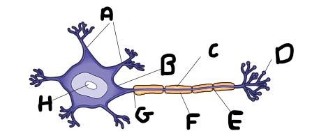
What is structure B?
soma

What is structure C?
schwann cell

What is structure D?
axon terminal

What is structure E?
node of ranvier

What is structure F?
myelin sheath

What is structure G?
axon

What is structure H?
nucleus
What are nerve impulses?
information that travels along neurons in the the form of electrical signals
What is known as action potential?
a nerve impulse
What does action potential arise from?
a change in the ion balance in the nerve cell which spreads rapidly from one end of the neuron to the other
Describe resting potential in a neuron.
Resting potential refers to a neuron that is not transmitting an action potential and is at rest, maintaining a negative potential inside the cell compared to the outside.
How does a neuron maintain its resting potential?
A neuron maintains its resting potential by actively transporting sodium and potassium ions across its membrane, using a sodium/potassium pump, which requires energy in the form of ATP.
Define the role of the sodium/potassium pump in neurons.
The sodium/potassium pump actively transports three sodium ions out of the neuron and two potassium ions into the neuron, helping to create a potential difference of −60mV.
Explain the significance of ion permeability in resting potential.
The cell membrane is more permeable to potassium ions, which contributes to the negative charge inside the neuron during resting potential.
What happens to the gated sodium ion channels during resting potential?
During resting potential, the gated sodium ion channels in the membrane are closed.
How is an action potential generated in a neuron?
An action potential is generated when a nerve impulse is stimulated by a receptor cell or another neuron, causing a quick change in the permeability of the axon membrane.
Describe the process of depolarization in neurons.
Depolarization is the wave-like change in the permeability of the axon membrane that occurs when an action potential is generated, resulting in the inside of the neuron becoming more positive compared to the outside.
What is the electrical potential during an action potential?
The electrical potential during an action potential results from ions moving across the neuron cell membrane when the correct channels are open in response to a stimulus.
How does the charge distribution change during an action potential?
During an action potential, the charge distribution changes as the inside of the neuron becomes more positive relative to the outside due to the movement of ions.