finger/thumb Image Critique
 Call Kai
 Learn
Learn Practice Test
 Spaced Repetition
 Match
Match Flashcards
 Knowt Play
1/8

There's no tags or description
Looks like no tags are added yet.
Name | Mastery | Learn | Test | Matching | Spaced | Call with Kai | Chat |
|---|
No analytics yet
Send a link to your students to track their progress
9 Terms
What are the 3 parts of the routine finger views?
PA, oblique, lateral
For a Finger PA, what would be the criteria for an acceptable image?
Evidence of proper collimation and presence of side marker placed clear of anatomy of interest
Entire digit from fingertip to distal portion of the adjoining metacarpal
No soft tissue overlap from adjacent digits
No rotation:
Equal concavity of both sides of the phalangeal bodies
Equal amount of soft tissue on both sides of the phalanges
Fingernail, if seen, centered over the distal phalanx
Open interphalangeal (IP) and MCP joint spaces
Bony trabecular detail and surrounding soft tissues
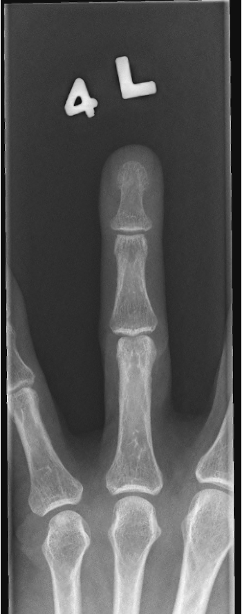
Is this image acceptable? If not, why?
YES!
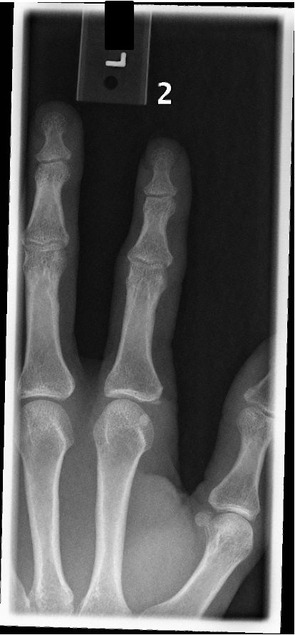
Is this image acceptable? If not, why?
Rotation
DIP and PIP are closed due to flexion
Insufficient collimation (entire 3rd digit is visualized)
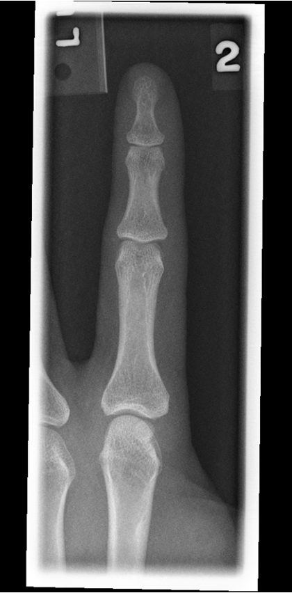
Is this image acceptable? If not, why?
Acceptable
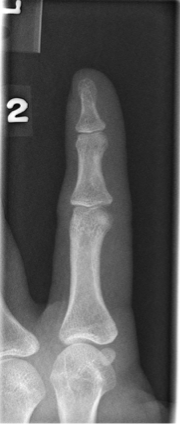
Is this image acceptable? If not, why?
No, Rotation
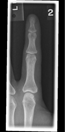
Is this image acceptable? If not, why?
Yes, acceptable
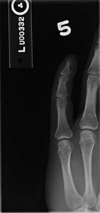
Is this image acceptable? If not, why?
DIP and PIP are closed due to flexion
Digit not centered to central ray (CR)
Insufficient collimation from side-to-side
For an Oblique PA, what would be the criteria for an acceptable image?
Evidence of proper collimation and presence of side marker placed clear of anatomy of interest
Entire digit, including the distal portion of the adjoining metacarpal
Digit rotated at a 45-degrees, demonstrated by the concavity of the elevated side of the phalangeal bodies
No superimposition of the proximal phalanx or MCP joint by adjacent digits
Open interphalangeal (IP) an MCP joint spaces
Bony trabecular detail and surrounding soft tissues