procedures for chest projection
 Call Kai
 Learn
Learn Practice Test
 Spaced Repetition
 Match
Match Flashcards
 Knowt Play
1/60

There's no tags or description
Looks like no tags are added yet.
Name | Mastery | Learn | Test | Matching | Spaced | Call with Kai | Chat |
|---|
No analytics yet
Send a link to your students to track their progress
61 Terms
projection is
the path of center ray from the x-ray tube to the image receptor
AP projection
when the CR enters the anterior surface and exits the posterior surface

PA projection
when the CR enters the posterior surface and exits the anterior surface
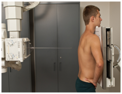
lateral projection
the patient or part of the radiographed is on the side, rotated a full 90 degrees from AP or PA
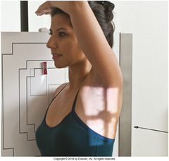
oblique projection
the patient or the part to be radiographed is rotated some degree between 0 and 90 degrees AP or PA

axial projection
there is a longitudinal angulation of the CR 10 degrees or more along the long axis of the body or the body part
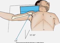
tangential projection
the CR skims between body parts to profile a bony structure and project it free of superimposition

view
describes the representation of an image as seen from the image receptor (opposite of projection)
method
is a radiologic procedure named after an individual
being upright, arms down, palms forward, and head and feet facing straight ahead is
anatomical position
recumbent position
lying down
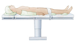
supine (dorsal) position
lying on the back
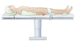
prone (ventral) position
lying face down
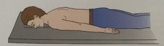
supine position with the head tilted lower is
trendelenburg
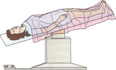
supine position with the head tilted higher is
fowler's
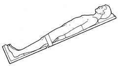
supine with the knees and hips flexed, thighs abducted and rotated laterally
lithotomy

recumbent oblique with the patient lying on the left side with the right knee and thigh flex is the
sim's (recovery) position
right lateral recumbent position
the patient is laying on their right side
left lateral recumbent position
the patient is laying on their left side
for right and left lateral position how is it named
it is named according to the side of the body closest to the image receptor
in an RAO (right anterior position) what surface of the body is closest to the image receptor
right anterior
in an LPO (left posterior) what surface of the body is closest to the image receptor
left posterior
all decubitus positions have what direction of central ray?
directed horizontally down
if the patient is in a dorsal decubitus position (lying on the back), what position is the patient lying in with the CR directed horizontally?
supine position or dorsal recumbent
if the patient is in a ventral decubitus position (lying on the stomach), what position is the patient lying in with the CR directed horizontally?
prone position or ventral recumbent
lateral decubitus is when?
the patient is recumbent and lying on the right or left side with the C.R. directly horizontally
abduction
is movement of a part away from the central axis of the body
adduction
is movement of a part towards the central axis of the body
extension
Straightening of a joint (increasing angle)
flexion
bending a joint (decreasing the angle)
eversion
turn the foot outward
inversion
turn the foot inward
supinate
palm up
Pronate
palms down
rotations described as rotation of a part toward the midline of the body
medial/interal
rotations described as rotation of a part away from the midline of the body
lateral/external
Circumduction
circular movement of a limb
to turn away from the regular course is called?
deviation
on a PA projection, where is the C.R. centered at?
T7 (thoracic vertebrae)
why should the patient roll their shoulder forwarded and have palms facing out?
to remove the scapula from the lung field
how many inches should the relaxed shoulders be from the top of the IR?
2 inches
what is one reason why chest radiography taken in an upright position?
1. the diaphragm will be at it's lowest point
what is the second reason why chest radiography taken in an upright position?
2. demonstrates air and fluid levels in the lungs
what is the third reason why chest radiography taken in an upright position?
3. minimizes the blood vessels and heart
why is it important to take chest radiographs in 2nd full inspiration?
this is where the lungs have the most volume; 10 ribs should show up on the x-ray
why should the chest radiographs be taken at 72" SID?
this reduces magnification and increases detail in the image
why is it preferable to do a PA projection of the chest rather than an AP?
this minimizes or decrease the magnification of the heart
in obese patients needing a chest x-ray what is needed to do?
IR needs to be transverse
if a patient has large breast, what is needed to be done to get the chest x-ray?
patient needs to move the breast upward and outward
on a good chest x-ray, how should the clavicle be positioned on a PA position?
the clavicle should be on the same horizontal plane and below the apices
on a good chest x-ray, how should the clavicle be positioned on a PA position?
manubrium should be superimposed on the T4
how should the patient's neck be positioned?
extended to ensure that the chin and neck are not superimposing on the upper lung region
when should exposure be taken for chest x-ray
2nd full inspiration
on a left lateral projection of the chest, what level should the C.R. be at?
T7 and 2 inches anterior to the mid-coronal plane
the shoulders, posterior ribs, and posterior pelvic wing should be ____ to ensure the patient is not leaning forward or backwards
perpendicular
to identify rotation on a lateral position one should for?
gastric air bubble and left hemidiaphragm
how to tell the left lateral chest x-ray was done correctly:
1. posterior ribs are superimposed
2. sternum is in profile and intervertebral foramina of the thoracic vertebrae are open
3. arms are removed from the field
situation: A PA chest radiograph reveals only 8 ribs seen above the diaphragm
solution: repeat the exposure on the 2nd full inspiration
situation: A chest radiograph reveals that the pendulous breasts of the patient are obscuring the base of the lungs
solution: have the patient left their breast up and out
An ambulatory patient comes to radiology with a clinical history of possible pneumonia. The patient complains of pain in the center of her chest. What positioning routine should be performed on this patient?
upright PA and left lateral
in lateral position, how would the midsagittal plane be to the IR?
parallel with shoulder