Looks like no one added any tags here yet for you.
diagnose; heart
Purpose of ECG Testing:
An ECG is often used (often in conjunction with other tests) to help _____ and monitor conditions affecting the _____
chest; palpitations; shortness
Purpose of ECG Testing:
It can be used to investigate symptoms of possible heart problems such as _____ pain, _____, dizziness, and _____ of breath
Arrhythmias; ischemia
Purpose of ECG Testing:
ECG can help detect
- _______
- Coronary Heart Disease
- Heart _____ (heart attacks)
- Cardiomyopathy
action potentials
ECG is an accumulation of _____
12; 2; 3; 6
A _____-lead ECG is most often used in the health field, however a ___-lead, ___-lead, ____-lead and many other variations can be used.
conductive pad; electrical
An ECG electrode is simply a ______ that is attached to the skin to record changes in _____ activity
electrical potential difference
Any pair of electrodes can measure the _____ between the two corresponding locations of attachment
bipolar lead
an ECG lead that consists of two surface electrodes of opposite polarity (one positive and one negative)
unipolar lead
an ECG lead that consists of one positive surface electrode and a reference point
electrical
Each lead gives an opportunity to look at the heart from a different ______ position
10
For the common 12-lead ECG, ______ electrodes are positioned on the body
positive
depolarization of the heart towards the positive electrode produces a ______ deflection
negative
depolarization of the heart away from the positive electrode produces a _____ deflection
towards
repolarization of the heart ______ the positive electrode produces a negative deflection
away
repolarization of the heart _____ from the positive electrode produces a positive deflection
0.04
Reading amplitude and time on ECG:
On the x-axis, the small box is _____ seconds
right arm to left arm
Lead I
right arm to left leg
Lead 2
left arm to left leg
Lead 3
left arm
the _____ lead can be positive or negative
right arm
the _____ is negative
left leg
the _____ is positive
true
T/F: electrodes go from negative to positive
II, III, aVF
Which leads look at the inferior wall of the heart?
V1, V2
Which leads look at the septum of the heart?
V3, V4
Which leads look at the anterior wall of the heart?
I, aVL, V5, V6
Which leads look at the lateral wall of the heart?
I, aVL, V5, V6
Which leads align with the left circumflex artery (Circ)?
II, III, aVF
Which leads align with the right coronary artery (RCA)?
V1, V2, V3, V4
Which leads align with left anterior descending artery (LAD)?
aVR
____ doesn't line up with anything
infraclavicular; medial; below
Electrode Placement for 12-Lead ECG:
Right arm
Right _____ fossae, _____ to the deltoid muscle, roughly 2cm ____ border of clavicle
fossae; deltoid; clavicle
Electrode Placement for 12-Lead ECG:
Left arm
Left infraclavicular ____, medial to the _____ muscle, roughly 2cm below border of _____
anterior axillary; costal
Electrode Placement for 12-Lead ECG:
Left Leg
In line with left _____ line, halfway between ____ margin and iliac crest
halfway; iliac crest
Electrode Placement for 12-Lead ECG:
Right Leg
In line with right anterior axillary line _____ between costal margin and ______
4th; right
Electrode Placement for 12-Lead ECG:
V1
_____ intercostal space ____ sternal edge
4th; left
Electrode Placement for 12-Lead ECG:
V2
_____ intercostal space ____ sternal edge
V2; V4
Electrode Placement for 12-Lead ECG:
V3
Midway between ____ and _____
5th; clavicular
Electrode Placement for 12-Lead ECG:
V4
_____ Intercostal space, mid-_____ line
Anterior axillary; V4
Electrode Placement for 12-Lead ECG:
V5
______ line in straight line with ____
Mid; V4; V5
Electrode Placement for 12-Lead ECG:
V6
_____-axillary line in straight light with _____ and _____
V4; V5; V6
Electrode Placement for 12-Lead ECG:
_____, ____ and ____ may need to be altered in female patients due to breast tissue, place electrode as close as possible to anatomical location directly under breast)
inferior; rightward
Electrode Placement for 12-Lead ECG:
Because of the altered location of the limb electrodes, Q waves in the _____ leads may be mased and there is mild _____ shift in the QRS axis.
Locate; hair; clean; water; abrasive; Explain; before; center; air
ECG Electrode Placement Protocol:
- _____ anatomical areas that will be used.
- Prepare skin by removing any excessive _____
- If possible _____ each site thoroughly with soap and ____ (preferable to alcohol wipe; alcohol dries the skin and can diminish electrical flow)
- Use ECG skin prep pad, paper or _____ tape (removes the stratum corneum to allow better electrical signals and scratches stratum granulosum to reduce motion artifact)
- ______ the electrode application procedure to the patient to decrease anxiety
- When only one patient is present, attach the lead wire to the electrode _____ placement
- Apply the electrode by pressing around the entire edge of the electrode _____ since it spreads the gel out and may create _____ pockets that contribute noise.
atrial depolarization
P wave represents ______
P-wave; Q-wave; atrial depolarization; ventricular activation
PR interval is actually the PQ interval. Measured from the start of the _____ to the beginning of the _____. So PR is actually a misnomer. Measures from the start of _____ to start of ______.
end; QRS; atrial; ventricular
PR segment is from the ____ of the P wave to the beginning of the _____ segment. It represents the the time delay between _____ and _____ activation.
ventricular depolarization; larger
QRS complex represents ______. Much _____ than the P wave because of the amount of muscle mass in the ventricle.
absolute refractory period; onset
ST segment represents the _______ (plateau of the action potential) of the ventricular contractile cells. Measures from J point to the _____ of T-wave. (Elevation or depression of ST is based off of PR segment)
ventricular repolarization
T Wave represents ______
depolarization; repolarization; arrhythmias
QT Interval represents the complete _____ and _____ on the ventricular tissue. A prolonged repolarization can lead to life-threatening ______.
upright; Q; 60-100
Sinus Rhythm:
- The P wave is _____ in leads I and II
- Each P wave is followed by a _____
- The heart rate is _____ beats/min
Tachyarrhythmia
______ is defined as an abnormal rhythm with a ventricular heart rate over 100bpm
Supraventricular
______ Tachycardia: Arrhythmia originating from above the AV node
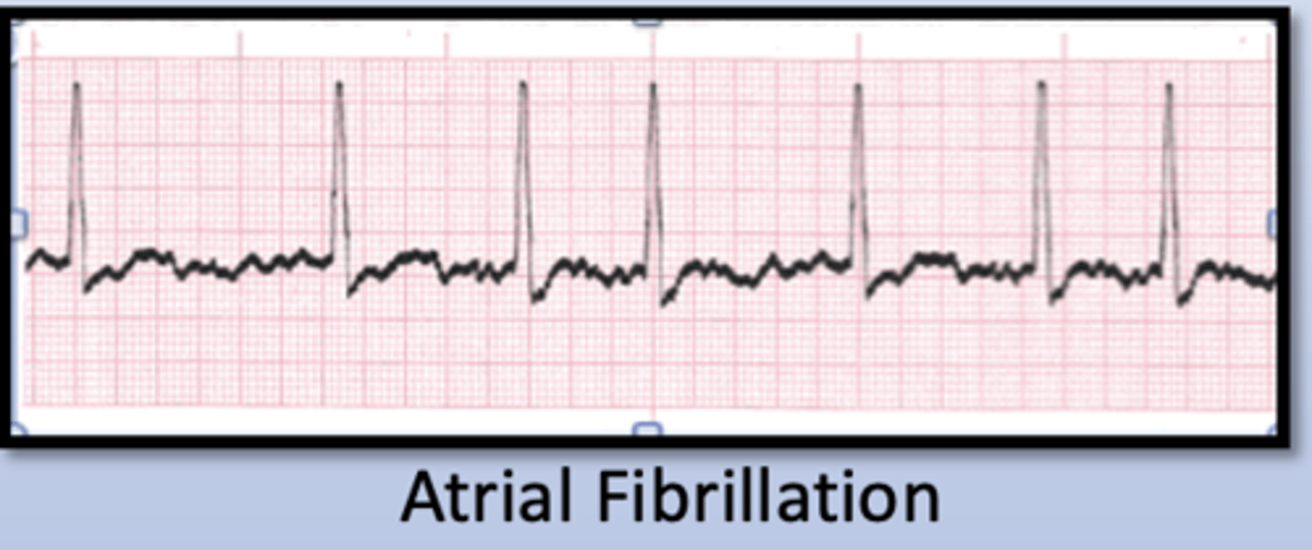
- Atrial Fibrillation
- Atrial Flutter
- Atrial tachycardia
Ventricular
______ Tachycardia
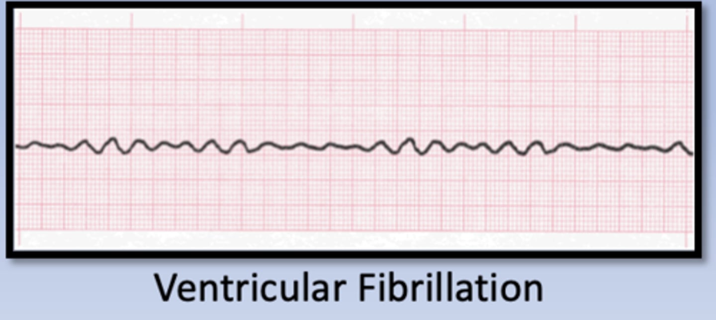
- ventricular fibrillation
- ventricular tachycardia
Bradyarrhythmia
_____ is defined as a heart rate below 60 beats and comprises multiple disorders
- Sinus bradycardia
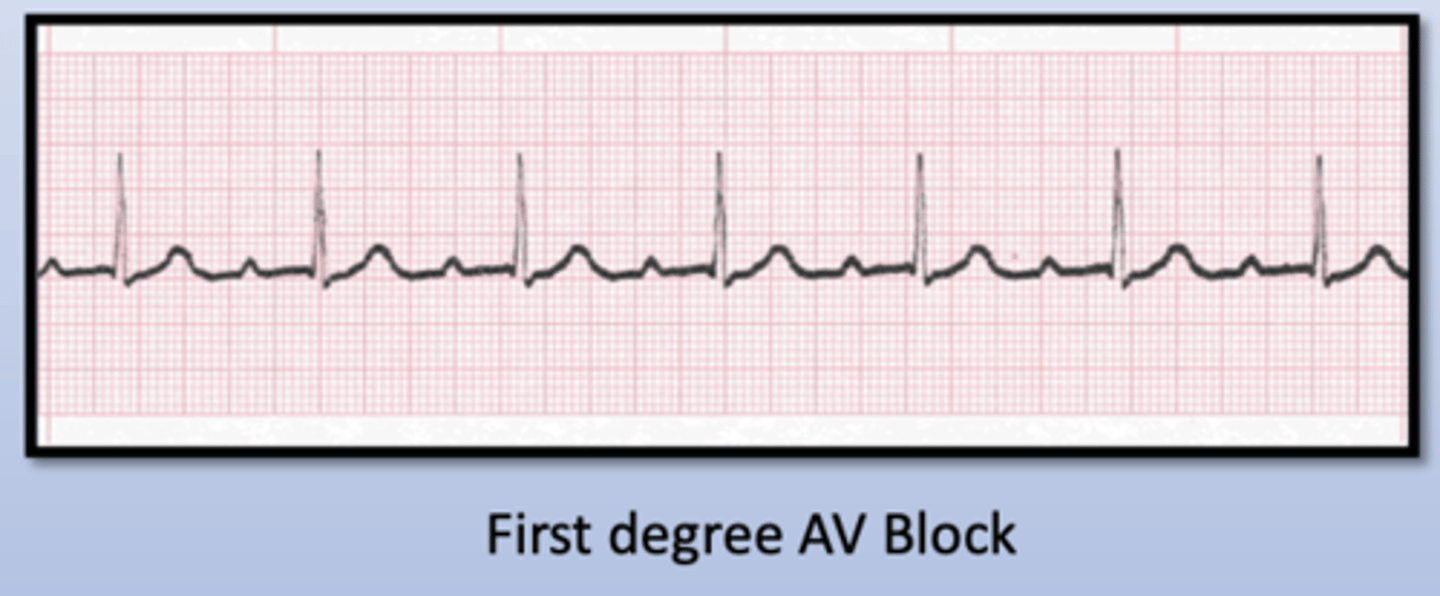
- First degree AV Block
- Second Degree AV Block
- Third Degree AV Block
- SA Node Exit Block
isoelectric
Normal ST segment is flat and ______.
height; baseline
ST segment deviation (elevation or depression) is measured as the _____ difference between the J point and the _____ (PR segment).
5
ST segment depression _____ mm or more is considered pathological.
Upsloping
_______ ST segment depression naturally occurs during exercise and should be considered normal
ischemia
Downsloping or horizontal ST segment depression is typical of _____.
elevation
ST segment _____ can be indication of ischemia as well