Spirochetes
 Call Kai
 Learn
Learn Practice Test
 Spaced Repetition
 Match
Match Flashcards
 Knowt Play
1/101

There's no tags or description
Looks like no tags are added yet.
Name | Mastery | Learn | Test | Matching | Spaced | Call with Kai | Chat |
|---|
No analytics yet
Send a link to your students to track their progress
102 Terms
Characteristics of spirochetes
Slender, helically coiled bacteria; motile by endoflagella; stain poorly with Gram stain; inactive biochemically; unstable in environment
How are spirochetes best visualized?
Dark-field microscopy, phase-contrast microscopy, or fluorescence microscopy
Three main genera of spirochetes
Leptospira, Brachyspira, Borrelia
Shape of Leptospira
Slender, coiled spirochetes with hook-shaped ends

Oxygen requirement of Leptospira
Obligate aerobes
How are Leptospira shed?
In urine of infected animals
Where do Leptospira survive in the environment?
Ponds, surface water, and moist soil
How are Leptospira species differentiated?
DNA homology
What is a serovar?
A serologically distinct variant within a species
Example Leptospira serovars
L. interrogans serovar canicola; L. interrogans serovar icterohemorrhagiae
Maintenance hosts of Leptospira
Rodents and small mammals
Leptospira infection in maintenance hosts
Usually subclinical with persistent urinary shedding
Incidental hosts of Leptospira
Develop disease ranging from mild to severe
Main routes of Leptospira transmission
Ingestion of contaminated water; entry through skin or mucosal abrasions
Leptospira serovar associated with cattle
L. borgpetersenii serovar hardjo and L. interrogans serovar hardjo
Leptospira serovar associated with pigs
L. interrogans serovar pomona
Leptospira serovar associated with dogs
L. interrogans serovar canicola
Major reproductive effects of leptospirosis in cattle and sheep
Abortions, stillbirths, agalactia
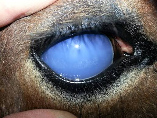
Major reproductive effect of leptospirosis in horses
Abortions and periodic ophthalmia
Major reproductive effect of leptospirosis in pigs
Abortions, reproductive failure, stillbirths
Clinical signs of leptospirosis in dogs
Renal disease in puppies; chronic disease in adults
younger animals are more severely affected
Why are many ingested Leptospira destroyed?
They are destroyed by gastric acid
How do Leptospira spread after entering the body?
Hematogenous spread
Main organs affected after Leptospira dissemination
Multiple organs, especially kidneys, CNS, and reproductive tract
Kidney lesion caused by Leptospira
Interstitial nephritis
Cause of renal damage in leptospirosis
Immune complex-mediated renal damage
Some Leptospira serovars produce what toxin?
Potent hemolysins
Clinical effects of Leptospira hemolysins
Anemia, icterus, hemoglobinuria
Vascular lesion associated with leptospirosis
Vasculitis causing hemorrhage
Reproductive consequences of Leptospira infection
Fetal death, resorption, abortion, birth of weak offspring
Type of immunity against Leptospira
Antibody-mediated immunity
Common eye finding in canine leptospirosis
Icterus
(Jaundice)

Major pulmonary lesion in canine leptospirosis
Hemorrhage
Liver lesion in canine leptospirosis
Swollen necrotic liver with acute hepatic hemorrhage
Kidney lesions in canine leptospirosis
Hemoglobinuric nephrosis and shrunken fibrotic kidneys
Kidney lesion in bovine leptospirosis
Dark swollen kidneys
Kidney lesion in equine leptospirosis
Interstitial nephritis
(a kidney disorder where the spaces between the tiny tubes (tubules) inside the kidneys become swollen and inflamed)
Direct diagnostic test for leptospirosis in kidneys
Direct fluorescent antibody stain
a lab technique that uses special antibodies attached to glowing dyes (fluorophores) to find and see specific targets like proteins or germs inside cells or tissue samples under a special microscope
Gold-standard serologic test for leptospirosis
Microscopic agglutination test (MAT)
What antigen does MAT use?
Live leptospires
MAT detects what?
Serovar-specific antibodies
Other serologic tests for leptospirosis
ELISA and CFT
Vaccines available for leptospirosis (what species)
Canine and bovine leptospiral vaccines
Human disease caused by Leptospira
Leptospirosis (Weil's disease, Fort Bragg fever, Swineherd's disease)
Human transmission of leptospirosis
Exposure to contaminated water or infected animals
Major clinical signs of human leptospirosis
Fever, anemia, jaundice, meningitis, nephritis
Mortality rate of human leptospirosis
Approximately 10-15%
Largest genus of spirochetes
Borrelia
Oxygen requirement of Borrelia
Microaerophilic
Generation time of Borrelia
≥18 hours
Borrelia are transmitted by what vectors?
Ticks
Borrelia burgdorferi causes what disease?
Lyme disease
Reservoirs of Borrelia burgdorferi
Mice, deer, and ticks
Major organs affected by Lyme disease
Joints, brain, eyes, heart
Mechanism contributing to Lyme arthritis
Immune complex formation
Clinical signs of Lyme disease in dogs
Fever, arthritis, renal, cardiac, and CNS complications
Clinical signs of Lyme disease in horses and cattle
Arthritis, encephalitis, nephritis, hepatitis
Which stage of Ixodes tick most commonly transmits Lyme disease?
Nymph
How long must a tick remain attached to transmit Lyme disease?
At least 36-48 hours
Characteristic skin lesion of Lyme disease in humans
Bull's-eye rash (erythema migrans)

Other human Lyme disease signs
Arthritis, headache, lymphadenopathy; cardiac and neurologic complications
When does IgM appear after Lyme infection?
About 2 weeks post-infection
When does IgG appear after Lyme infection?
≥30 days post-infection
Tests used to diagnose Lyme disease
IFA, ELISA, confirmed by Western blot
Borrelia coriaceae causes what disease?
Epizootic bovine abortion
(An epizootic is a disease outbreak that quickly affects a large number of animals of the same kind in the same place at the same time)
Vector of Borrelia coriaceae
Tick
Ornithodoros coriaceus
Clinical findings of Borrelia coriaceae infection
Asymptomatic cows, late-term abortion, weak calves
Oxygen requirement of Brachyspira
Anaerobic
Habitat of Brachyspira
Intestines of swine

Brachyspira hyodysenteriae causes what disease?
Swine dysentery (bloody scours)
An intestinal infection that causes severe diarrhea containing blood
Reservoirs of Brachyspira hyodysenteriae
Rodents and carrier swine
Transmission of swine dysentery
Fecal-oral transmission
Major virulence factors of Brachyspira hyodysenteriae
β-hemolysin and cytolysin
Age group most affected by swine dysentery
Weaner and grower pigs
Main site of swine dysentery lesions
Large intestine and rectum
Characteristic lesion of swine dysentery
Mucohemorrhagic enteritis
Chronic lesion of swine dysentery
Diphtheritic inflammation of the colon, cecum, and rectum
Bartonella henselae characteristics
Short Gram-negative aerobic rods; fastidious; slow-growing
Natural reservoir of Bartonella henselae
Healthy cats (oropharynx)
Does Bartonella henselae usually cause disease in cats?
No
Human disease caused by Bartonella henselae
Cat scratch disease

Cat-to-cat transmission of Bartonella henselae is through what
By *Ctenocephalides felis* (cat flea)
Is there direct cat-to-cat transmission of Bartonella henselae?
No
Human transmission of cat scratch disease
Cat bites, scratches, contact with flea feces, or cat saliva
Incubation period of cat scratch disease
1-3 weeks
Clinical signs of cat scratch disease
Papular lesions and chronic lymphadenitis
Is cat scratch disease usually self-limiting?
Yes (resolves on its own without needing specific medical treatment)
Diagnosis of cat scratch disease
History, clinical signs, serology, PCR
Treatment for cat scratch disease
Tetracyclines
Characteristics of Coxiella burnetii
Gram-negative, rickettsia-like, obligate intracellular bacterium producing an endospore-like structure
Environmental resistance of Coxiella burnetii
Resistant to heat, sunlight, and many chemical disinfectants
Human disease caused by Coxiella burnetii
Q fever

Main reservoirs of Coxiella burnetii for humans
Cattle, sheep, and goats
Routes of Coxiella burnetii transmission
Arthropods, inhalation, ingestion
Infection in nonpregnant ruminants
Usually inapparent; organisms shed in urine, feces, milk, and other body fluids
Infection in pregnant ruminants
Massive shedding in placental discharges; sporadic abortions in sheep and goats (rarely cattle)
Main human sources of Q fever
Inhalation of organisms and unpasteurized milk
Pathogenesis of Q fever
Replication in vascular endothelium, respiratory epithelium, serosal epithelium, and renal epithelium causing widespread vasculitis, infarction, and diffuse granulomas
Clinical signs of Q fever in humans
Fever, chills, headache, interstitial pneumonia, endocarditis, hepatitis
Diagnosis of Q fever
ELISA and IFA for C. burnetii antibodies