S1.6 Muscular System
1/10
Earn XP
Description and Tags
The ATTI exam is not going to dive deep into the name of the varies muscles. Instead, it focuses on the muscle tissues. And the mechanism of the muscle contraction, especially the
Name | Mastery | Learn | Test | Matching | Spaced | Call with Kai |
|---|
No analytics yet
Send a link to your students to track their progress
11 Terms
The 3 types of muscle tissues?
Which ones of them is/are Involuntary/Voluntary?
Cardiac Muscle (Involuntary)
Smooth Muscle (Involuntary)
Skeletal Muscle (Voluntary)
Cardiac Muscle
Describe the muscle type.
Which Nervous System control the Cardiac Muscle?
(Involuntary)
Specialized, Organized type of tissue that only exists in the heart.
Striations:
Controlled by Autonomic Nervous System
aka No Conscious control

Smooth Muscle
Describe the muscle type.
Which Nervous System control the Smooth Muscle?
Where can you find them? (4)
(Involuntary)
Narrow, Spindle shaped cells with a single centrally located nucleus.
Non-Striated = In anatomy, "non-striated" refers to muscle tissue that lacks the characteristic striped appearance (striations) seen under a microscope.
Controlled by Autonomic Nervous System
aka No Conscious control
Can be found in
Digestive system
Arteries and Veins
Bladder
Eyes

Skeletal Muscle
Describe the muscle type.
What can be found in the human body?
(Voluntary)
Highly organized tissues that attach to bones or skin to produce movements.
Straintions Mutiple nuclei per fiber
Voluntary control:
By the somatic nervous system
Conscious control of movement
Found in:
Tongue
Diaphram
Upper Esophagus
What is Striations in Cardiac Muscle?
How many Nucleus Per Fiber in Cardiac Muscle?
What is a Intercalated Disc?
Definition:
1. Striations in Cardiac Muscle: Striations are the light and dark bands observed in cardiac muscle tissue under a microscope. They are caused by the regular arrangement of contractile proteins (actin and myosin) into functional units called sarcomeres.
Nucleus Per Fiber in Cardiac Muscle: Cardiac muscle fibers (cardiomyocytes) are typically uninucleated (containing one nucleus per cell), though some can have two nuclei.
Intercalated Disc: Intercalated discs are specialized cell junctions primarily found in cardiac muscle. They connect individual cardiac muscle cells, allowing for rapid electrical communication (via gap junctions) and strong mechanical adhesion (via desmosomes), enabling synchronized contraction of the heart.
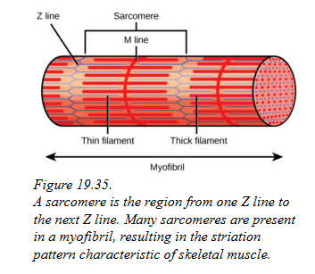
What is a Sarcomere?
A sarcomere is the basic contractile unit of muscle fibers. It is the smallest functional unit within a muscle that is responsible for muscle contraction.
When a muscle contracts, the sarcomeres shorten as the actin filaments slide over the myosin filaments (called the sliding filament theory).
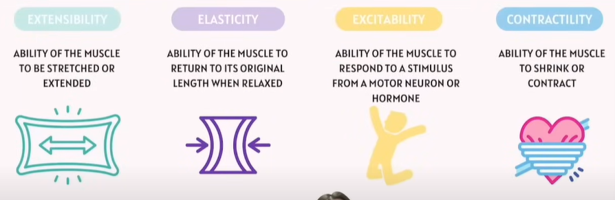
All muscular tissue shared similar characteristics
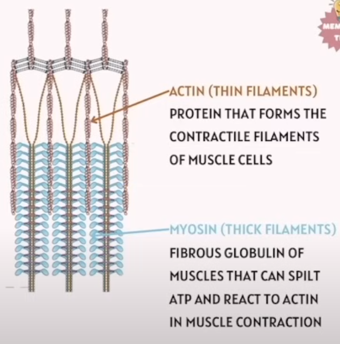
What is Actin, Myosin, and Sarcomeres
Which one has thin and which has thick filament?
Actin: (Thin filaments)
Protein that forms the contractile filaments of muscle cells.
Actin = Thin
Myosin: (Thick Filaments)
Fibrous globulin of muscles that can split ATP and React to Actin in muscle contraction.
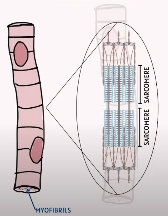
What is the relationship between Myofibrils & Sarcomeres
Term | Definition | Relationship |
|---|---|---|
Myofibrils | Long, cylindrical organelles are found inside muscle fibers (cells). They are made up of repeating units called sarcomeres. | Myofibrils are like strings of sarcomeres lined up end-to-end along the length of the muscle cell. They run parallel throughout the cell and are responsible for contraction. |
Sarcomeres | The smallest functional contractile units within myofibrils. Each sarcomere extends from one Z-line to the next. | Sarcomeres are the building blocks of myofibrils When all sarcomeres in a myofibril shorten, the entire muscle fiber contracts. |
🔑 In simple words:
Myofibril = a long chain of sarcomeres.
Sarcomere = unit within a myofibril responsible for muscle contraction.
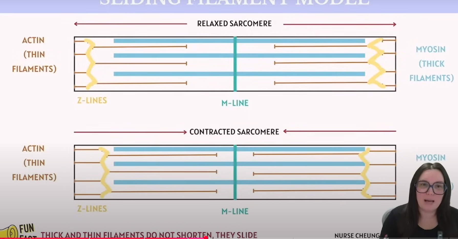
How does the sliding filament model work?
Where do the actin and myosin connect to?
Actin connects with the Z line
Myosin connects with the M line
Key Note for ATTI:
Thick and Thin filaments do not shorten; they simply slide.
Striations
What does striation mean in regarding to Muscle?
Which muscle type has the striation character?
Striations: 条纹 /straɪˈeɪ.ʃən/
Striations are the striped or banded patterns seen in certain types of muscle tissue under a microscope, caused by the arrangement of muscle fibers.
There are two main contexts where you might encounter striations:
In Skeletal Muscle:
Skeletal muscle fibers have a highly organized structure of actin and myosin proteins, which create repeating units called sarcomeres. These sarcomeres are arranged in such a way that they create visible stripes or striation patterns in the muscle fibers.
These striations are important because they reflect the muscle's ability to contract in a coordinated manner, enabling voluntary movements like walking or lifting.
In Cardiac Muscle:
Cardiac muscle, which makes up the heart, also has striations, similar to skeletal muscle, due to the sarcomere structure. These striations help with the heart’s pumping action.
Why "Striations" Matter:
In skeletal muscles, the striations reflect the precise alignment of muscle fibers, allowing for efficient force production and contraction.