Lab Exam #3 Review
1/39
Earn XP
Description and Tags
BIO 047
Name | Mastery | Learn | Test | Matching | Spaced |
|---|
No study sessions yet.
40 Terms
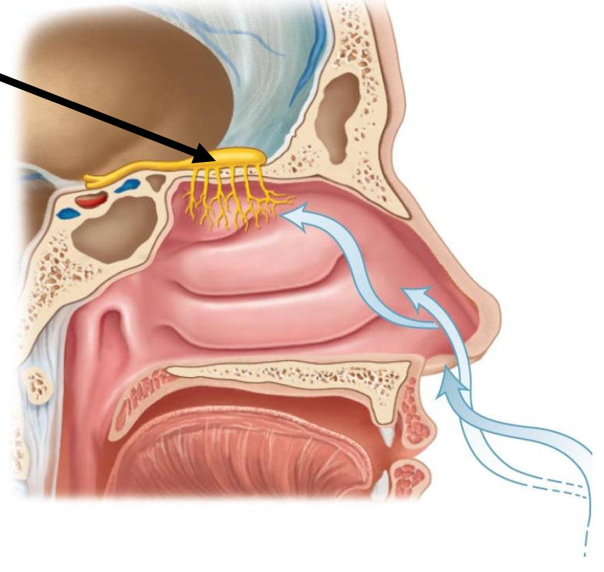
The arrow is pointing to the structure known as
a. Olfactory tract
b. Olfactory bulb
c. Olfactory nerve
d. Olfactory epithelium
b. Olfactory bulb
Where are the receptors for smell located?
a. Olfactory tract
b. Olfactory bulb
c. Olfactory nerve
d. Olfactory epithelium
d. Olfactory epithelium

The black arrow is pointing to
a. Inferior rectus
b. Medial rectus
c. Lateral rectus
D. Superior oblique
c. Lateral rectus
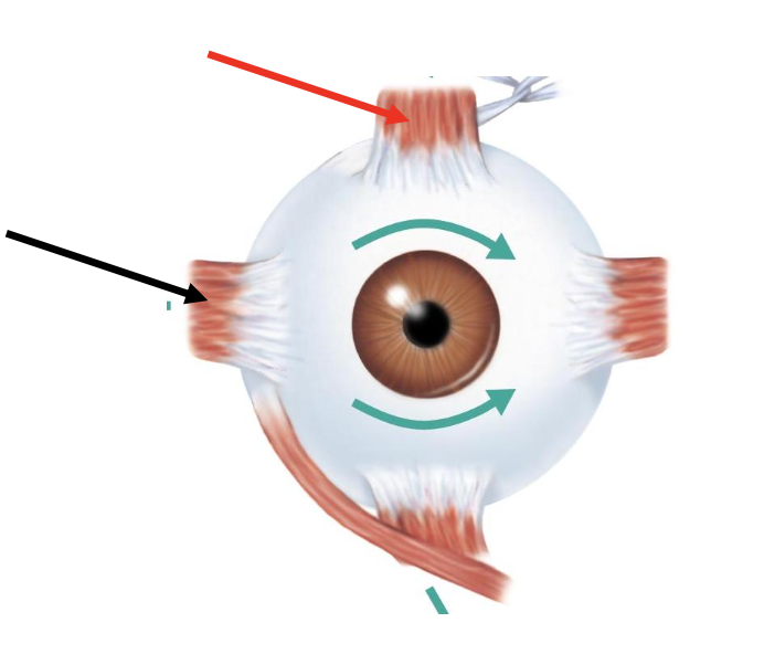
The red arrow is pointing to
a. Inferior oblique
B. Inferior rectus
C. Superior rectus
D. Superior oblique
C. Supeiror rectus
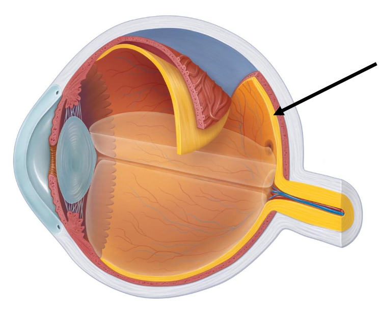
What structure is the arrow pointing to?
a. Sclera
B. Retina
C. Choroid
D. Optic disc
B. Retina
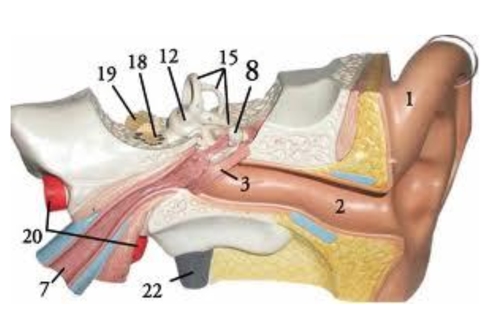
Label #15 on the ear model refers to the _____ that plays the role of _____.
a. Vestibule, hearing
b. Semicircular canals, maintaining static equilibrium
c. Vestibule, maintaining static equilibrium
d. Semicircular canals, maintaining dynamic equilibrium
d. Semicircular canals, maintaining dynamic equilibrium
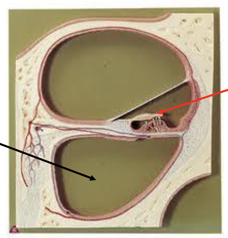
Identify the structure the black arrow is pointing to.
a. Scala media
b. Scala tympani
c. Scala vestibuli
d. Membranous labyrinth
b. Scala tympani
Identify the structure the red arrow is pointing to.
a. Vestibular membrane
b. Basilar membrane
c. Tectorial membrane
d. Modulus
c. Tectorial membrane

What cell is the arrow pointing to?
a. Neutrophil
b. Basophil
C. Lymphocyte
D. Monocyte
b. Basophil
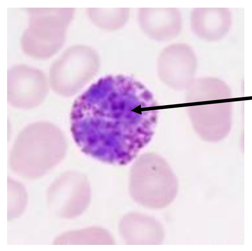
The cell from the photo contains cytoplasmic granules?
a. True
b. False
a. True

What strucuture is labeled #2?
a. Sclera
B. Fovea centralis
C. Optic nerve
D. Optic tract
a. Sclera
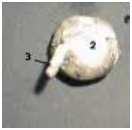
What structure from the photo is labeled #3?
a. Sclera
b. Fovea centralis
C. Optic nerve
D. Optic tract
C. Optic nerve

The black arrow is pointing to
a. Thyroid gland
b. Cricoid cartilage
c. Hyoid bone
d. Thyroid cartilage
c. Hyoid bone
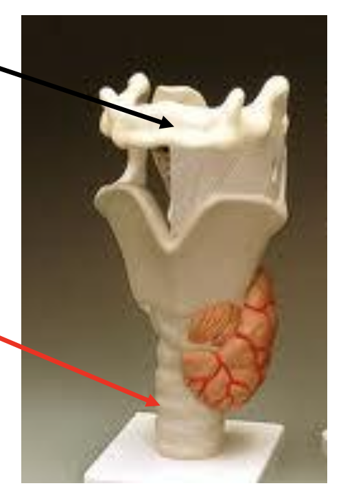
The red arrow is pointing to
a. Laryngeal prominence
b. Thyroid cartilage
c. Tracheal cartilage
d. Epiglottis
c. Tracheal cartilage

The arrow is pointing to
a. Nucleus
b. Mitochondrion
c. T tubule
D. Intercalated disc
D. Intercalated disc
Structure indicated by the arrow on the picture is
a. Ganglion cell
b. Bipolar cell
c. Rod cell
D. Cone cell
D. Cone cell
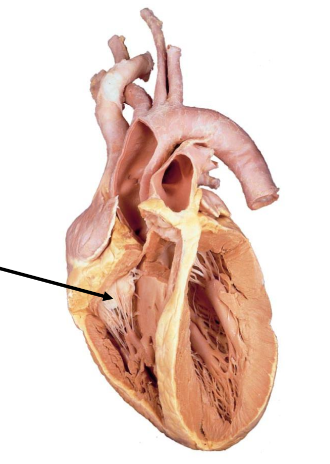
What structure is the arrow pointing to?
a. Papillary muscle
b. Tricuspid valve
c. Auricle
D. Aorta
b. Tricuspid valve
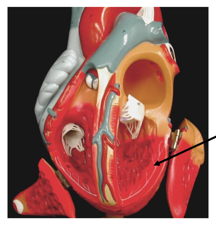
Black arrow is pointing to
a. Coronary artery
b. Cruca
c. Purkinje fibers
d. Chordae tendinae
c. Purkinje fibers
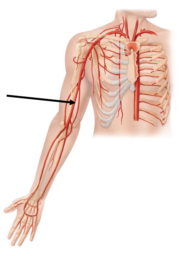
The arrow is pointing to
a. Brachiocephalic trunk
b. Brachial artery
c. Radial artery
d. Ulnar artery
b. Brachial artery
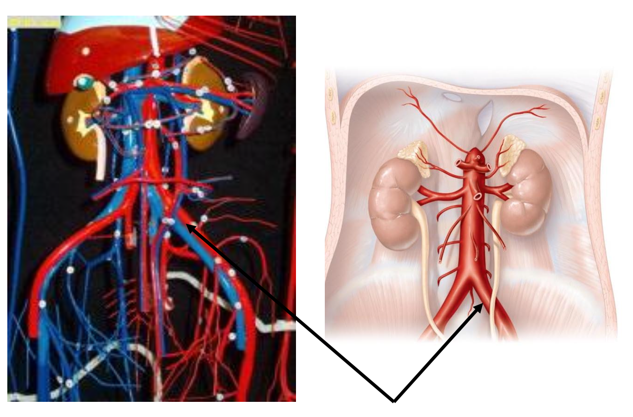
The arrow on the vascular tree is pointing to
a. Gonadal artery
b. Renal artery
c. Superior mesenteric artery
d. Common iliac artery
d. Common iliac artery
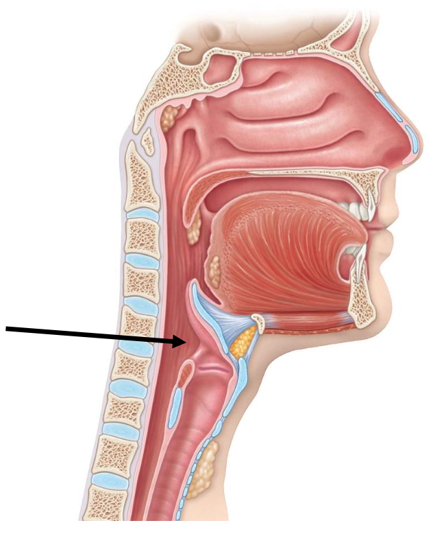
The arrow is pointing to
a. Nasopharynx
b. Oropharynx
c. Larynx
d. Laryngopharynx
d. Laryngopharynx
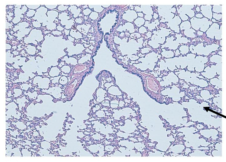
The photo is a micrograph of a part of the lung. The arrow is pointing to
a. Bronchiole
b. Alveolar duct
c. Alveolus
d. Pleura
c. Alveolus
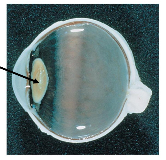
The arrow is pointing to
a. Cornea
b. Conjunctiva
C. Lens
d. Aqueous humor
C. Lens
The lens functions to
a. Maintain a normal intraocular pressure
b. Focus light on retina
c. Hold the lens
D. Allow light to enter into eye
b. Focus light on retina
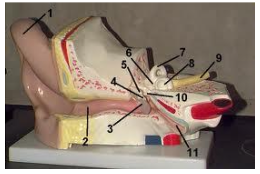
What structure is labeled #11?
a. Pinna
b. External auditory meatus
c. Auditory tube
d. Tympanic membrane
c. Auditory tube
What is the function of the auditory tube?
a. To produce cerumen
b. To hold the auditory ossicles
c. To pass on the sound waves
d. To equalize pressure
d. To equalize pressure
What structure is label #1?
a. Sclera
b. Cornea
c. Lens
d. Iris
b. Cornea
What structure is label #11?
a. Retina
b. Cornea
c. Lens
d. Vitreous humor
c. Lens

The black arrow is pointing to
a. Platelet
b. Monocyte
c. Lymphocyte
d. Neutrophil
c. Lymphocyte
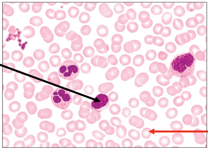
The structure indicated by the red arrow contains
a. Nucleus
b. Nucleus and other organelles
c. Hemoglobin and nucleus
d. Hemoglobin
d. Hemoglobin

Identify the structure (projection) the arrow is pointing to on the picture
a. Tonsil
b. Fungiform papilla
c. Vallate papilla
d. Epiglottis
b. Fungiform papilla
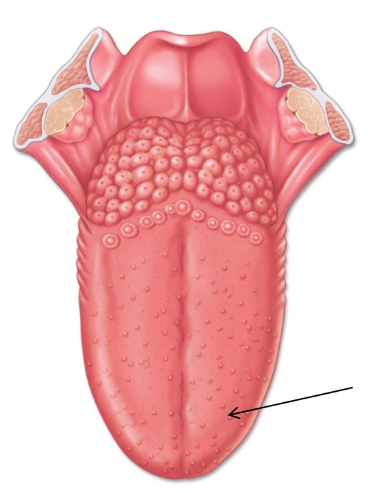
Fungiform papilla contains taste buds found on the apical surface
a. True
b. False
True
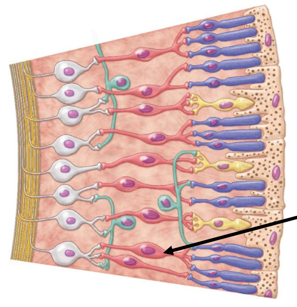
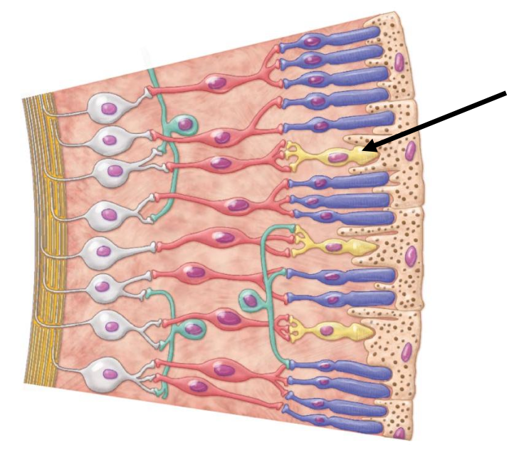
Structure indicated by the arrow on the picture is
a. Ganglion cell
b. Bipolar cell
d. Rod cell
d. Cone cell
b. Bipolar cell
The cone cells are responsible for color vision.
a. True
b. False
a. True
The arrow is pointing to the
a. Aorta
b. Superior vena cava
c. Pulmonary trunk
d. Pulmonary vein
a. Aorta
Blood from the aorta receives blood from the
a. Left atrium
b. Left ventricle
c. Pulmonary arteries
d. Inferior vena cava
b. Left ventricle
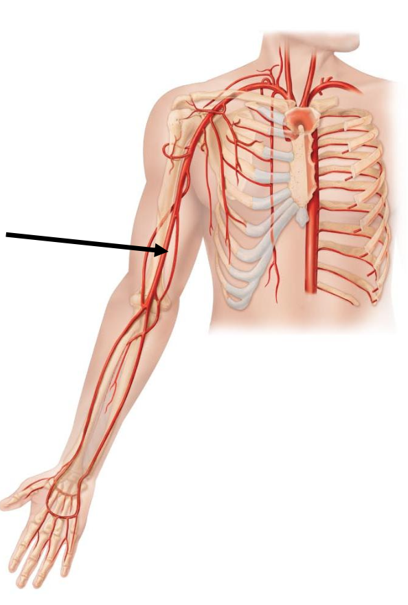
The arrow is pointing to
a. Brachiocephalic trunk
b. Brachial artery
c. Radial artery
d. Ulnar artery
b. Brachial artery
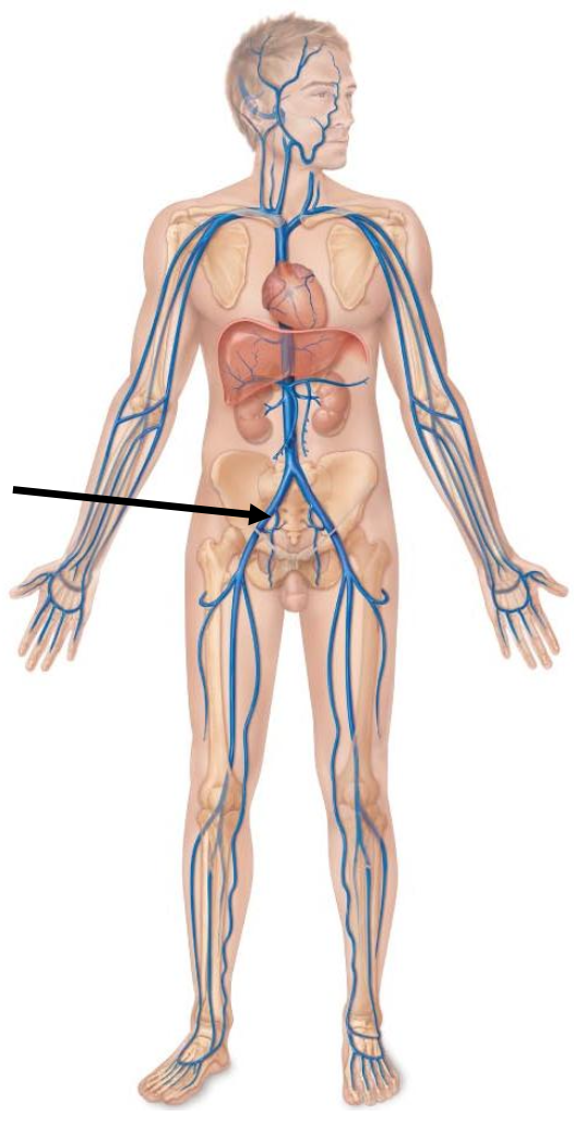
The arrow is pointing to
a. Femoral vein
b. External iliac vein
c. Great saphenous vein
d. Common iliac vein
b. External iliac vein

Which structure harbors the receptors for hearing?
a. #2
b. #6
c. #7
d. #8
d. #8
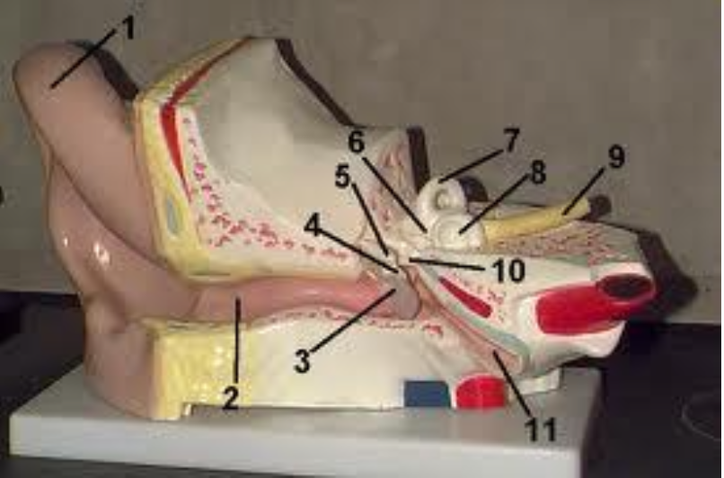
Which structure harbors the receptors for dynamic equilibrium?
a. #2
b. #6
c. #7
d. #8
c. #7