Bontrager Chapter 1- Part 2
1/64
There's no tags or description
Looks like no tags are added yet.
Name | Mastery | Learn | Test | Matching | Spaced | Call with Kai |
|---|
No analytics yet
Send a link to your students to track their progress
65 Terms
What are the exposure factors?
Kilovoltage (kV)
Milliamperage (mA)
Exposure time (ms)
SID
What is kilovoltage?
Controls the energy ( penetrating power) of the X-ray beam.
What is the kilovoltage peak? kVp
The maximum electrical potential used to create the X-ray photons within the tube.
What is milliamperage?
Controls the number of X-rays produced
What is exposure time?
Controls the duration of the exposure.
What are the four image quality factors?
Density/brightness
Contrast/adjacent shades
Resolution/detail
Distortion
What is density?
The amount of blackness on the processed radiograph.
What is the primary controlling factor of film density?
mAs
It controls density by controlling the quantity of X-rays emitted from the tube and the duration of the exposure.
What is SID?
Source to image receptor distance
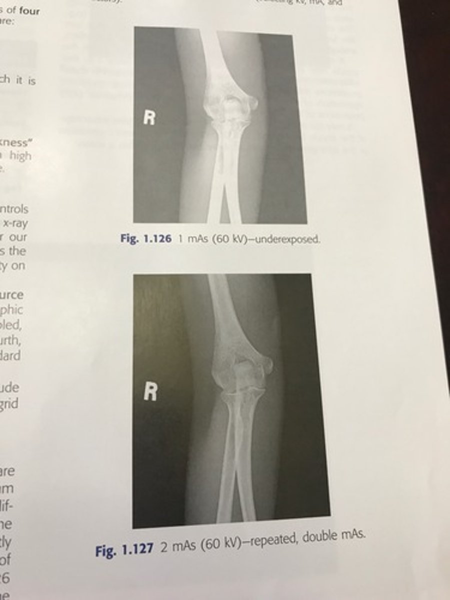
What is the general rule for under or over exposed repeat images?
Minimum of 25-30% change in mAs is required to make a visible difference.
* sometimes even doubling mAs is necessary
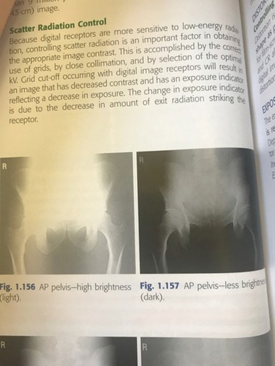
Examples of over/under exposed
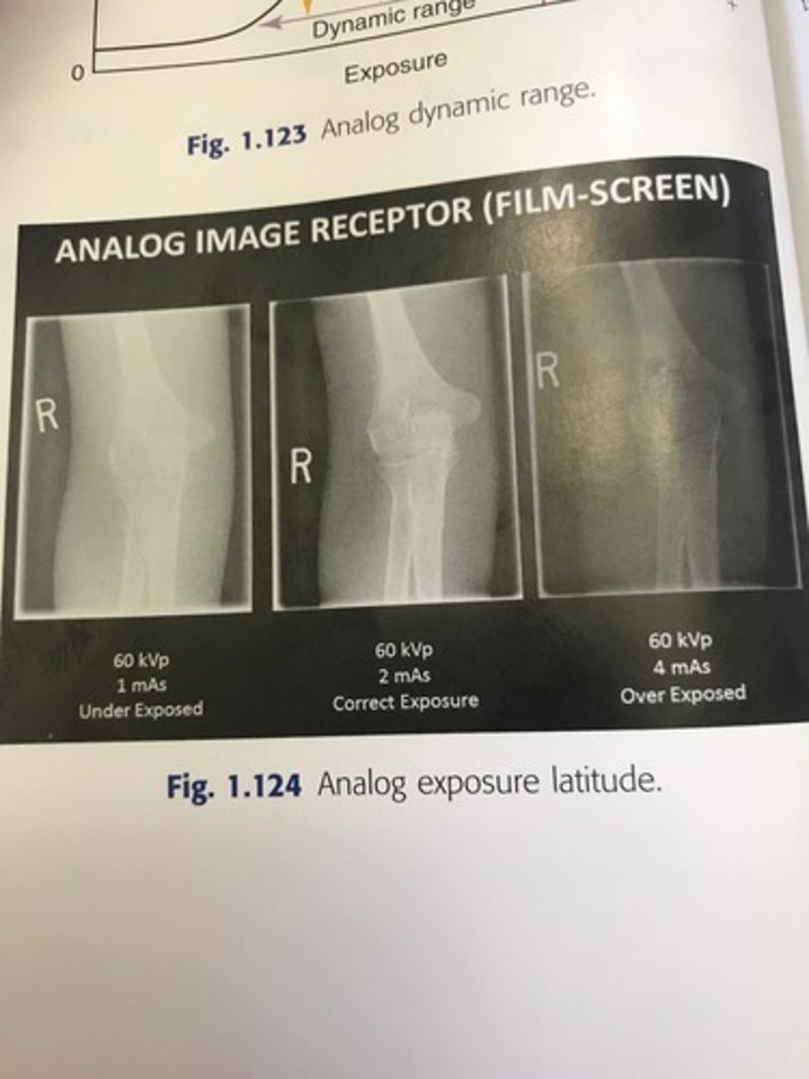
Example of over/under exposed
What is the anode heel effect?
The intensity of radiation emitted from the cathode end of the tube is greater than that emitted at the anode end.
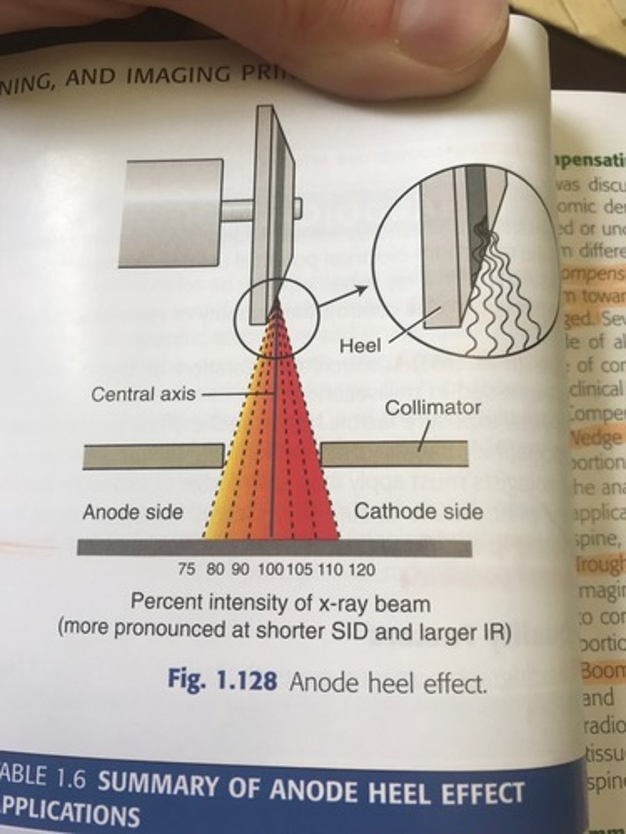
How should you position the patient using the anode heel effect?
The thicker portion of the part should be positioned at the cathode, thinner part under anode.
anode heel effect image

What is a compensating filter?
It filters out a portion of the primary beam toward the thin or less dense part of the body that is being imaged.
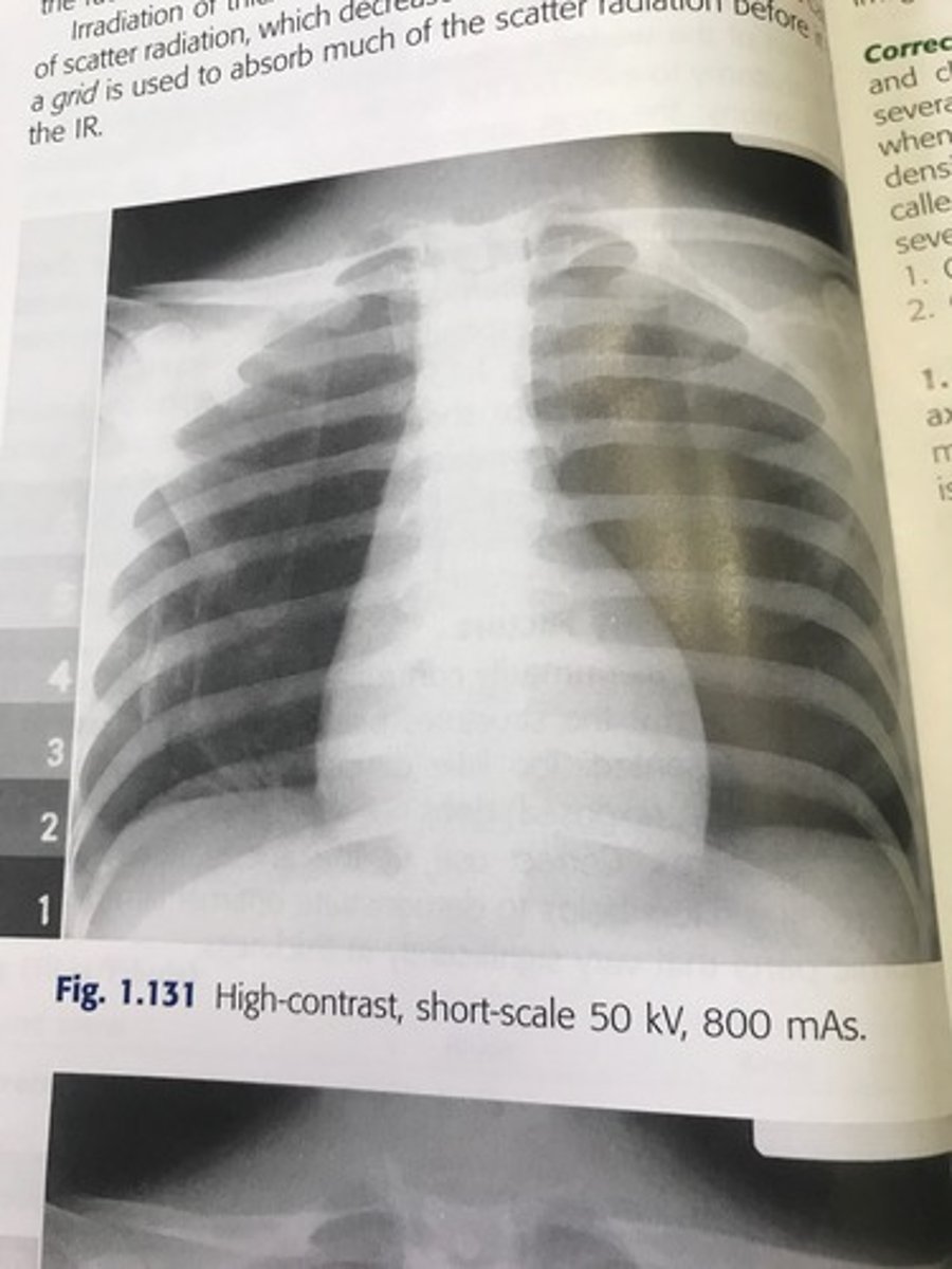
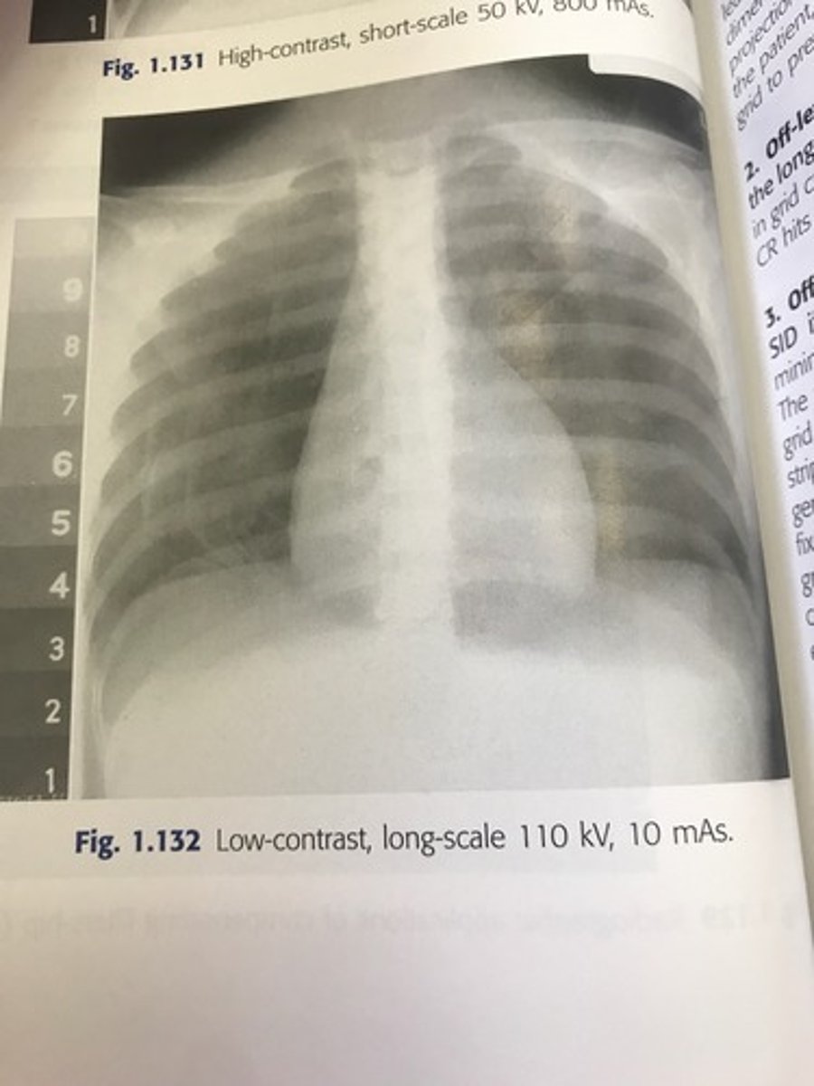
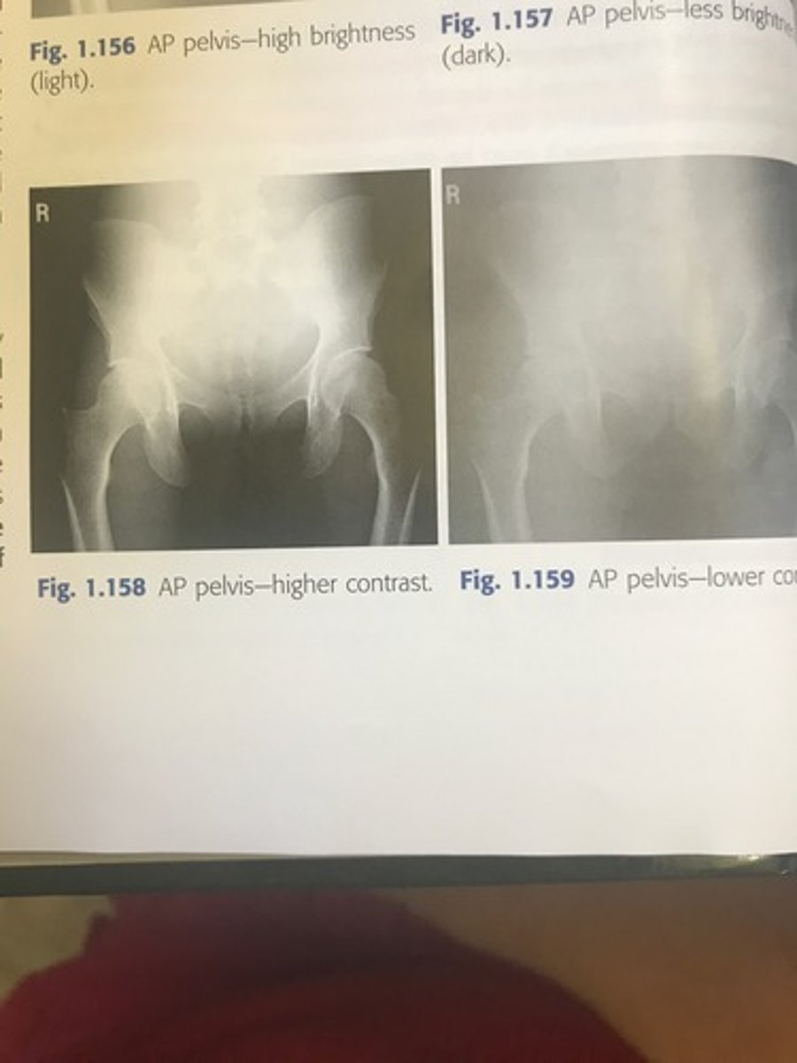
What is radiographic contrast?
Differences in density level between adjacent structures of a radiographic image. Controlling factor is kVp
When the density difference is large the contrast is what?
High- short scale
When the density difference is small the contrast is what?
Low-long scale
Why is radiographic contrast important?
It allows you to see anatomic detail. It allows you to visualize a quality image.
When is it recommend that you use a grid?
On any body part that is thicker than 10 cm. Grid absorbs scatter radiation
What is spatial resolution?
Recorded sharpness of structures on the image.
Spatial resolution is controlled by what?
Geometric factors
Film screen system
Motion
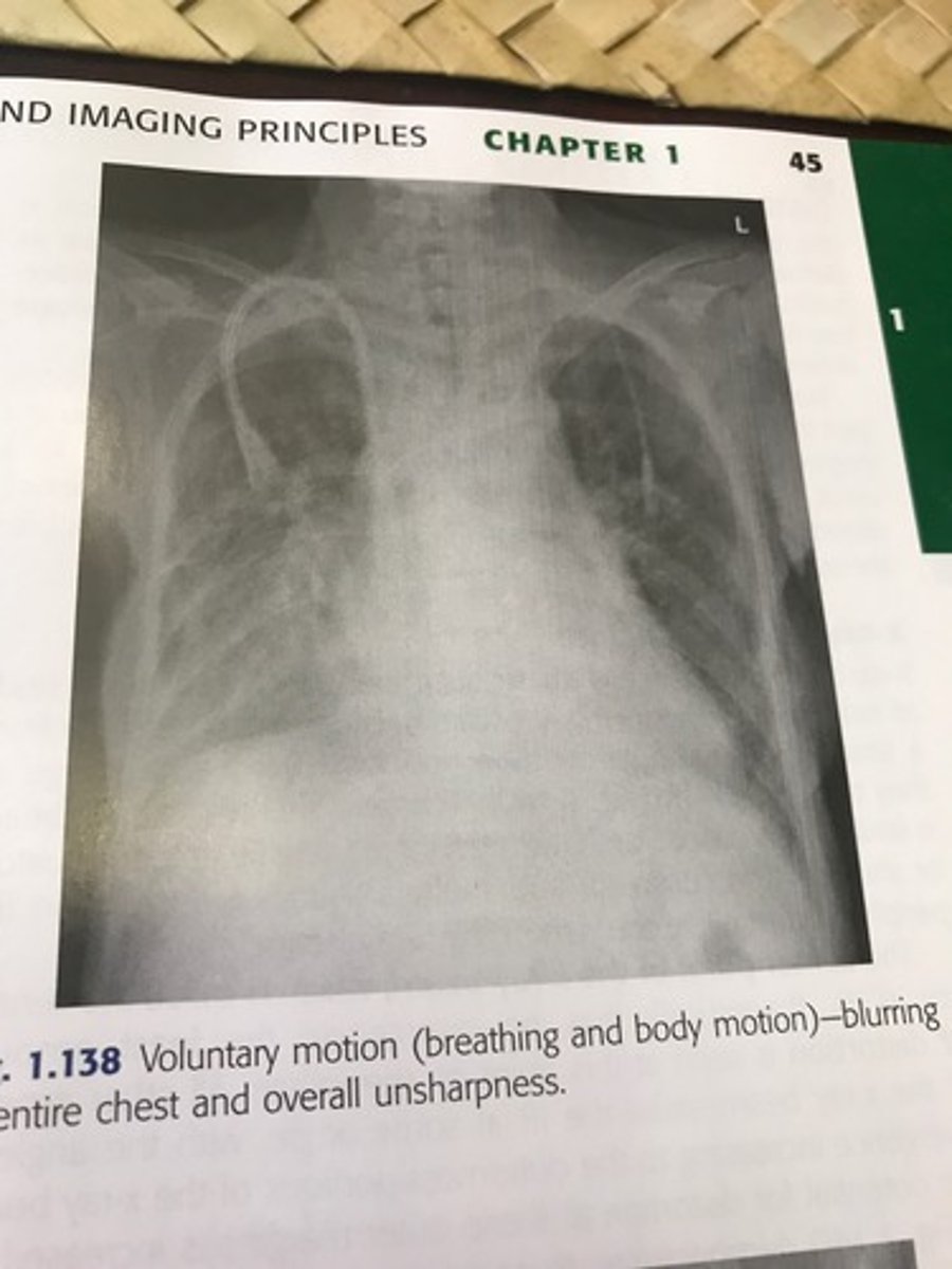
What is the greatest deterrent to image sharpness?
Motion.
Voluntary and involuntary
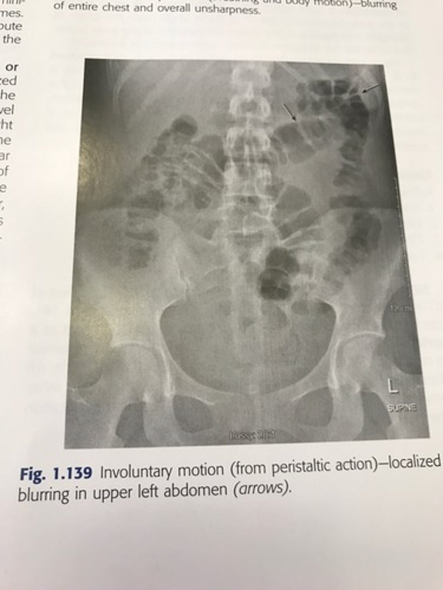
Voluntary motion vs. involuntary
Voluntary includes breathing or movement of body parts.
Involuntary includes peristaltic action, tremors, or the chills.
Voluntary motion image
Involuntary motion image
4 ways to control image quality
1) small focal spot
2) short exposure time
3) IR speed
4) SID & OID
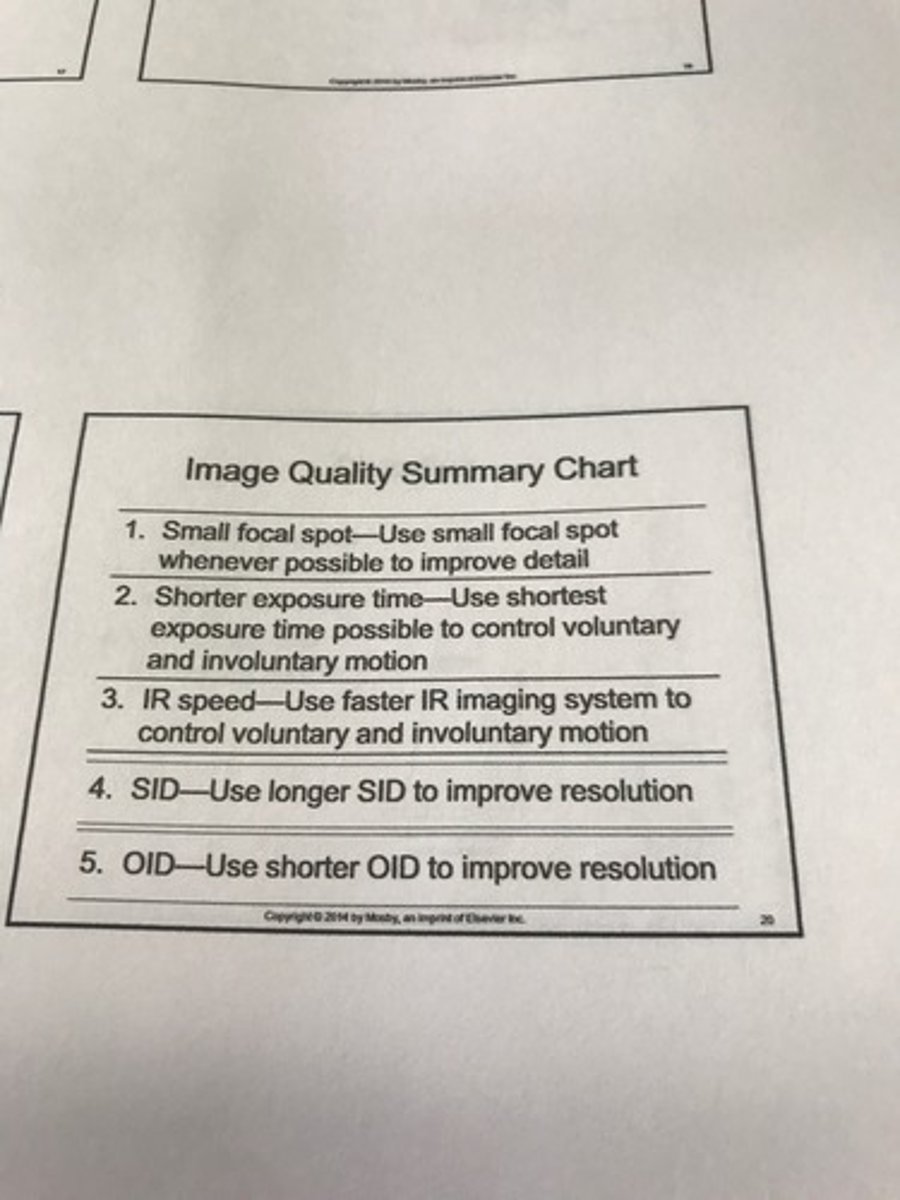
What do you need to get the best image?
1- small focal spot
2- long SID
3- shirt OID
What is distortion?
a misrepresentation of the size or shape of an object.
What are the 4 primary controlling factors of distortion?
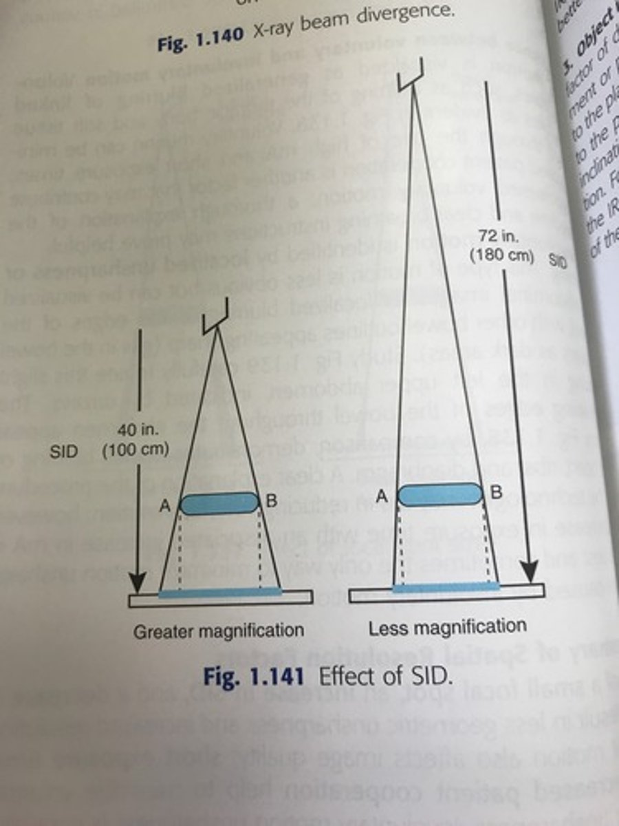
1) source image receptor distance (SID)
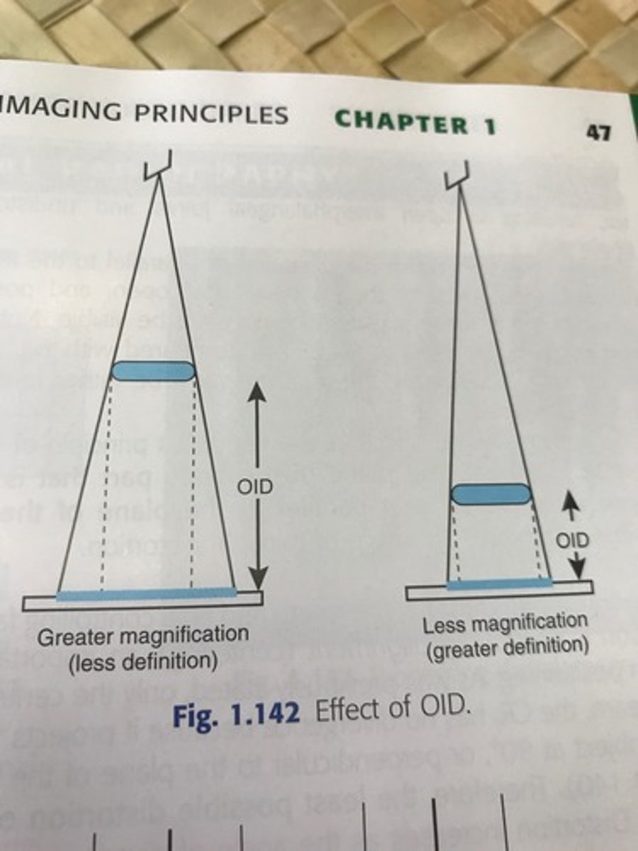
2) object image receptor distance (OID)
3) object image receptor alignment
4) central ray alignment
Increase in SID results in what?
Less magnification and reduced distortion
Decreased OID results in what?
Less magnification
Greater definition
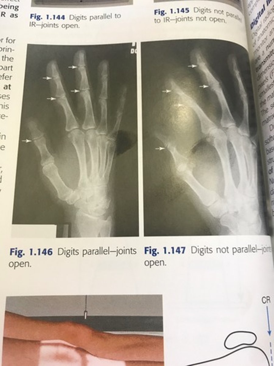
What happens when the body part being imaged isn't parallel to the image receptor?
Distortion!
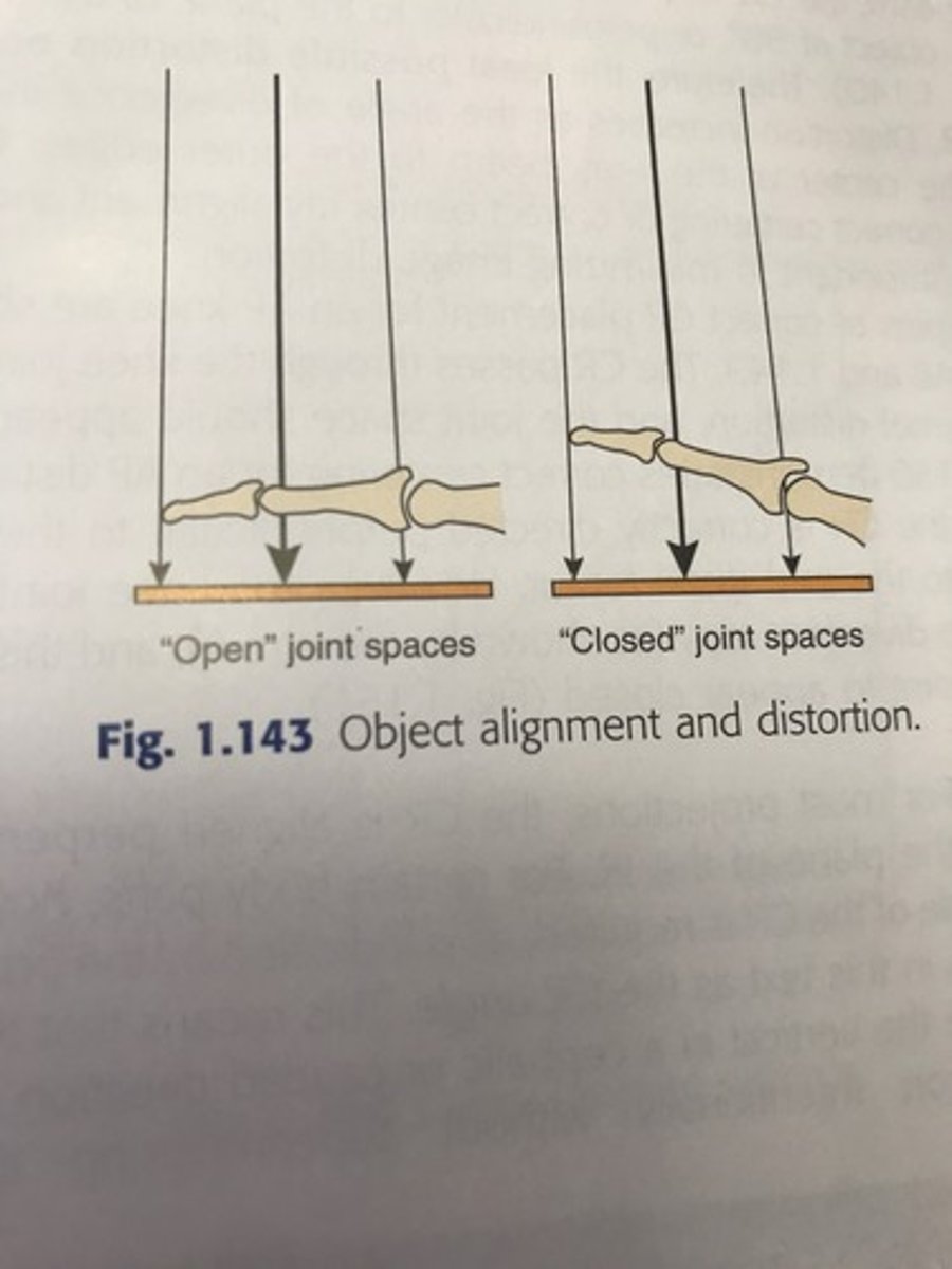
The least possible distortion occur where?
Central ray.
It is important to have correct positioning
How is a digital image formed?
By a matrix of pixels.
Pixel is the smallest unit, they make up rows and colums which forms the matrix.
The higher number of pixels you have makes what better?
Resolution!
What are the image quality factors in digital radiography?
1- brightness
2- contrast resolution
3- spatial resolution
4- distortion
5- exposure indicator
6- noise
What is brightness?
Intensity of light representing individual pixels in an image
What is spatial resolution?
Recorded sharpness (detail) of structures on an image.
What are the controlling factors for spatial resolution?
Pixel size, display matrix. Dependent on the display capabilities of the monitor.
What is contrast resolution?
Differences in brightness between light and dark areas of an image
What is distortion in digital radiography?
Misrepresentation of object shape or size.
What are the controlling factors for distortion?
SID
OID
CR alignment
*same as analog
What is an exposure indicator?
A numeric value that is representative of the exposure the image receptor received.
How do you find out if your exposure indicator number is valid?
Each institution will have a chart that your number must fall between to be an acceptable image. Varies on who makes the equipment
What is noise?
Random disturbance that obscures or reduces image clarity.
What is post processing?
Changing or enhancing the electronic image to improve diagnostic quality.
-Algorithms applied to improve diagnostic quality of image.
What are post processing options?
1- windowing
2- smoothing
3- magnification
4- edge enhancement
5- equalization
6- subtraction
7- image reversal
8- annotation
What was one of the first applications of computers in radiography?
Computed tomography
What does PACS stand for?
What is it?
Picture- digital medical images
Archiving- electronic storage of images
Communication- routing and displaying of images
System- specialized computer network that manages the complete system.
What does PACS do?
Sends images to different areas of the hospital
What is RIS?
Radiology Information System
What is DICOM?
The current standard that ensures all manufactures and types of equipment are able to communicate and transmit images effectively.
What are the 3 most effective ways to protect patients and staff from radiation?
1- time
2- distance
3- sheilding
What are the SI units of radiation?
Air kema
Gray
Sievert
What is personal monitoring?
Refers to the measurement of the amount of radiation dose received by occupationally exposed individuals
What does OSL stand for?
optically stimulated luminescence
Worn at waist or chest level
What are the ALARA principles?
1- always wear personal monitor
2- don't restrain patients
3- optimal exposure factors
4- cardinal rules of radiation
What is the most important method to prevent unnecessary exposure?
Avoid repeat radiographs
What are some ways to avoid repeat radiographs?
Give clear instructions
Accurate positioning and exposure factors
How does collimation protect the patient?
Limits the size and shape of X-ray field to area of interest. Use four side collimation. Remember to position according to divergence of beam.
What are the two types of collimation?
Manual
PBL: positive beam limitation (auto collimation)
What are the two types of shielding?
Contact: attaches to patient
Shadow: attaches to collimator to cast shadow on important areas of patient
When are flat gonadal shields mainly used?
When a patient is in a recumbent position. They are vinyl covered lead shields that are cut into various shapes and sizes
Reduces dose 50-90%