Neuro 480- Midterm 3- Chapter 52 -Patterning the Nervous System
1/14
There's no tags or description
Looks like no tags are added yet.
Name | Mastery | Learn | Test | Matching | Spaced |
|---|
No study sessions yet.
15 Terms
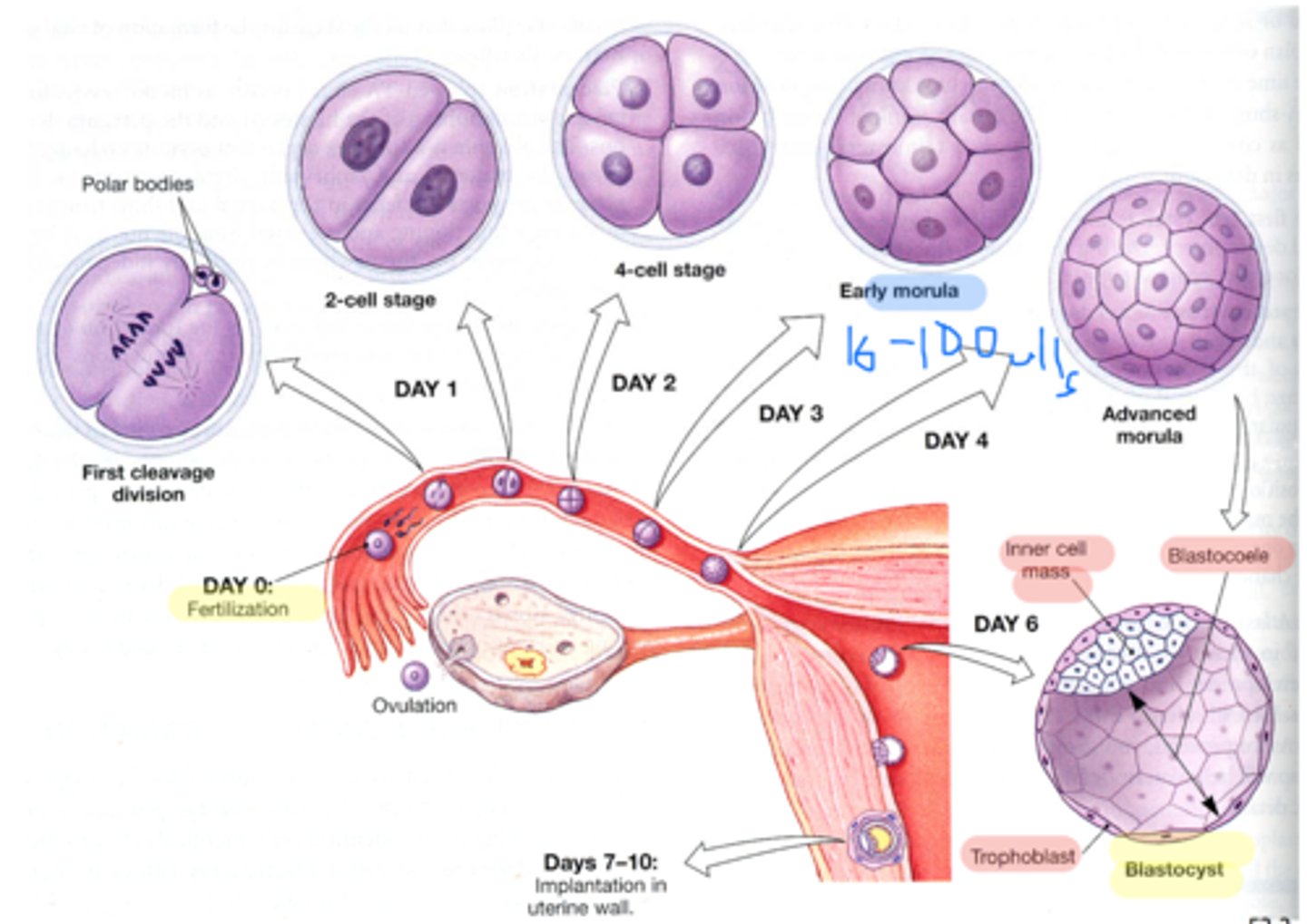
Describe the events occurring during very early development (fertilization to day 10 post-day fertilization). Be sure to include the following terms in your discussion: morula, blastocyst, trophoblast, inner cell mass, implantation, bilaminar disc, epiblast and hypoblast.
The first week of human development begins with fertilization of the egg by sperm forming the first cell, the zygote. Cell division leads to a ball of cells, the morula. Further cell division and the formation of a cavity in the ball of cells forms the blastocyst.
Trophoblast-a layer of tissue on the outside of a mammalian blastula, supplying the embryo with nourishment and later forming the major part of the placenta.
Inner Cell mass- is the mass of cells inside the primordial embryo that will eventually give rise to the definitive structures of the fetus.
Implantation- a process in which a developing embryo, moving as a blastocyst through a uterus, makes contact with the uterine wall and remains attached to it until birth
Bilaminar disc- In week 2 of implantation, the trophoblast penetrates deeper into the endometrium, and the blastocyst changes morphologically. The inner cell mass produces a bilaminar embryonic disk composed of epiblast (future embryonic ectoderm and mesoderm) and embryonic endoderm.
Epiblast- The epiblast is derived from the inner cell mass and lies above the hypoblast. The epiblast gives rise to the three primary germ layers (ectoderm, definitive endoderm, and mesoderm)
Hypoblast- The hypoblast is a tissue type that forms from the inner cell mass. It lies beneath the epiblast and consists of small cuboidal cells. Extraembryonic endoderm is derived from hypoblast cells.
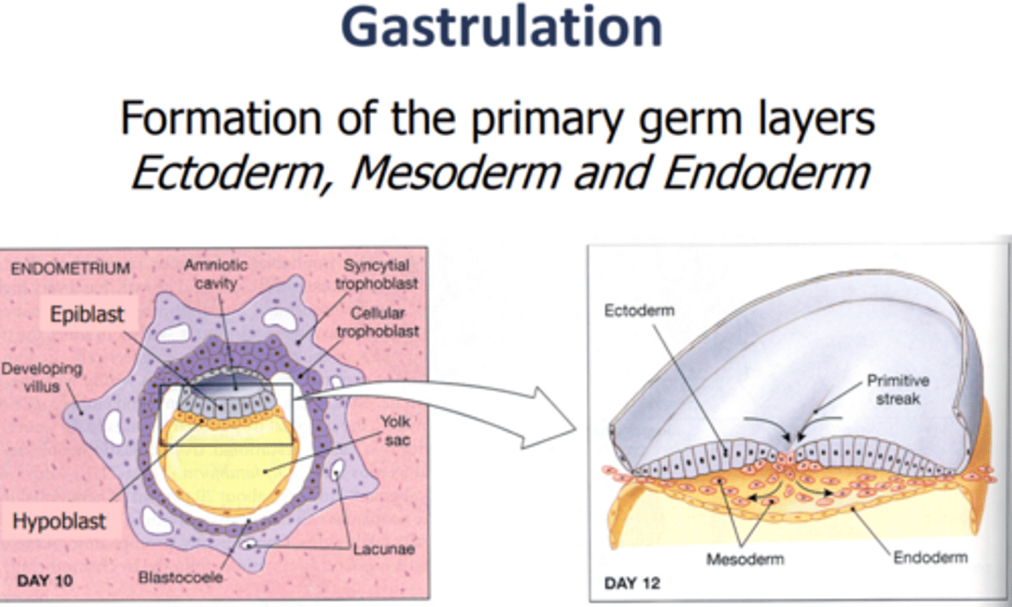
Explain how the primary embryonic germ layers (ectoderm, mesoderm, and endoderm) form during grastrulation.
This image shows the process of gastrulation. Gastrulation occurs when a blastula, made up of one layer, folds inward and enlarges to create a gastrula. A gastrula has 3 germ layers--the ectoderm, the mesoderm, and the endoderm. Some of the ectoderm cells from the blastula collapse inward and form the endoderm.
Gastrulation is the process in which the ICM (inner cell mass) in converted into the trilaminar embryonic disc, which is comprised of the three germ layers (ectoderm, mesoderm and endoderm).
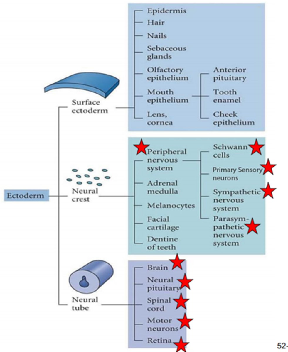
Know that ectoderm gives rise to the epidermis and the major structures of the CNS and PNS
Know that ectoderm gives rise to the epidermis and the major structures of the CNS and PNS
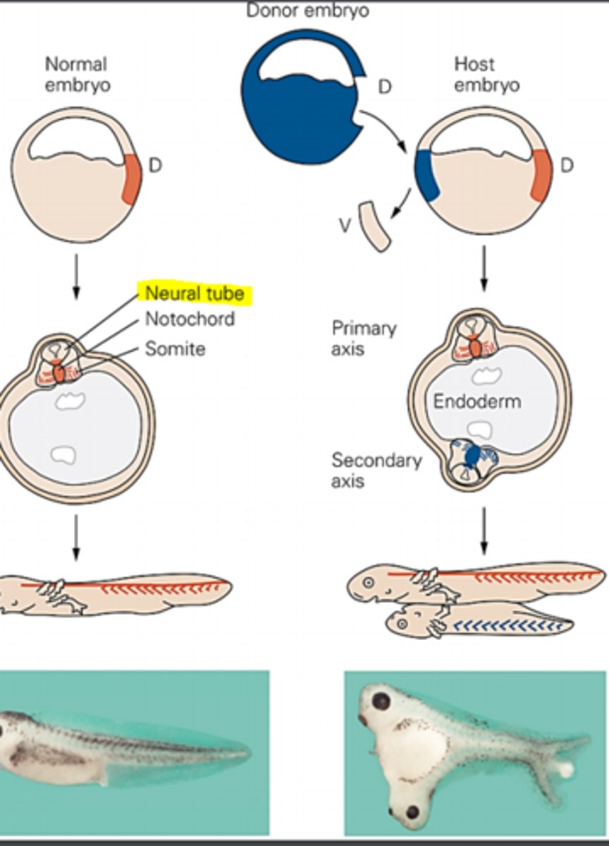
Describe Hans Spemann and hilde mangold's experiment that led to the discovery of the organizer region.
Signals from the organizer region induce a second neural tube- These guys made the remarkable discovery that specific signals from a specialized group of cells are responsible for triggering the formation of the nerual plate. This special group of cells is called the organizer region. Working with amphibian embryos they showed the organizer activity is restricted to a region of the embryo called the dorsal lip of the blastopore. They grafted the dorsal blastopore lip from an early gastrula stage embryo into a region of a host embryo. Signals from grafted cells induce a second embryonic axis, which includes a virtually complete neural tube. As the embryo matures the secondary neural tube develops into a complete nervous system.
Probably less important- the donor tissue was from a pigmented embryo, whereas the host tissue was unpigmented, permitting the fate of grafted cells to be monitored by their characteristic pigmentation.
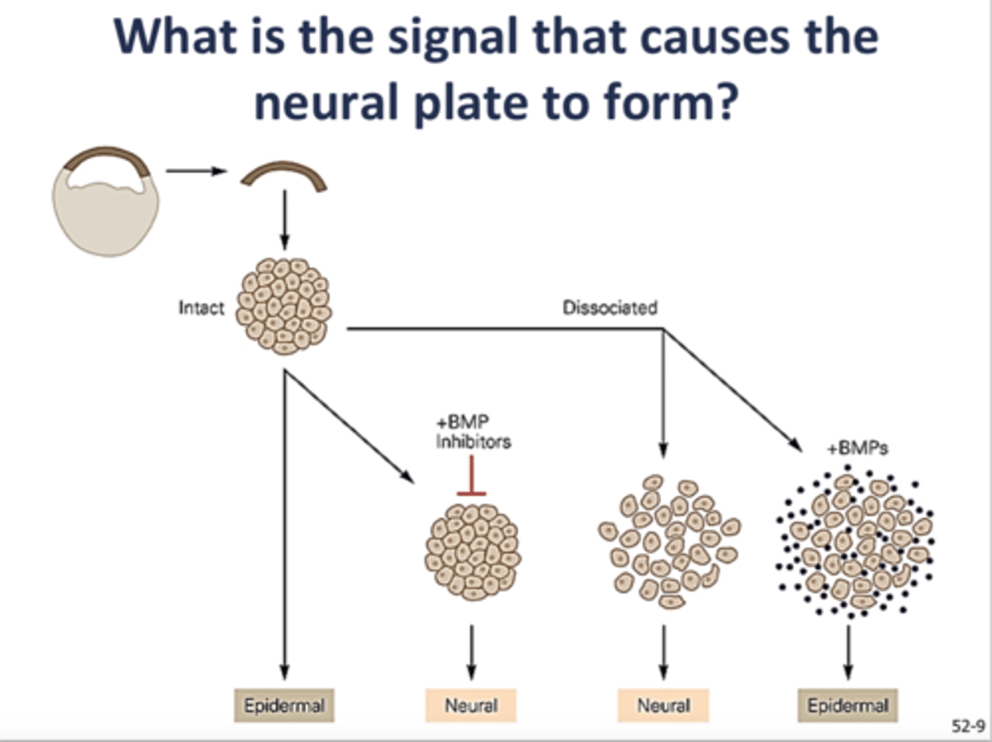
Explain how the neural plate forms (termed primary neural induction). Include the roles of the organizer region and the following molecules: BMP, chordin, noggin, and follistatin
Signals from a specialized portion of mesoderm (called the organizer) cause the overlying ectoderm to become the neural plate.
Ectodermal cells acquire neural or epidermal character depending on the presence of absence of BMP signaling. When ectodermal cell aggregates are exposed to BMP signaling they differentiate into epidermal tissue. When BMP signaling is blocked, either by dissociating ectodermal tissue into single cells or by addition of BMP inhibitors (chordin, noggin, and follistan) to ectodermal cell aggregates, the cells differentiate into neural tissue.
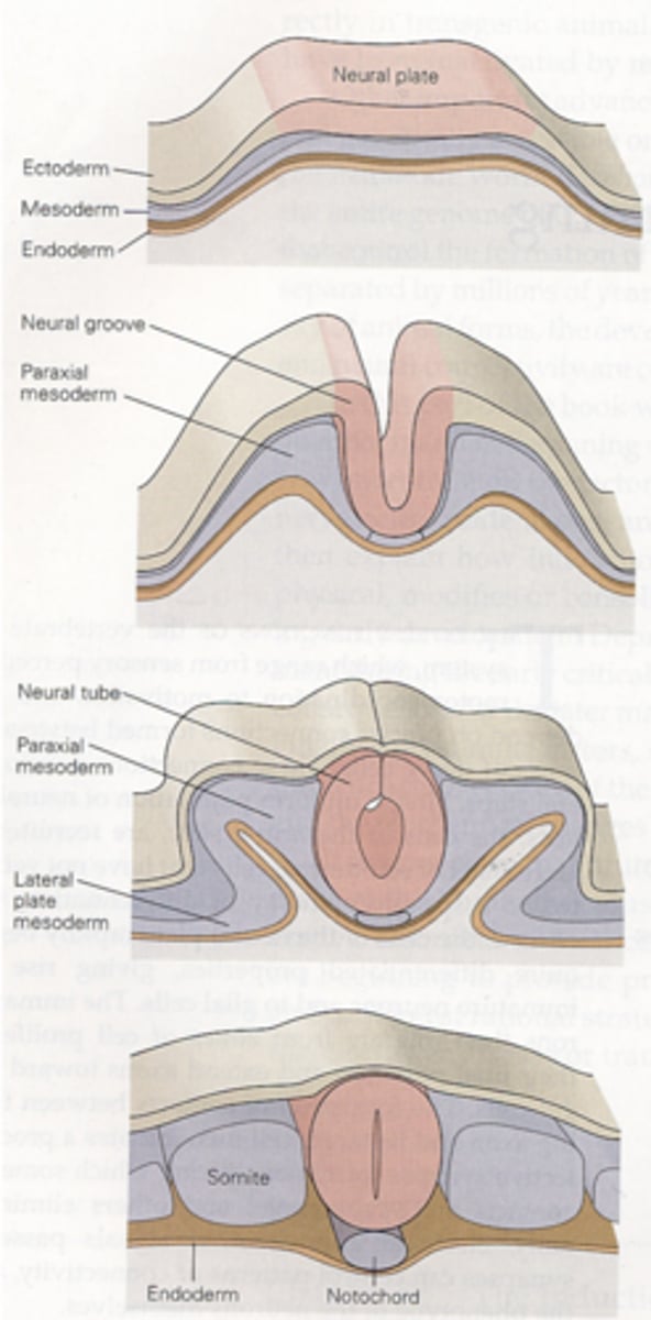
Describe the anatomy of neurulation. Include the terms ectoderm, neural plate, neural groove, neural tube and neural crest in your answer.
Neurulation is the process by which the neural plate becomes the neural tube. The neural tube is the precursor the the central and peripheral nervous systems.
The process: the ectoderm gives rise to the neural plate. The neural plate buckles at its midline giving rise to the neural groove. Closure of the dorsal neural folds forms the neural tube. The caudal region of the neural tube becomes the spinal cord and the rostral region the brain.
Neural crest cells?
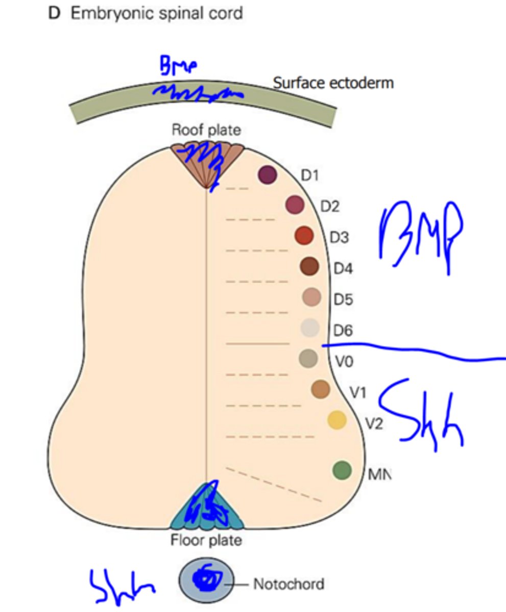
After closure of the neural tube in early embryos, molecules named sonic hedgehog (Shh) and bone morphogenetic protein (BMP) are involved in dorsal-ventral patterning of the nervous system. Explain where each of these molecules is expressed. Discuss how gradients of these two molecules result in the mature dorsal-ventral pattern found in the spinal cord.
Sonic hedgehog (Shh) is expressed in the floor plate and notochord. There is a dorsal (high) to ventral (low) gradient of Shh signaling in the neural tube, which gradient controls cell identity. It gives rise to 5 major classes of ventral neurons: the interneurons V0-V3 and motor neurons (MN).
BMP is expressed in the ectoderm and the roof plate of the neural tube. It induces formation of sensory interneurons in the dorsal spinal cord.
As a result of Shh and BMP signaling the dorsal horn of the spinal cord is sensory nuclei and the ventral horn is somatic motor nuclei and the lateral horn contains visceromotor nuclei.
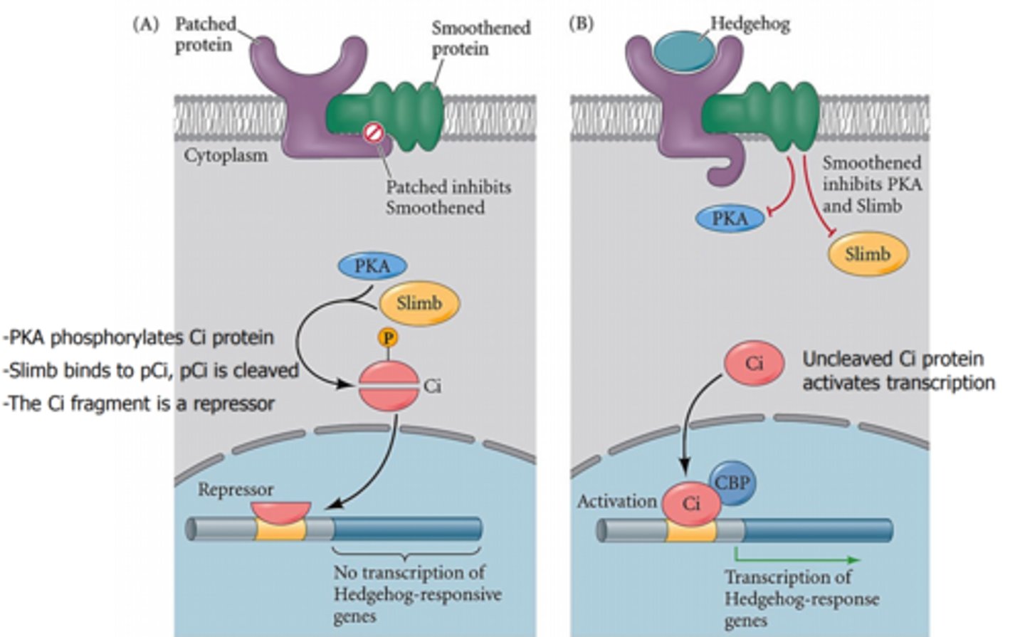
Explain the molecular mechanism by which sonic hedgehog changes gene expression in target cells. Include the roles of Shh, patched, smoothened, Ci, transcriptional activators, transcriptional repressors and Shh responsive genes.
Shh binds to a ligand-binding transmembrane region called patch. and a signal-transducing subunit called smoothened. This binding activates smoothened and starts a cascade.
Normally, while smoothened is not activated, a set of transcription factors known as Gli proteins are proteolytically processed and bind Shh target genes preventing their activation. However, when smoothened is activated, this proteolytic processing is inhibited and and Gli transcription factors go and direct the expression of Shh target genes. Thus Gli can be both a transcription activator and repressor depending on the presence of Shh. The major Gli target genes are genes that encode for yet more transcription factors. Some of these transcription factors are activated only when when Shh is at a particular concentration threshold which is what creates the pattern of different progenitor domains.
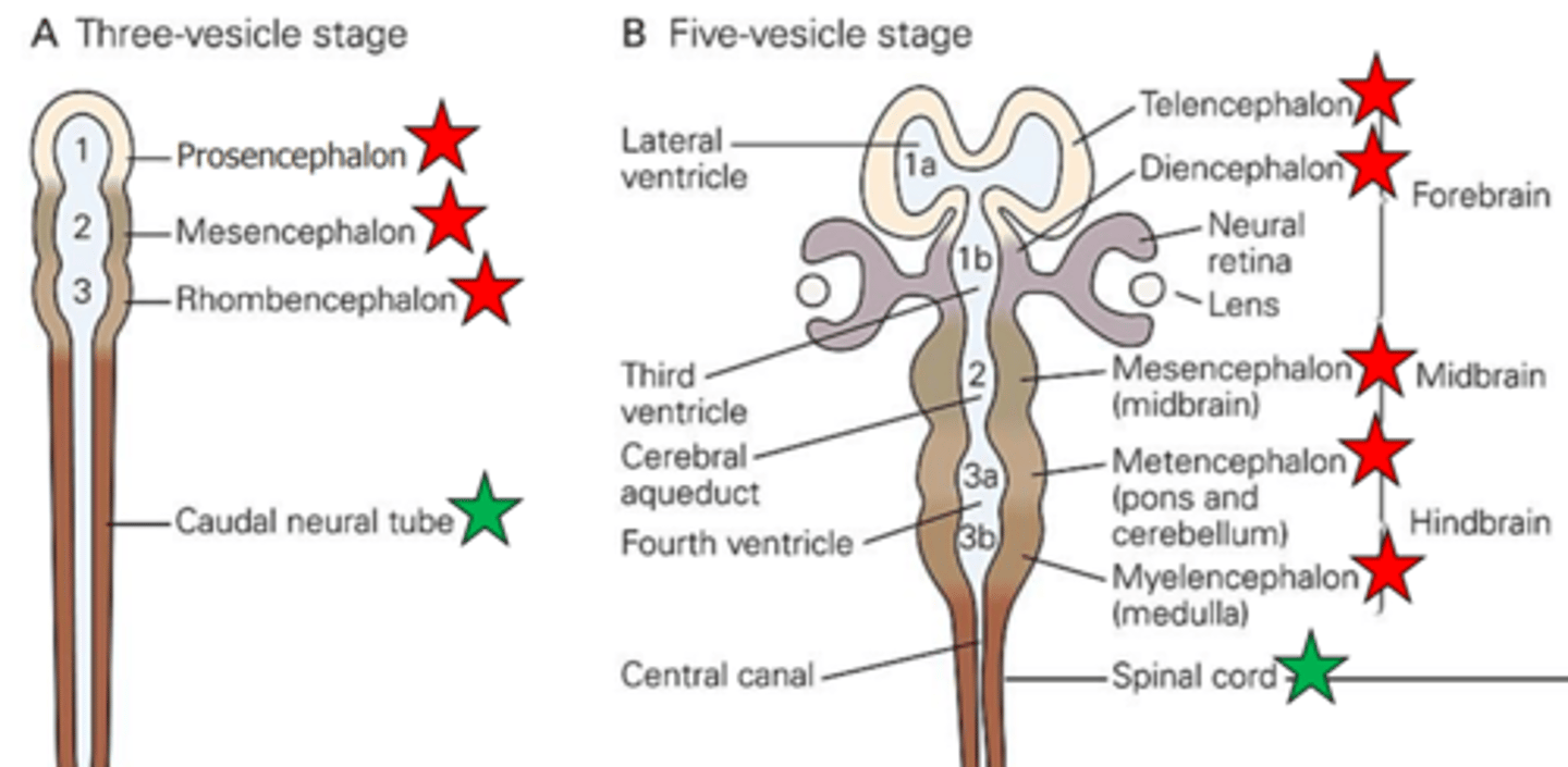
Know the important events that occur as the neural tube develops into the brain and spinal cord. Also be able to identify each structure or region on a diagram.
1. The prosencephalon (forebrain) divides, forming the telencephalon and diencephalon. The telencephalon then develops to form the cerebral hemispheres. The diencephalon develops to form the thalamus, hypothalamus, and retina.
2. The mesencephalon (midbrain) develops into the midbrain
3. The rhombencephalon (hindbrain) divides, forming the metencephalon and myelencephalon develops into the medulla.
4. The caudal neural tube becomes the spinal cord.
5. The fluid filled center of the neural tube becomes the brain ventricles and the central canal of the spinal cord.
Explain how Wnt signaling influences the rostrocaudal pattern of the neural plate
Just as Shh and BMP are factors that affect signaling dorsal-ventrally of the neural plate, Mnt is a factor that affects the development of the neural plate rostral (anterior)- caudal (posteriorly).
The net level of Wnt signaling activity is low at rostral levels of the neural plate and increases progressively in the caudal direction. This activity gradient arises because the mesoderm that flanks caudal regions of the neural plate expresses high levels of Wnt, whereas the endoderm that underlies the rostral region of the neural plate is a source of secreted proteins that inhibit Wnt signaling.
In response to this Wnt signaling gradient and other signals, cells in anterior and posterior regions of the neural plate begin to express different transcription factors.
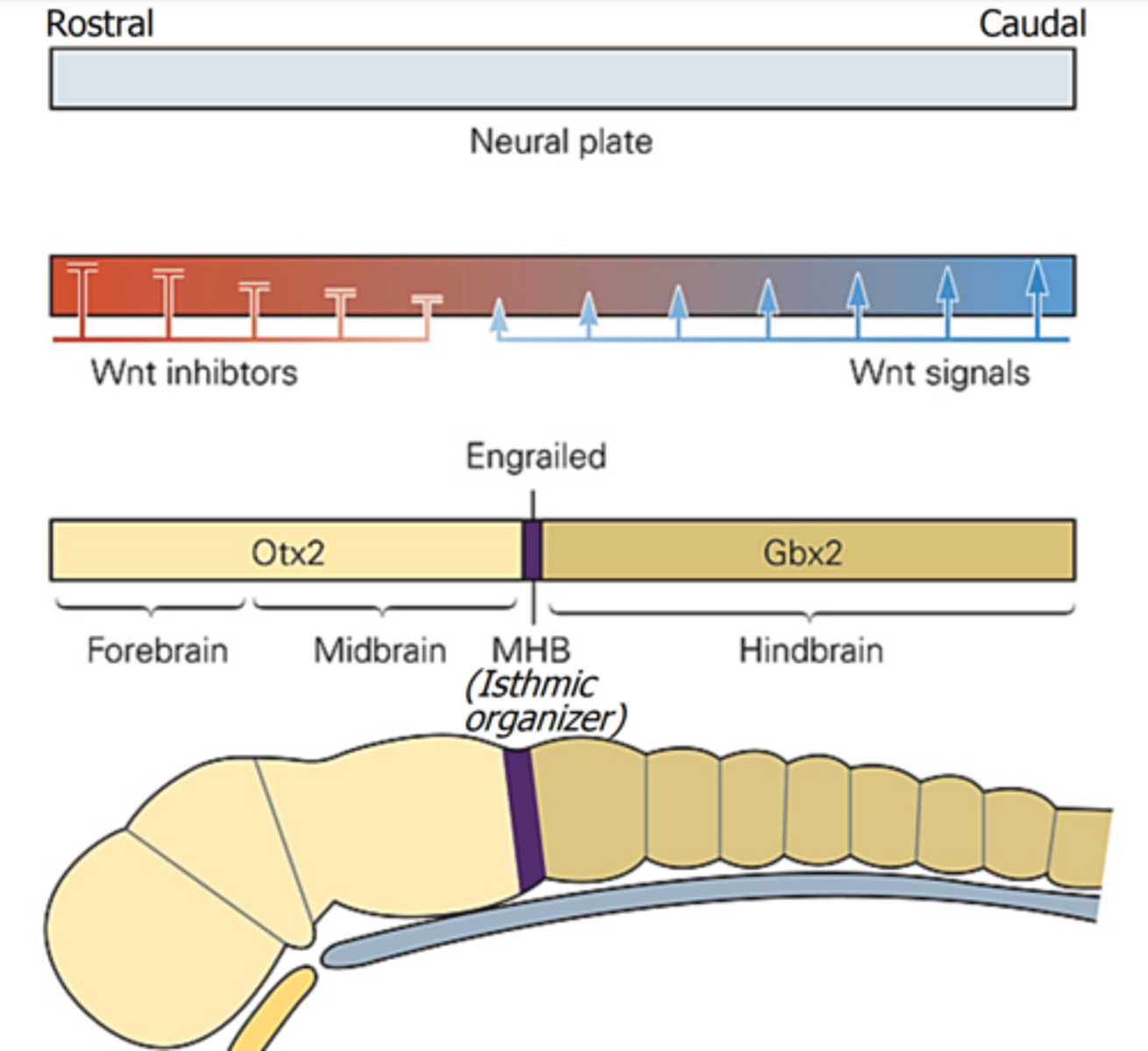
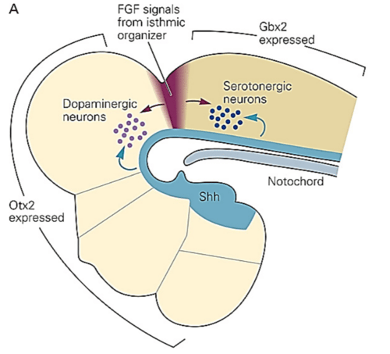
Explain how the midbrain and hindbrain are patterned. Include the roles of the isthmic organizer at the midbrain-hindbrain boundary, secreted molecules (FGF and Shh), and transcription factors (Otx2 and Gbx2). What causes the production of dopaminergic neurons and serotonergic neurons?
In response to the gradient of Wnt rostral-caudally, Otx2 is expressed anteriorly and Gbx2 is expressed posteriorly. This affects the individual patterning of the midbrain and forebrain. The intersection of these two transcriptional factor domains marks the midbrain-hindbrain boundry, also known at the isthmic organizer.
The spread of FGF8 from the isthmic organizer into the midbrain domain marked by Otx2 expression induces differentiation of dopaminergic neurons, whereas its spread into the hindbrain domain marked by Gbx2 expression triggers the differentiation of serotonergic neurons. FGF acts in concert with Shh signals from the ventral midline to specify the identity of position of the previously mentioned neurons.
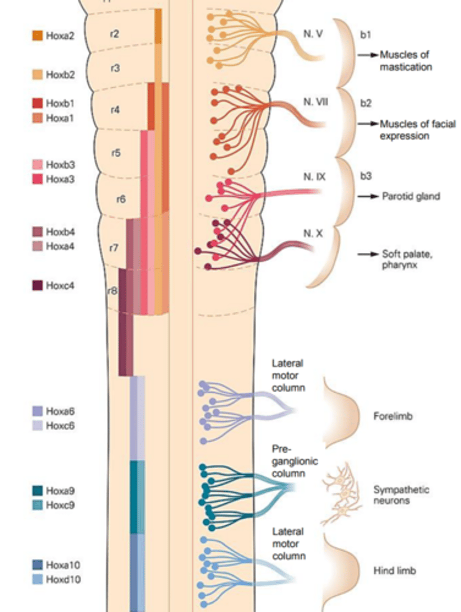
Briefly explain what rhombomeres are
A rhombomere is a transiently divided segment of the developing neural tube. The rhombomeres appear as a series of slightly constricted swellings in the neural tube, caudal to the cephalic flexure.
Textbook says: fundamental building blocks of the hindbrain, they are compartamental units that are arrayed along the rostrocaudal axis of the hindbrain.
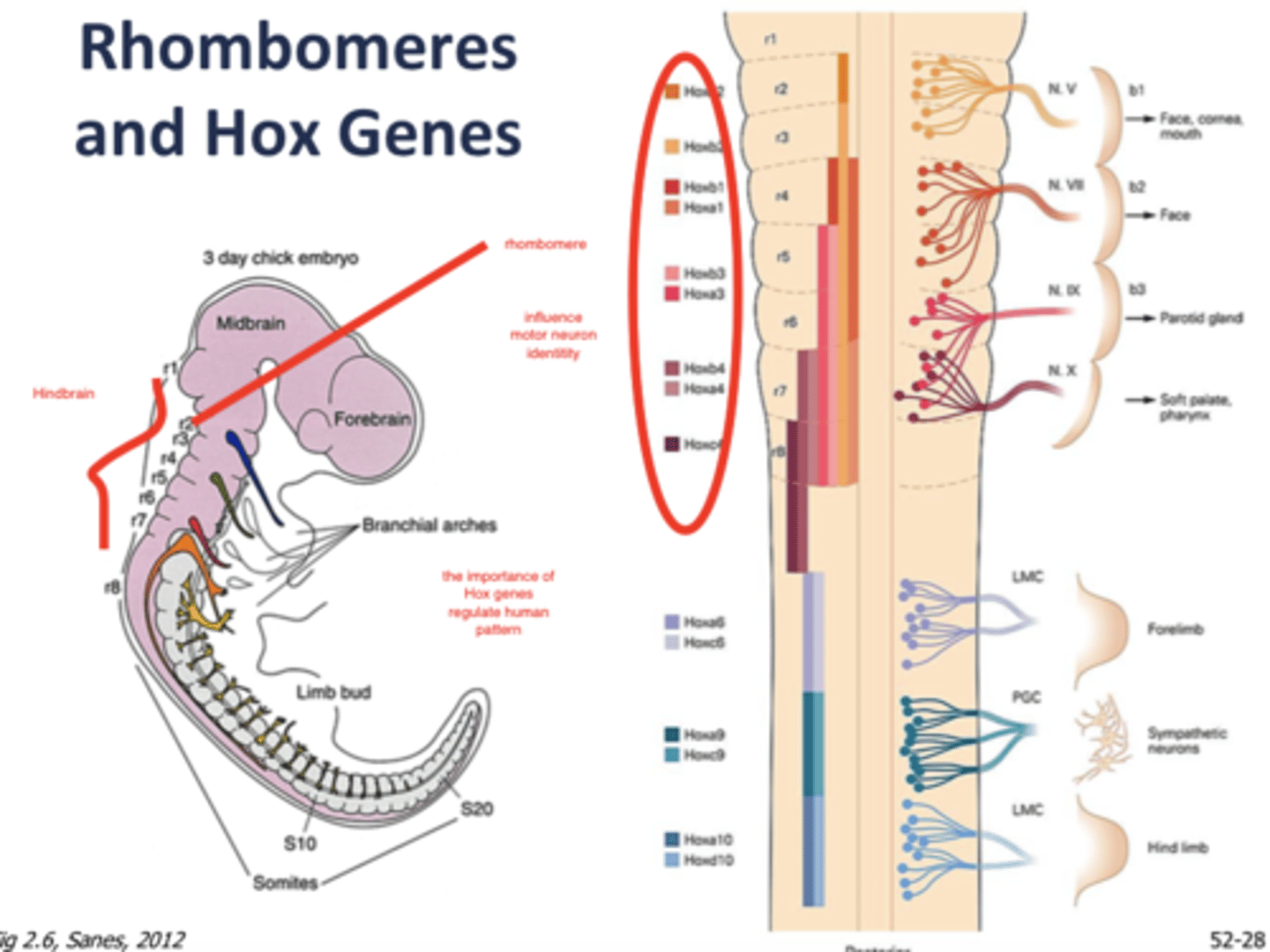
Explain the basic rostrocaudal pattern of Hox gene expression. Explain what Hox genes do in the hindbrain and spinal cord.
What are hox genes? They are a family of genes that encode transcription factors that regulate developmental processes in organisms.
Rostrocaudal pattern: Members of the Hox gene family are expressed in overlapping domains (rhombomeres) along the rostrocaudal axis of the developing midbrain, hindbrain, and spinal cord. Specific hox genes control the identity of neurons in individual rhombomeres. For example, Hoxb1 is expressed at high levels in rhombomere 4, the domain that gives rise to facial motor neurons, but is absent from rhombomere 2, the domain that gives rise to trigmeninal motor neurons.
Hox genes in hindbrain and spinal cord- Hox gene expression controls motor neuron identity in the hindbrain (think back to the mutants they made). Hox genes control the identity and projection of spinal motor neurons. Hox genes also organize the hindbrain and spinal cord (because of the rhombomeres and individual hox genes in them?)
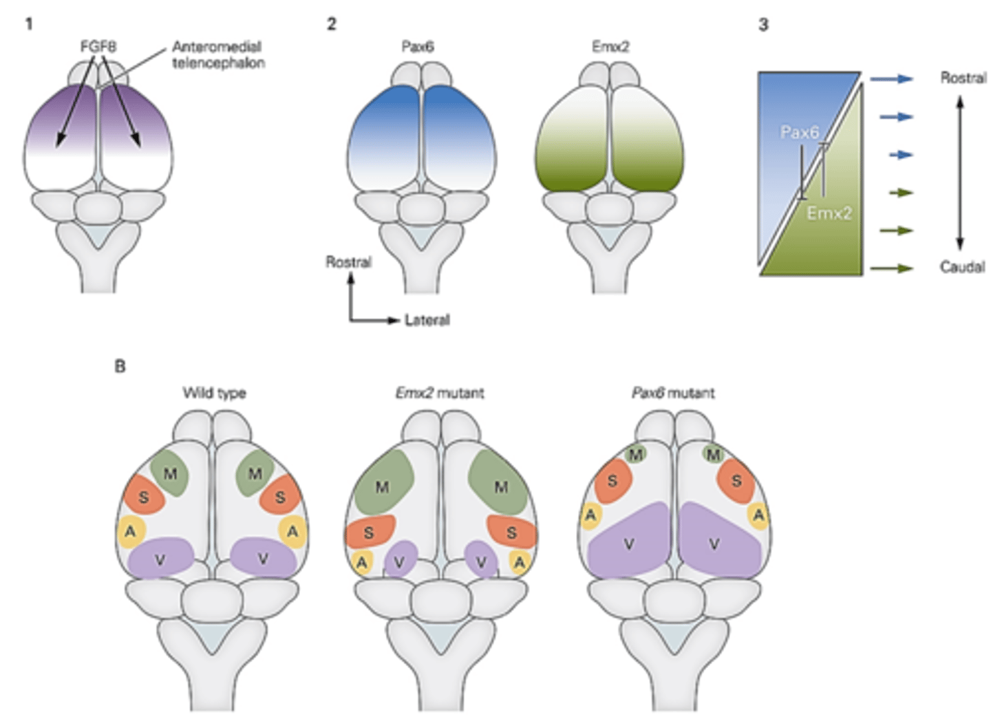
Explain how the developing forebrain is patterned anteroposteriorally (rasutrocaudally). Be sure to include the following in your answer: FGF8 gradient, Pax6 gradient, Emx2 gradient, motor cortex, somatosensory cortex, auditory cortex and visual cortex.
Two transcription factors, Pax6 and Emx2 have inverse anteroposterior gradients, Pax6 being high anteriorly and Emx2 being high posteriorly.
FGF signals are also high anteriorly and work to promote Pax6 and repress Emx2 expression.
Motor areas develop in the anterior region and visual areas in more posterior regions. These gradients of transcriptional factors lead to the development of the different functional areas of the brain.
Mutations: Genetic elimination of Emx2 function results in expansion of the motor areas and contraction in auditory and visual areas. Conversely, elimination of Pax6 function results in an expansion of the visual areas and a contraction of motor and auditory areas.
Give two examples of how the cortex retains plasticity during development
Plasticity is when the nervous system is sensitive to experience or input from the environment
Two examples of how the cortex retains plasticity during development:
1) Sensory input regulates the organization of 'barrels' in the developing somatosensory cortex in rodents.You get a group of cells in a barrel formation. Each cell receiving input from a single whisker of the snout of an animal. If prospective visual cortical tissue is transplanted into the somatosensory cortex around the time of birth, barrels form in the transplanted tissue with a pattern that closely resembles that of the normal somatosensory barrel field suggesting that afferent input superimposes aspects of neocortical patterning on the basic features of the protomap.
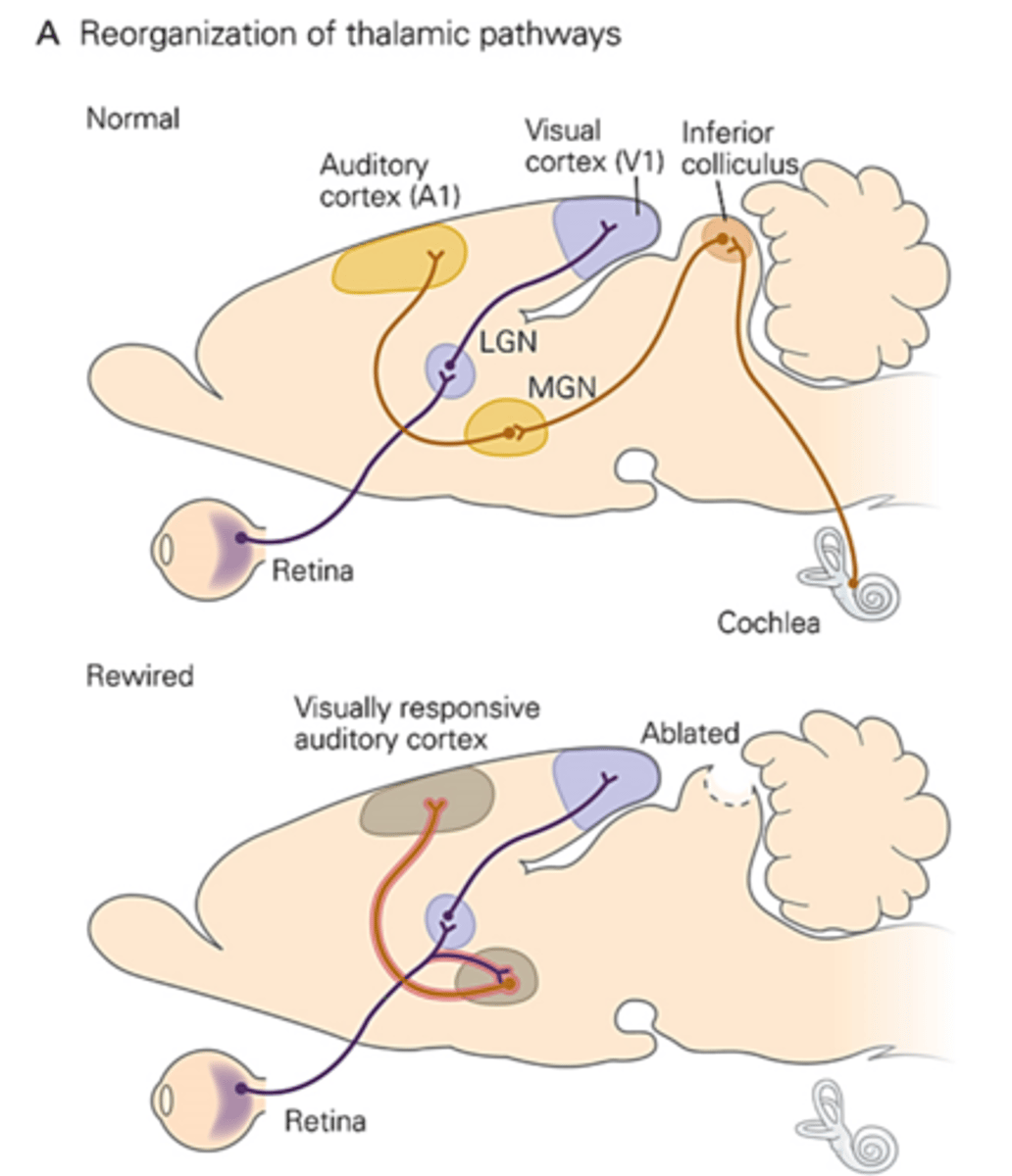
2) Rerouting thalamocortical input can recruit cortical areas for new sensory functions- If the inferior colliculus (receives auditory information from the cochlea) is ablated, then visual axons from the retina will innervate the MGN (should have been inervated by auditory information from the inferior colliculus) and then will go to the auditory cortex, now making the auditory cortex responsive to visual stimuli.