Intracellular Accumulations
1/23
There's no tags or description
Looks like no tags are added yet.
Name | Mastery | Learn | Test | Matching | Spaced |
|---|
No study sessions yet.
24 Terms
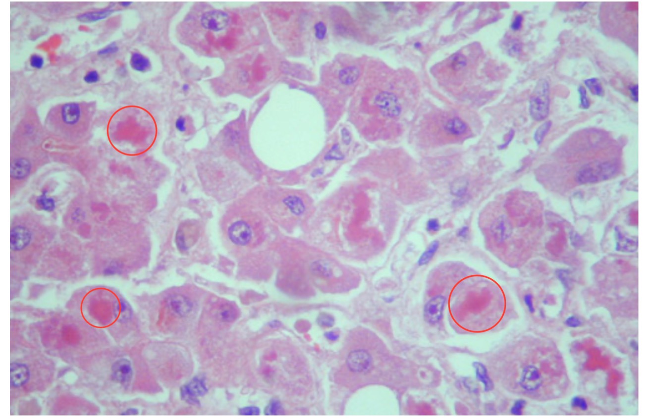
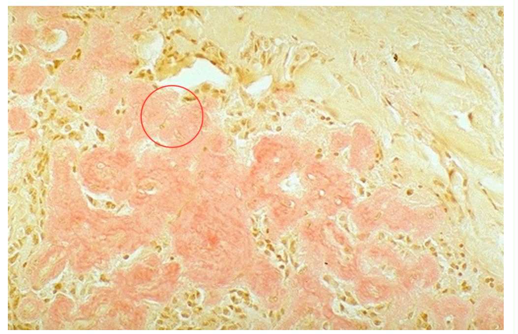
Histological Section of the Liver containing cytoplasmic organelle damage. Identify the red globular material encircled. Identify type of intracellular accumulation.
Mallory Bodies. Protein Accumulation
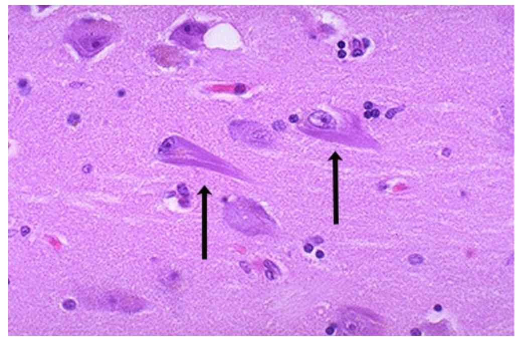
Histological view of neurons in a patient with Alzheimer’s disease. Identify the manifestation pointed by the black arrows.
Neurofibrillary tangles
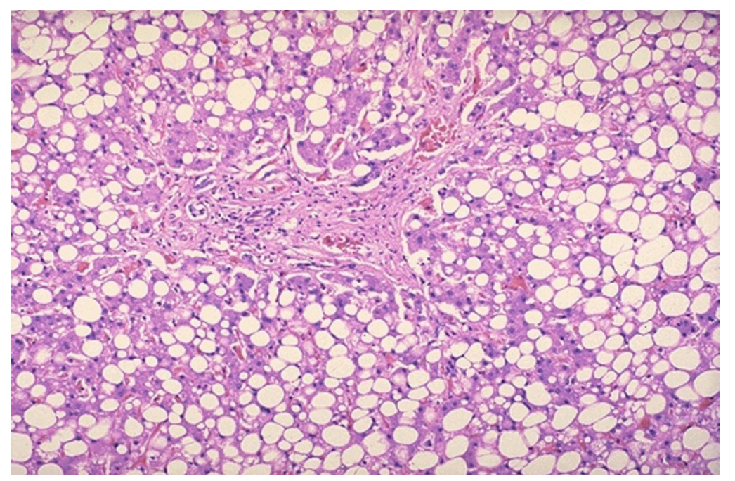
Histological view of the liver full of white or clear droplets. Identify type of intracellular accumulation. Identify the condition and the causes of this condition.
Fat or Lipid Accumulations. Steatosis or Fatty Liver Disease. Causes: Metabolic Diseases (DM Type II, Metabolic Syndrome, & Obesity) & Chronic Alcoholism
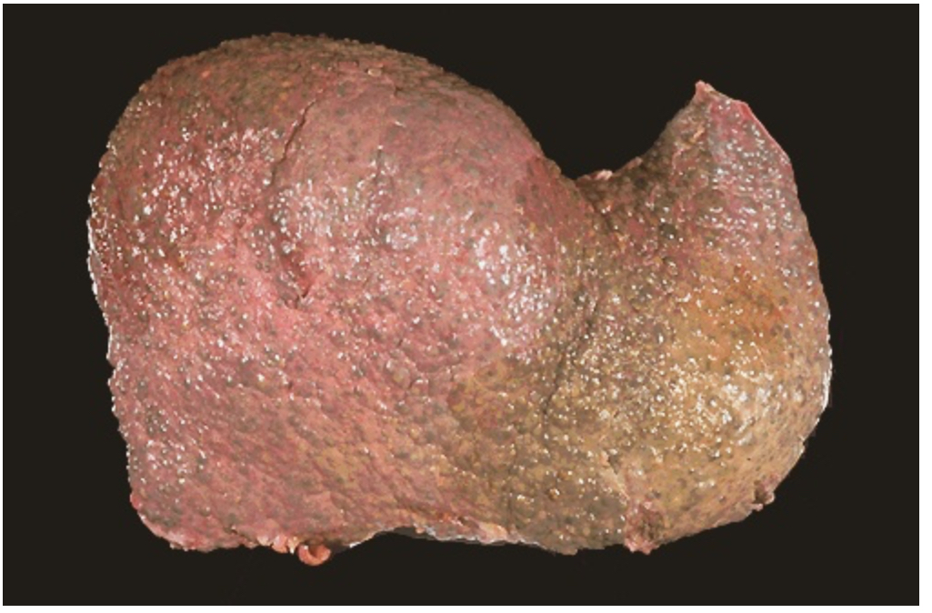
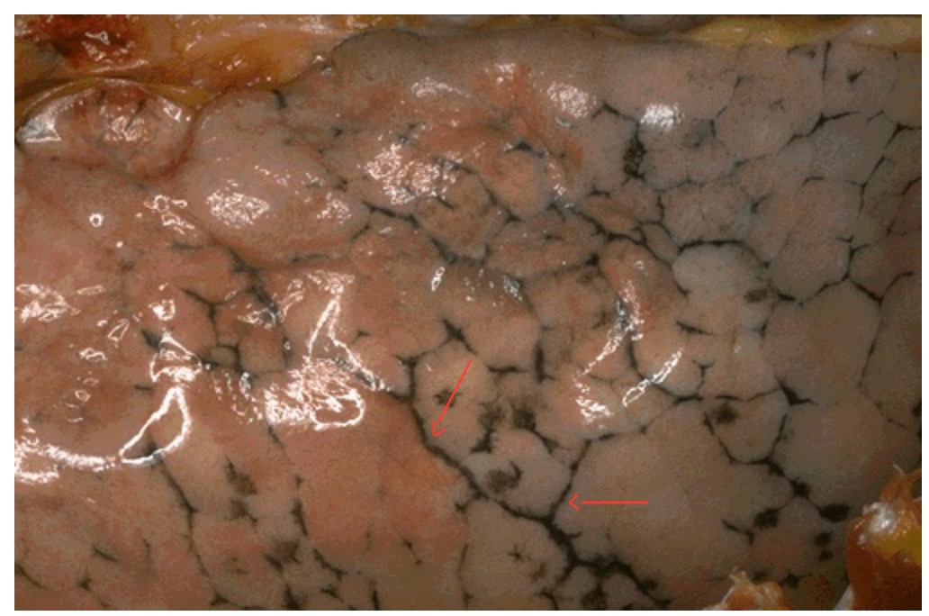
Gross anatomy of a Liver with Injury caused by chronic alcoholism. This leads to ______ and regeneration of the hepatocytes in nodules. The firm, nodular appearance of the liver is called ______
Fibrosis; Cirrhosis
The Congo Red Stain reveals amorphous orange-red deposits of ______. Identify intracellular accumulations.
Amyloid. Protein accumulations
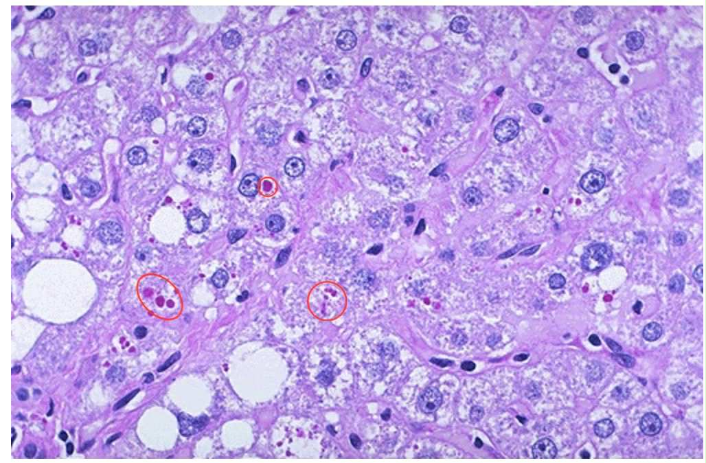
Cellular injury can lead to accumulation of a specific product. The red globules seen in PAS reaction are accumulations of what specific product?
Alpha-1-antitrypsin
A histological section of the spleen with Gaucher disease. The large pale cells pointed are accumulated storage product. What enzyme is deficient in this condition?
Glucocerebrosidase enzyme
Histological view of the liver with yellow-brown pigments.
What is the yellow-brown pigment called?
Why does it accumulate?
What does it illustrate?
Lipofuscin (lipochrome)
Wear and tear with aging
Autophagocytosis (Autophagy)
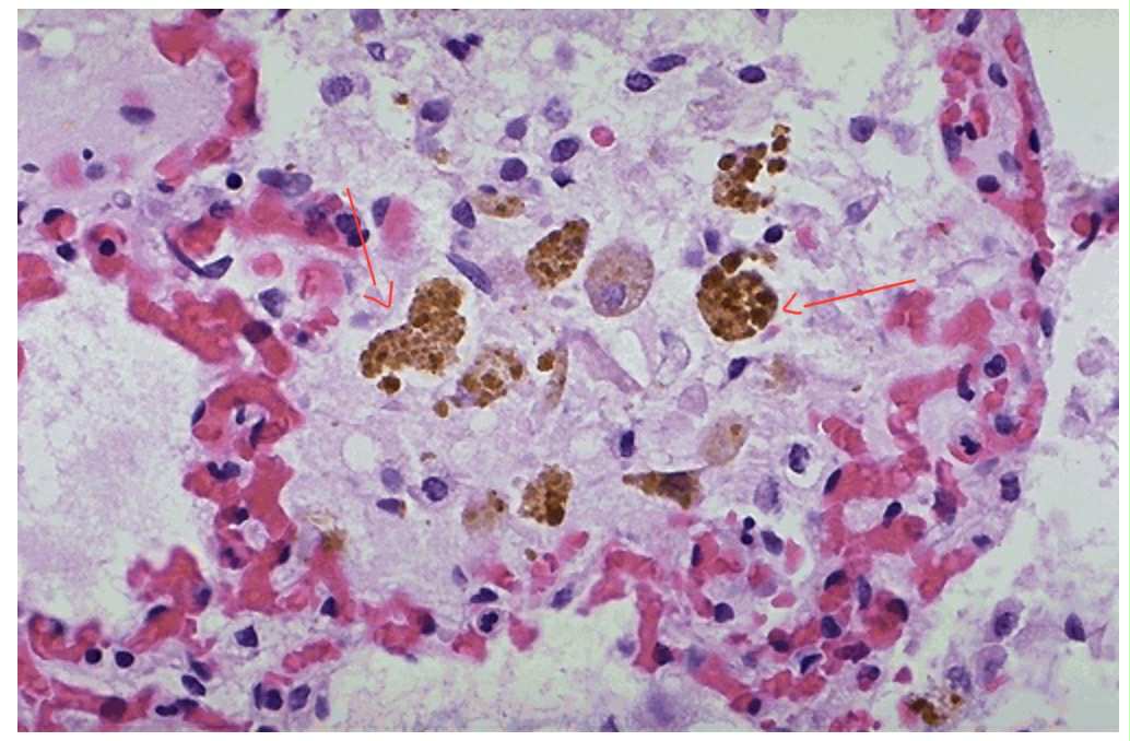
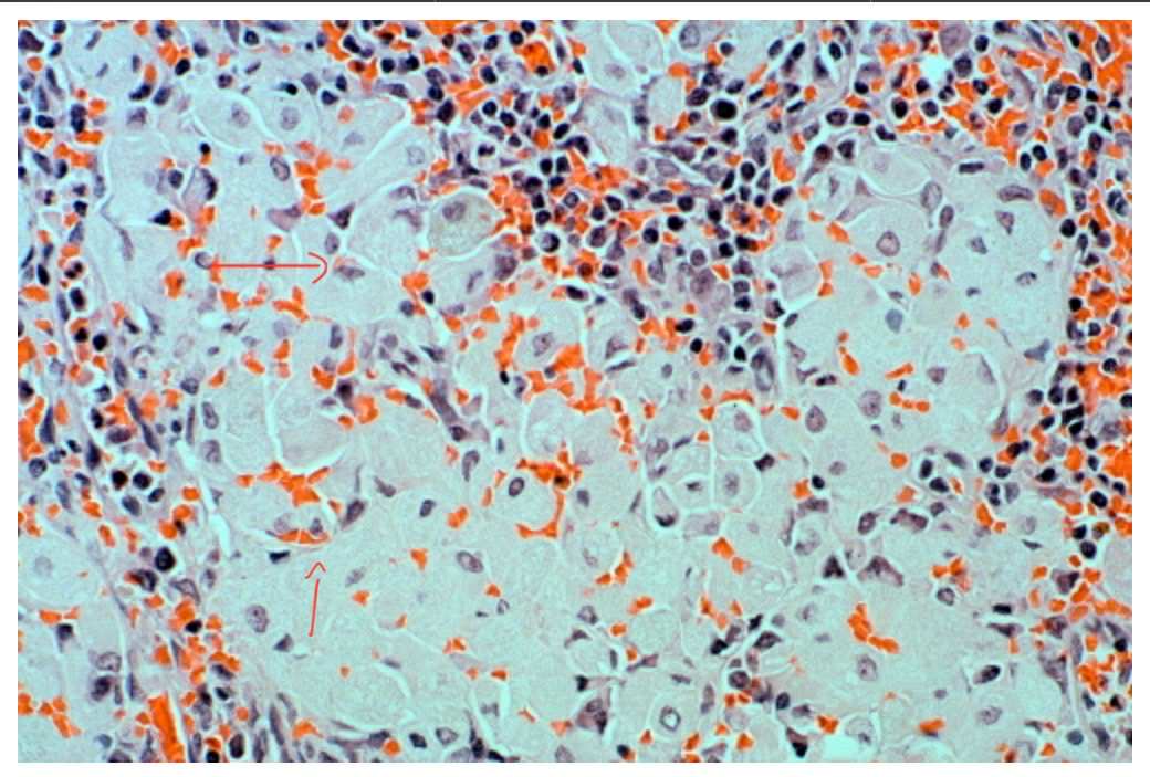
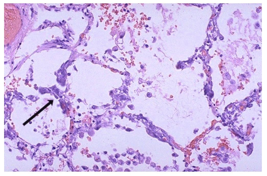
Histological section of an alveolus with macrophages. Identify what the brown coarsely granular material is. What results from this accumulation?
Hemosiderin. Breakdown of RBCs and release of iron in heme
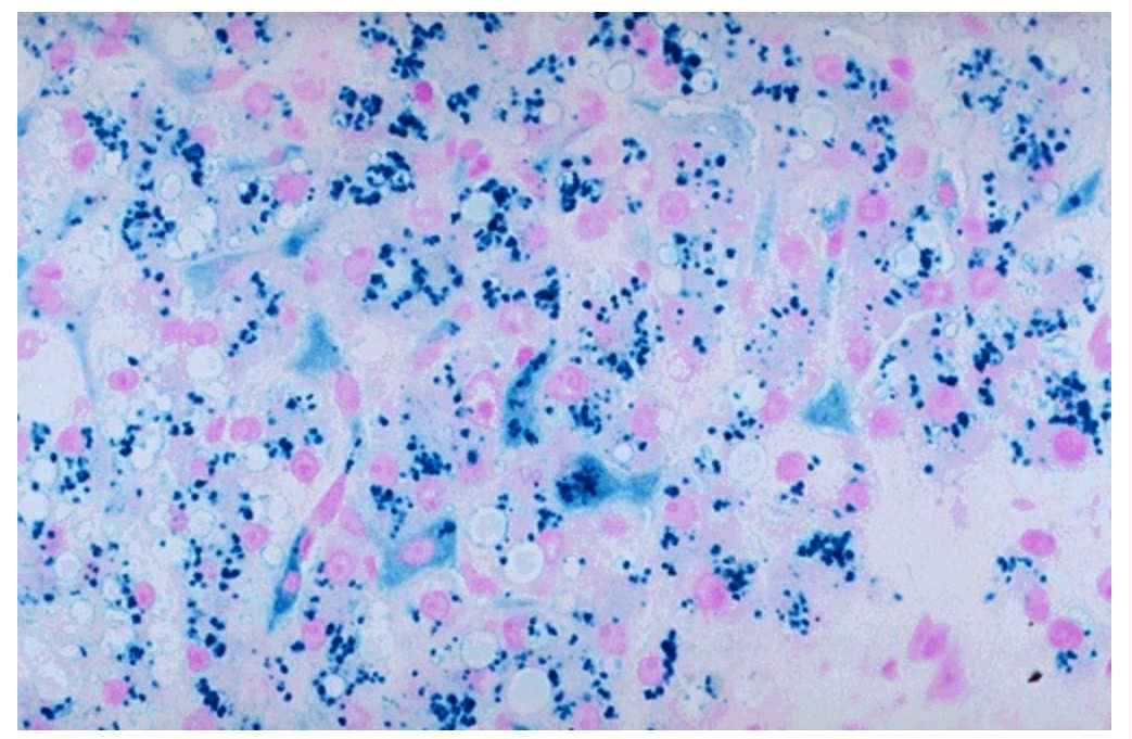
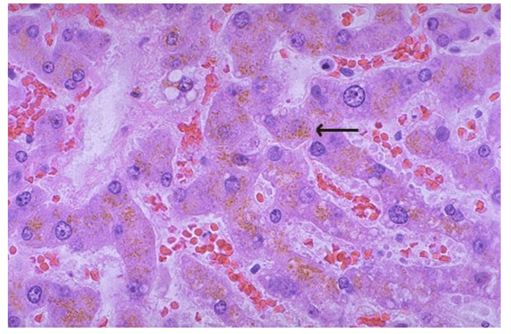
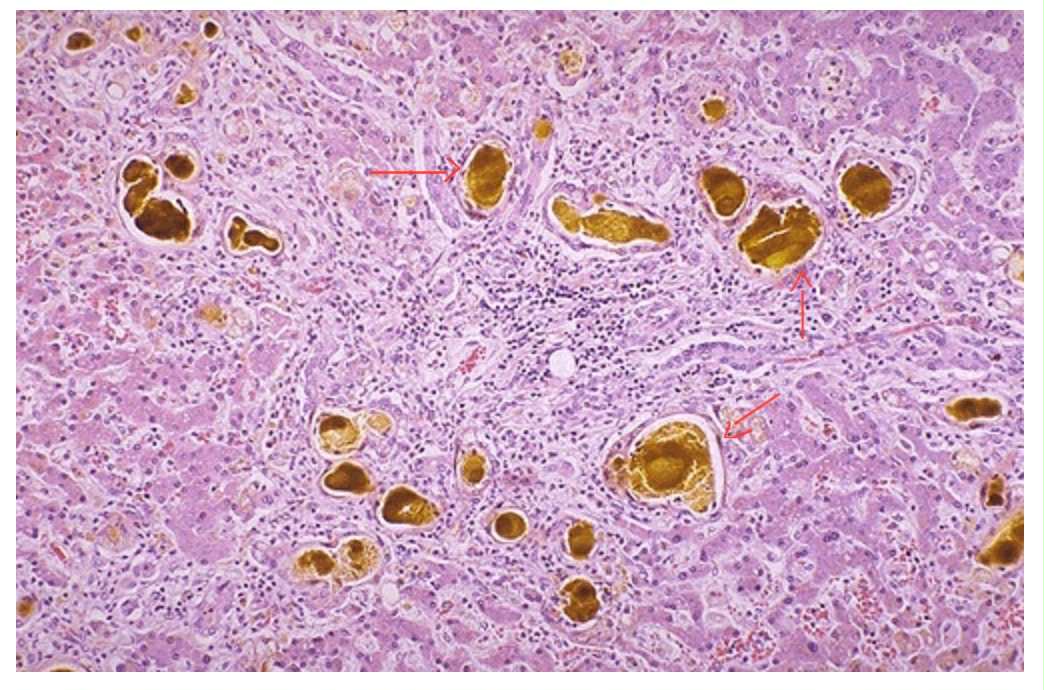
Prussian blue reaction in this histological slide of the liver with iron stain. Identify the pink pigments found in the cytoplasm of the hepatocytes and the Kupffer cells.
Hemosiderin
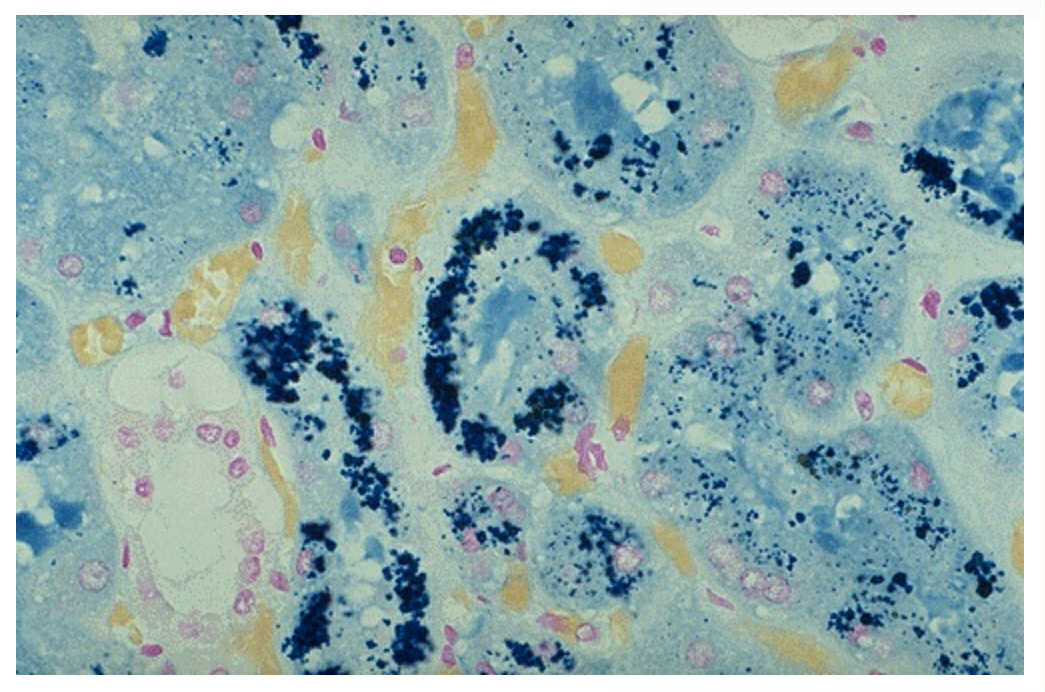
Prussian blue iron stain on a section of the kidney. The renal tubules contain large amounts of hemosiderin. This is observed in what kidney disorder?
Chronic Hematuria
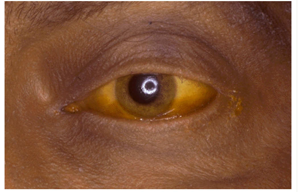
Sclera of the eye is yellow because the patient has _____ or ______. What laboratory test finding is most likely present?
Jaundice; Icterus. An elevated total serum bilirubin.
Yellow-green globular material is seen within the small bile ductules.
What is the name of the pigment?
This condition is a problem with excretion of bile or an obstruction to the flow of bile. The result would show the patient exhibiting jaundice.
Bilirubin
Hepatic cholestasis
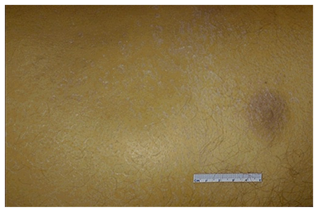
Where is the easiest site to detect icterus/jaundice?
Sclera
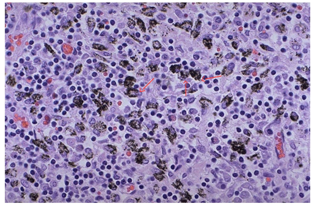
What are these black streaks seen in between lobules of the lung pleural surface?
Who are people that show more of these pigments in their lungs?
Anthracotic pigments
Smokers
This condition is an accumulation of carbon from breathing dirty air.
What are the cells in this hilar lymph node that contains the dark pigment?
Anthracosis
Macrophages
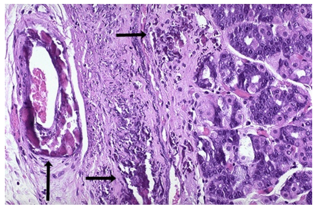
What are these bluish-purple deposits found in the submucosa, the artery, and the stomach wall?
What is the condition called?
Calcium deposits
Dystrophic calcification
This is a lung of a patient with high serum calcium level (hypercalcemia). What type of calcification is this?
Metastatic Calcification
Give the BMI values of the following:
Underweight
Normal
Overweight
Obese
<18.5 kg/m2
18.5-24.9 kg/m2
25-29.9 kg/m2
>30 kg/m2
Enumerate the methodologies you can measure with estimation of total body fat.
Dual Energy X-ray Absorptiometry (DEXA)
Bioelectrical Impedance Analysis (BIA)
Skin fold thickness
Hydrodensitometry
It was originally developed for measurement of bone mineral density. The energy absorbed by different tissues can be measured as differences in attenuation (brightness of the image obtained).
DEXA
It can provide an estimate of body fat based upon the relationship between the volume of the human body, as a conductor of a weak electric current, the subject's height, and the impedance (resistance to current travel through tissues).
BIA
Employed as a simple method for estimation of body fat. Estimates may take into account age, gender, nutritional status, genetic background, and levels of physical activity.
Skin fold thickness
Involves obtaining measurement of body composition by having the subject immersed in water and measuring the subject's weight in air and weight while immersed. Calculations are based upon assumptions regarding fat free mass (tissues such as muscle and bone) and fat mass, which have different densities.
Hydrodensitometry