Session 5.2: The Arm and Elbow
1/64
There's no tags or description
Looks like no tags are added yet.
Name | Mastery | Learn | Test | Matching | Spaced | Call with Kai | Chat |
|---|
No analytics yet
Send a link to your students to track their progress
65 Terms
Anterior compartment
Flexor compartment
Muscles act on the shoulder/elbow to flex them
Posterior compartment
Extensor compartment
Muscles act to extend the elbow
All the muscles in the anterior compartment of the arm are innervated by...
The musculocutaneous nerve
All the muscles in the posterior compartment of the arm are innervated by...
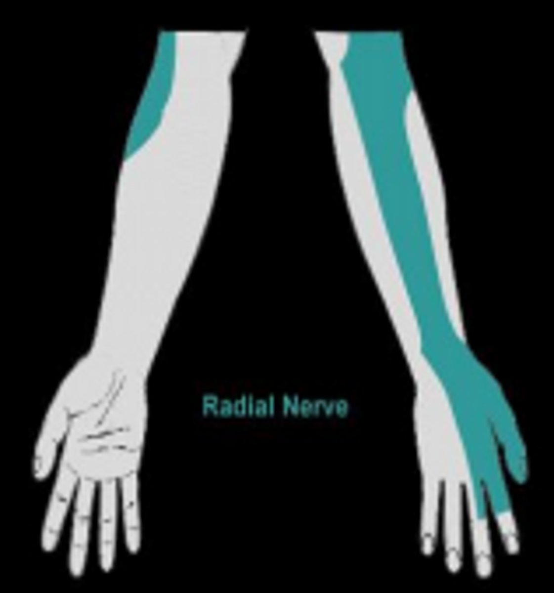
The radial nerve
Surgical neck of the humerus is prone to...
Fracture
Risk of axillary nerve damage

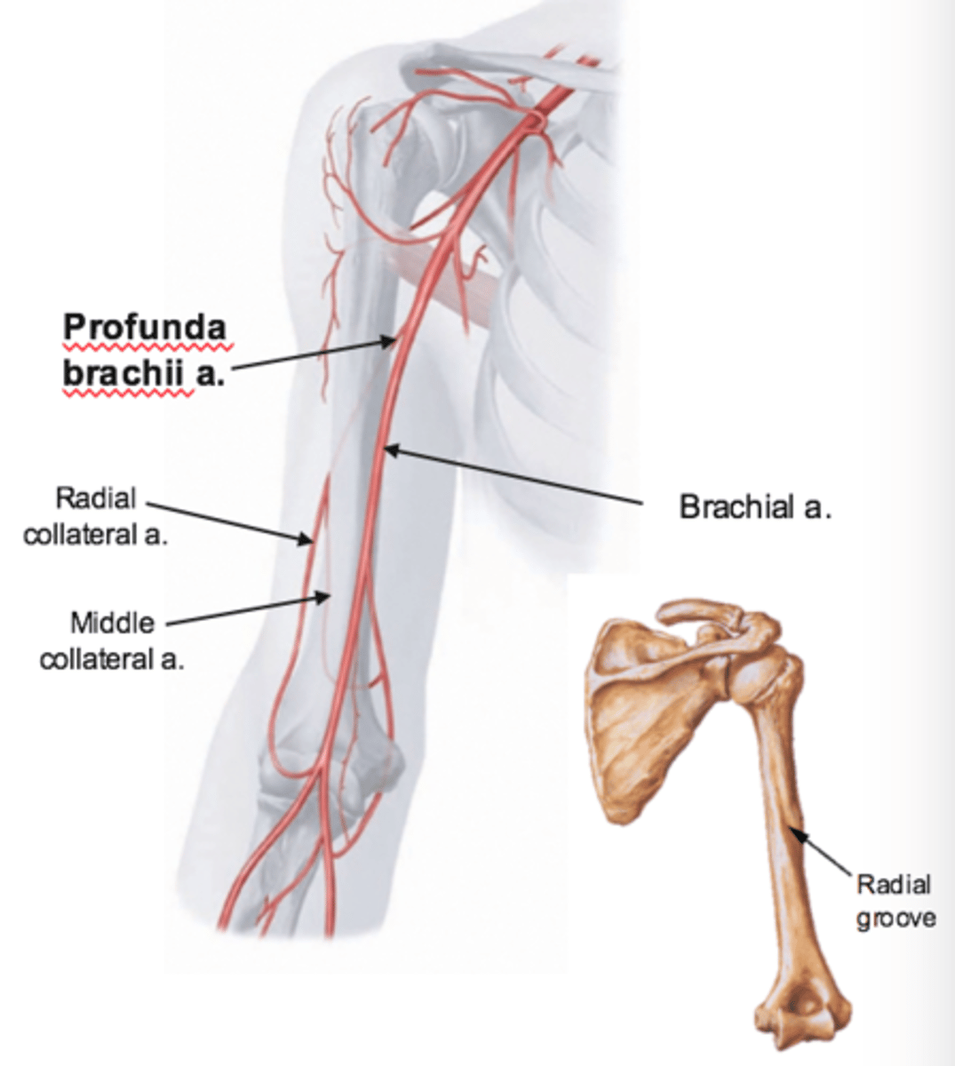
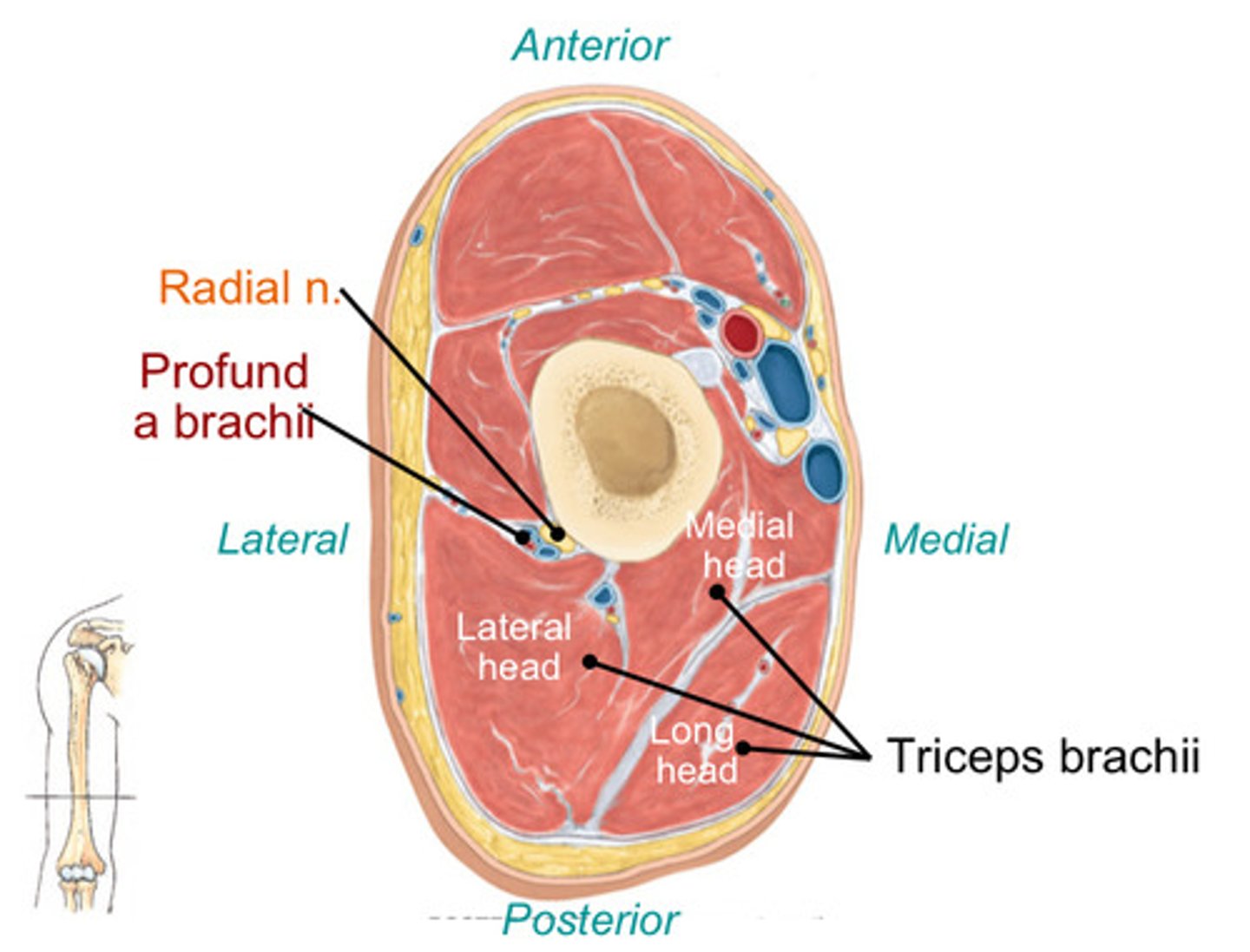
Shaft of the humerus is prone to...
Mid-shaft fractures through the spiral groove (posteriorly) can damage the radial nerve & profunda brachii artery
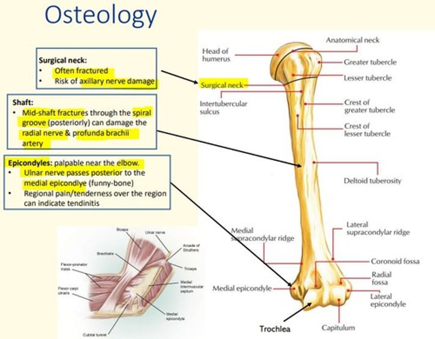
What nerve passes posterior to the medial epicondyle (funny bone) of the humerus?
Ulnar nerve
Regional pain/tenderness over this region can indicate tendoinitis

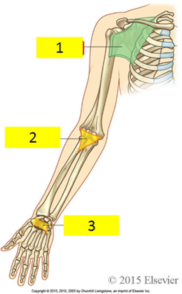
Areas of transition in the upper limb
Axilla, cubital fossa, carpal tunnel
Compartments of the arm
Anterior: flexion of shoulder and elbow
Posterior: extension of shoulder and elbow
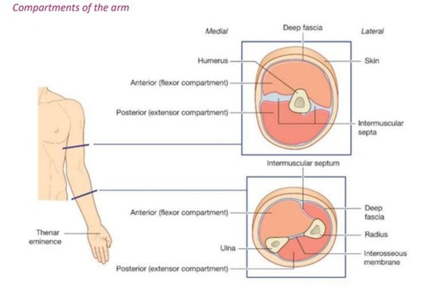
The compartments of the arm are enclosed in a ___ ___ with two ___ ___, attached to the humerus bone.
The compartments of the arm are enclosed in a deep fascia with two fascial septa, attached to the humerus bone.

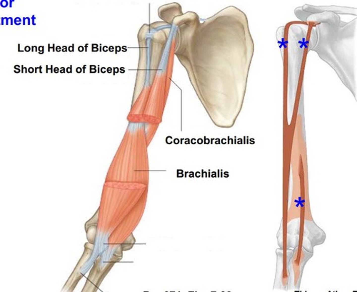
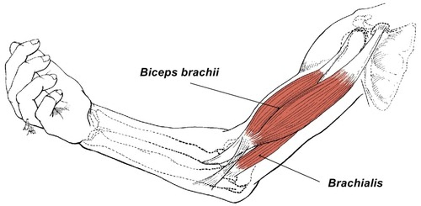
What are the muscles in the anterior compartment of the arm
Biceps brachii
Corachobrachialis
Brachialis
What is the blood supply to the anterior compartment of the arm
Brachial artery
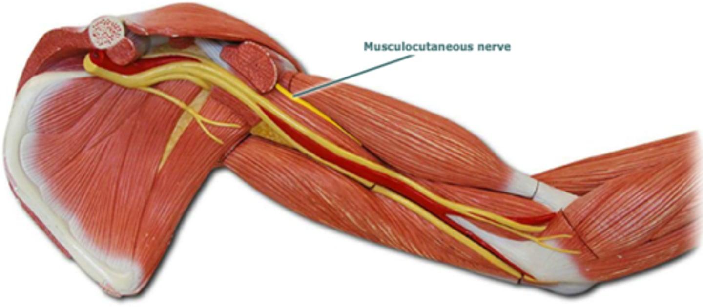
What is the nerve supply to the anterior compartment of the arm
Musculocutaneous nerve
Biceps brachii
Flexes forearm and supinates forearm
Innervated by musculocutaneous nerve (C5 - C7)
Corachobrachialis
Flexes and adducts arm
Innervated by musculocutaneous nerve (C5 - C7)
Brachialis
Flexes forearm in all positions
Innervated by musculocutaneous nerve (C5 - C6) and radial nerve (C5 - C7)
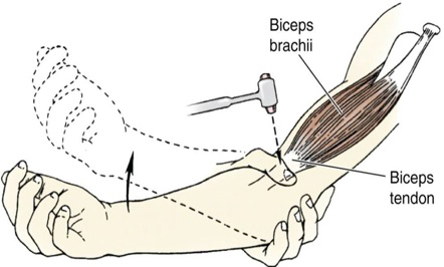
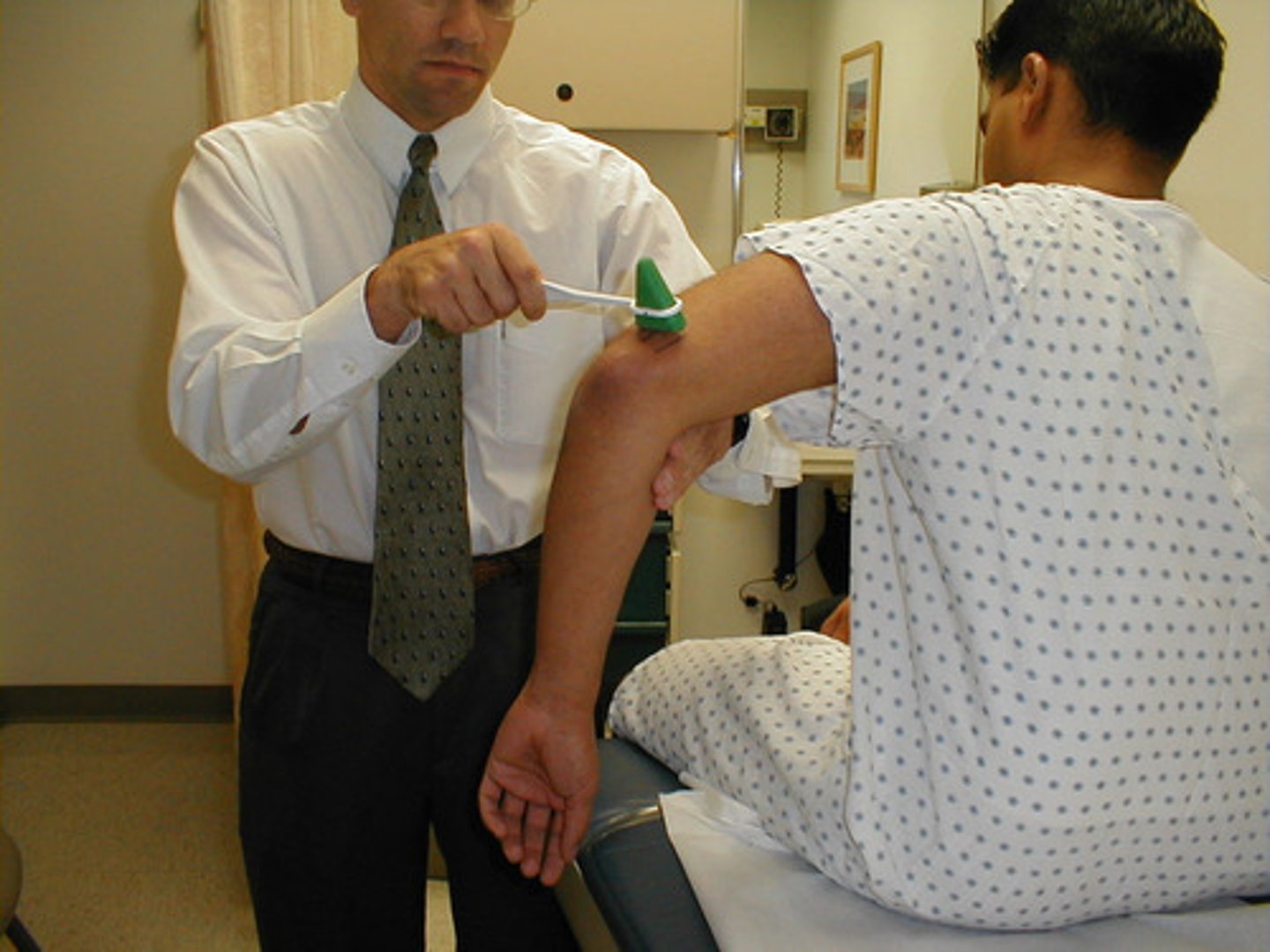
Testing for the C5, C6 reflex
Tap the biceps tendon
What structures pass through the anterior compartment of the arm?
- Musculocutaneous nerve
- Median nerve
- Ulnar nerve
- Brachial artery
- Basilic vein
- Radial nerve (lower end of the compartment)
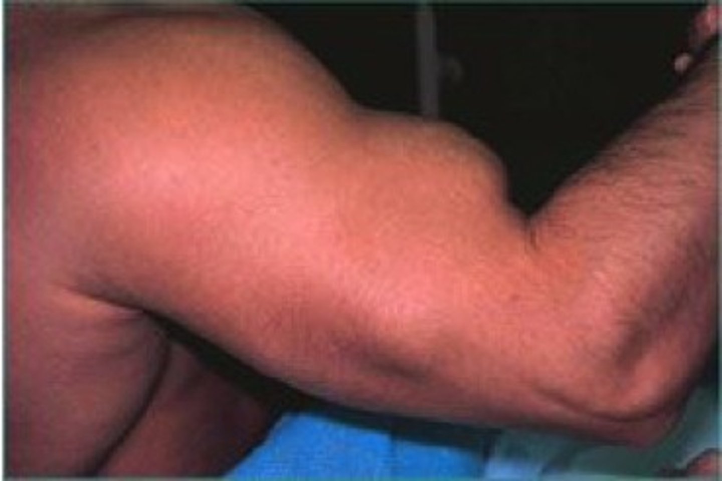
Popeye deformity
Rupture of biceps long head tendon
Torn from attachment at supraglenoid tubercle of scapula
Detached muscle belly forms a ball
Occurs due to wear/tear
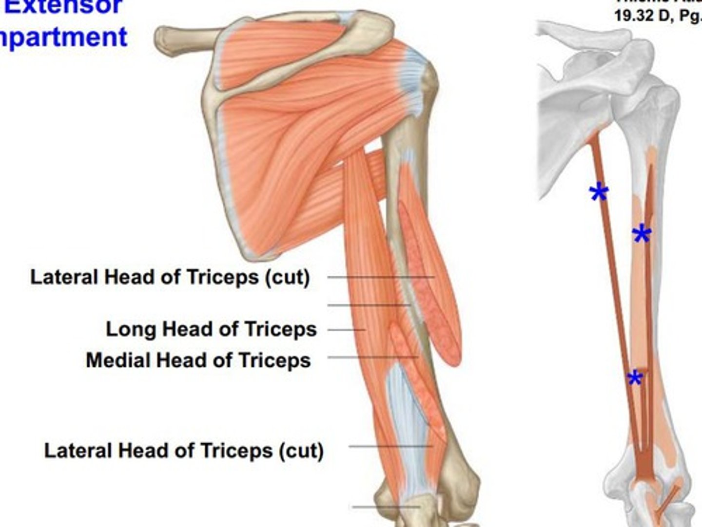
What are the muscles in the posterior compartment of the arm
Triceps brachii
Anconeus
What is the blood supply to the posterior compartment of the arm
Profunda brachii
Ulnar collateral arteries
What is the nerve supply to the posterior compartment of the arm
Radial nerve
Triceps brachii
Extends forearm at elbow
Innervated by the radial nerve (C6 - C8)
Anconeus
Extends forearm at elbow; stabilises elbow joint
Innervated by the radial nerve (C7 - T1)
What structures pass through the posterior compartment of the arm?
- Radial nerve
- Ulnar nerve
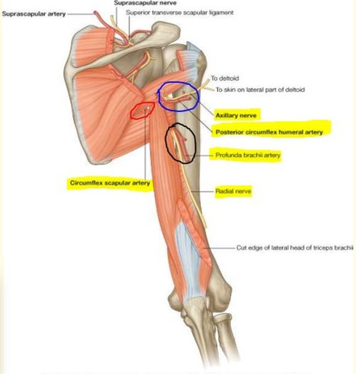
Posterior Compartment of the Arm
What structures pass through the quadrangular space?
Axillary nerve
Posterior circumflex arteries

Posterior Compartment of the Arm
What structures pass through the triangular interval & spiral groove?
Radial nerve
Profunda artery

Posterior Compartment of the Arm
What structures pass through the triangular space?
Circumflex scapular arteries
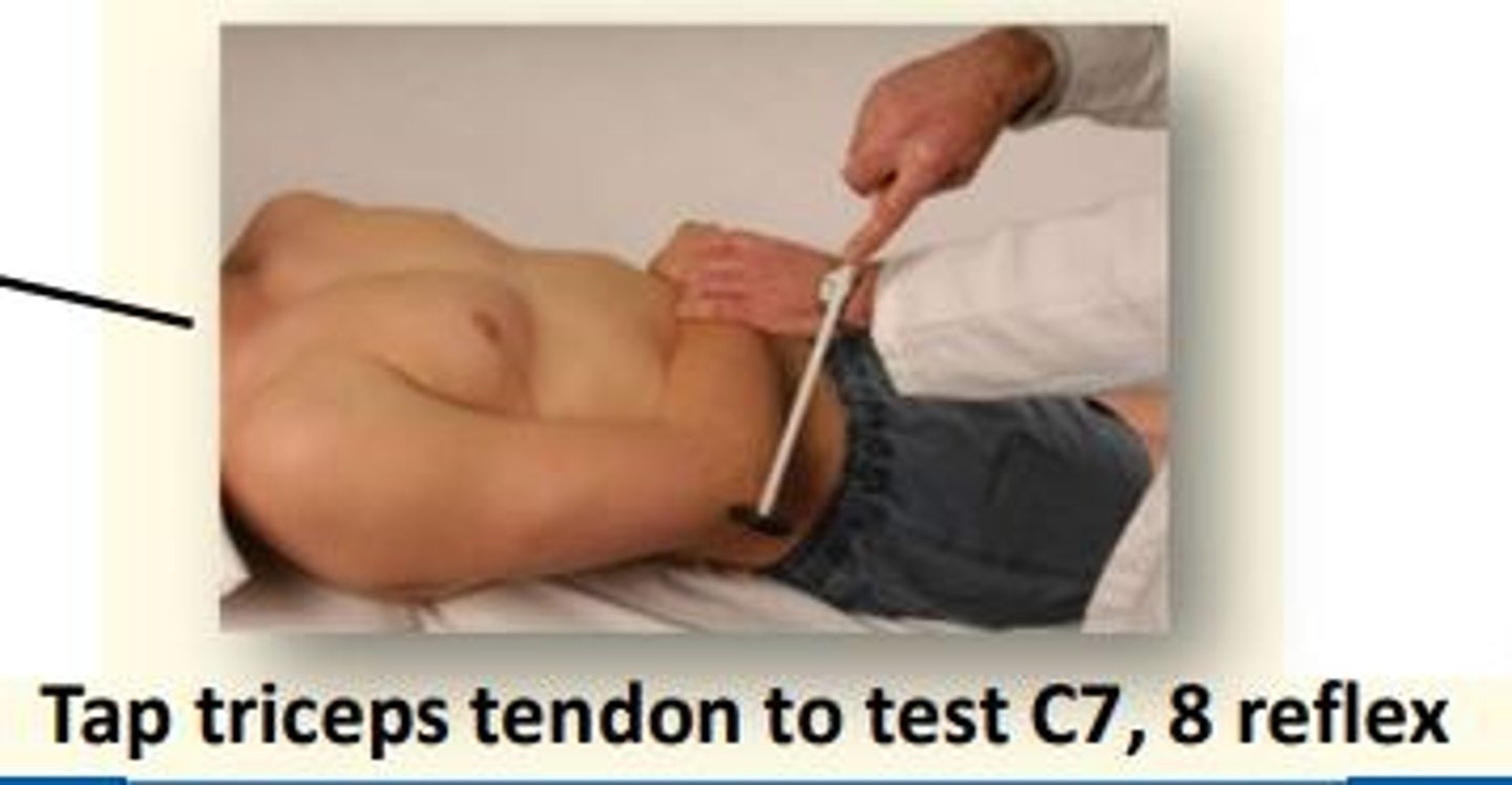
Testing the C7, 8 reflex
Tap the triceps tendon
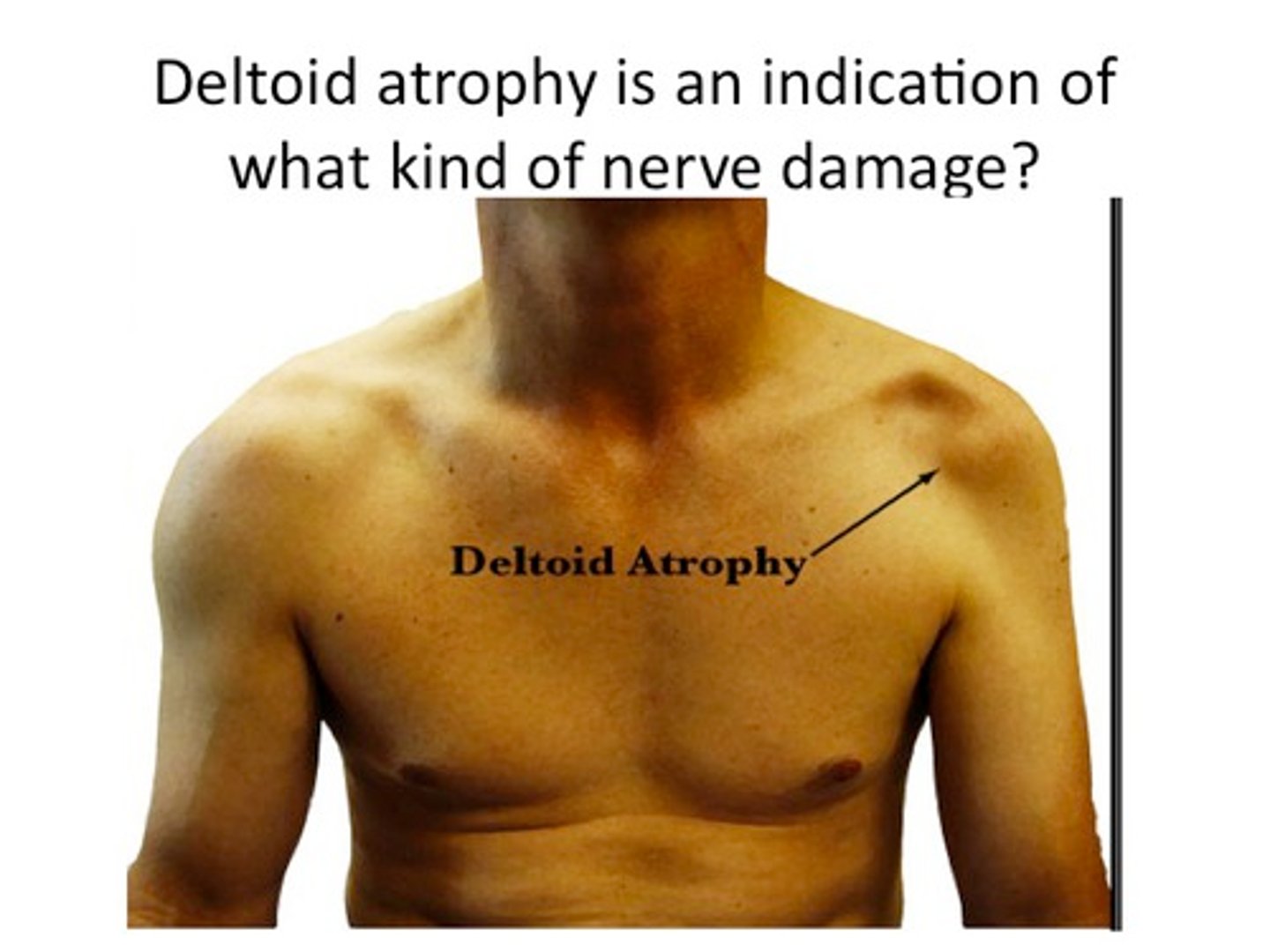
Axillary nerve
Mixed sensory & motor nerve (C5, 6)
Innervates teres minor & deltoid muscles
Sensation to skin above deltoid (regimental badge area) and shoulder joint
Axillary nerve damage
The axillary nerve can become damaged when the shoulder dislocates
Damage to the axillary nerve leads to weak abduction (deltoid) and weak external rotation (teres minor).
Musculocutaneous nerve
Mixed sensory & motor nerve (C5-C7)
Innervates BBC (biceps brachii, brachialis, corachobrachialis)
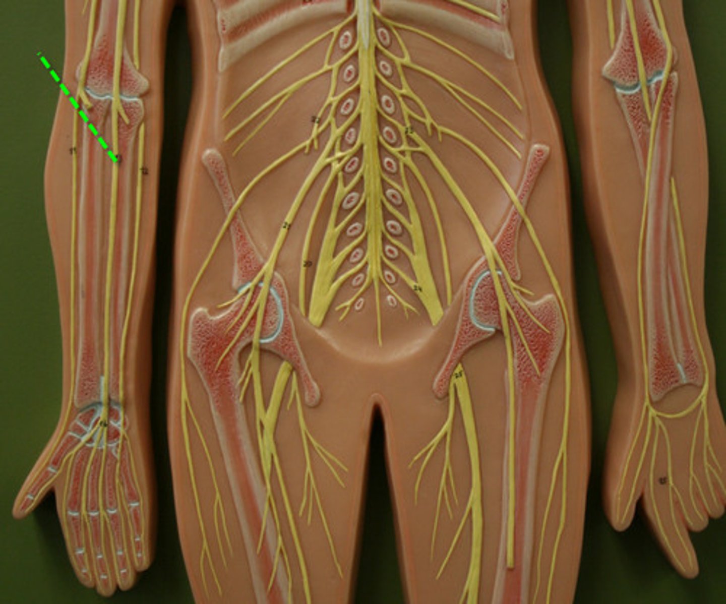
Radial nerve
Mixed sensory & motor nerve (C5-T1)
Radial nerve lies under brachioradialis and divides into superficial radial nerve (dosrum of hand) and posterior interosseous nerves (extensors of forearm)

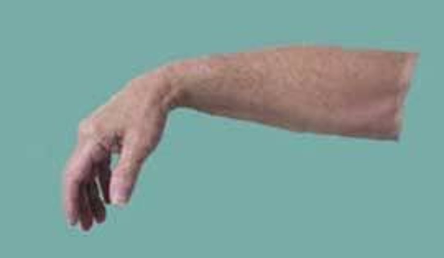
Radial nerve damage
Radial nerve can become damaged by humeral fractures involving the spiral groove (mid-shaft).
This leads to wrist drop & reduced grip strength
Anaesthesia of the 1st dorsal web space
Median nerve
Mixed sensory & motor nerve (C6-T1)
Travels with the brachial artery
Gives off the anterior interosseous nerve below cubital fossa which supplies deep muscles of the flexor compartment.
Median nerve continues down the forearm and gives off the palmar cutaneous branch just before the wrist - which innervates thenar eminance.
Ulnar nerve
Mixed sensory & motor nerve (C8, T1)
Descends medially under medial epicondyle of elbow to enter forearm. Innervates the flexor carpi ulnaris and half of the flexor digitorum profundus.

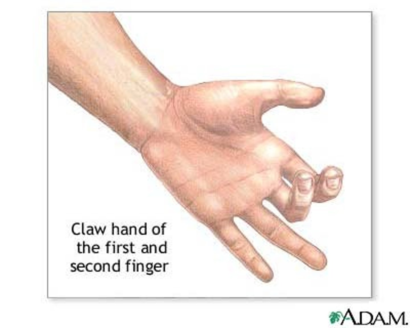
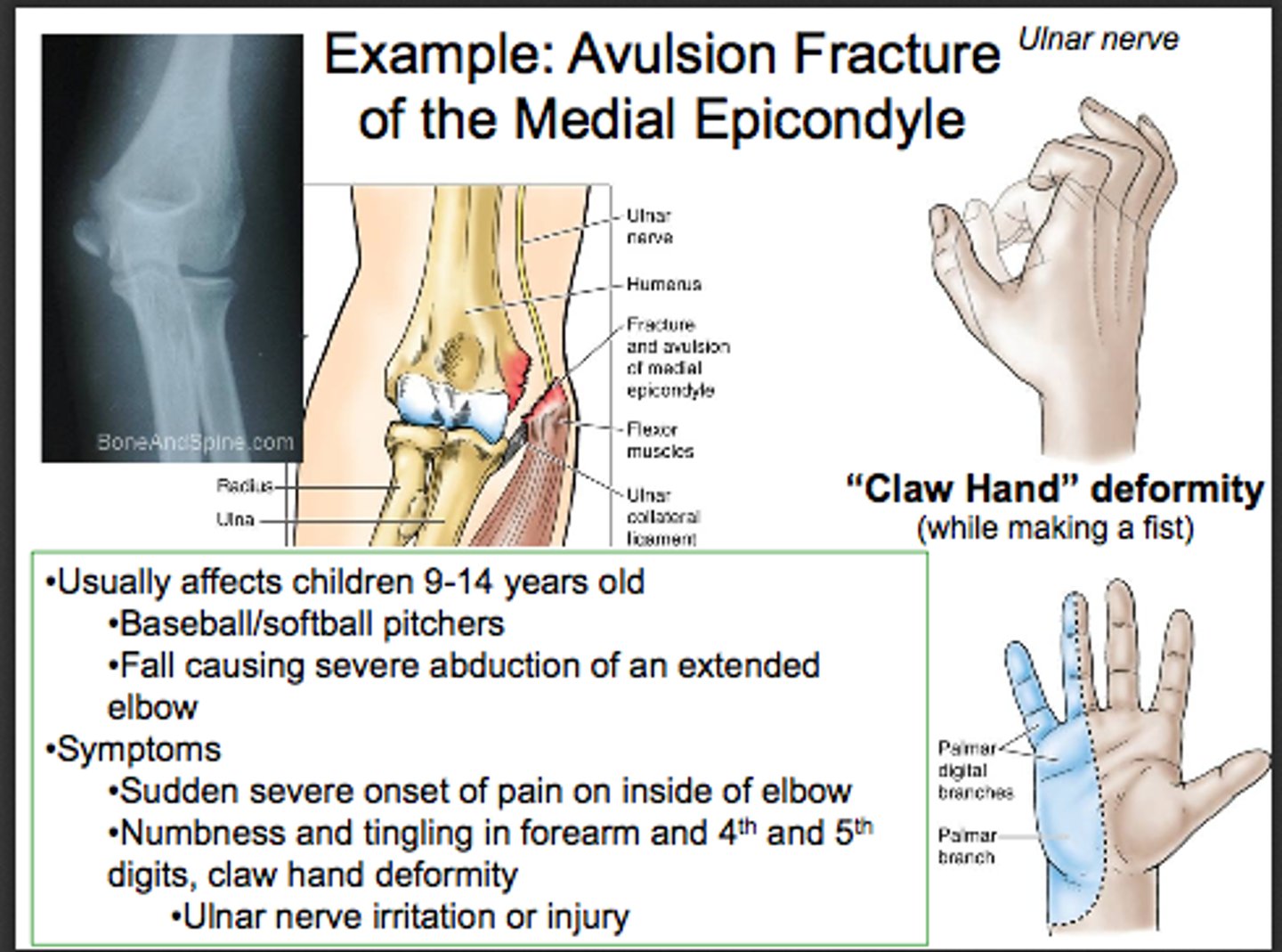
Ulnar nerve injury
Claw hand deformity
Small muscles of hand wasted (except thenar eminance and lateral lumbricals which are innervated by the median nerve).
Myotome
Group of muscles innervated by a single spinal nerve
Myotome testing is an essential part of the neurological examination when suspecting radiculopathy
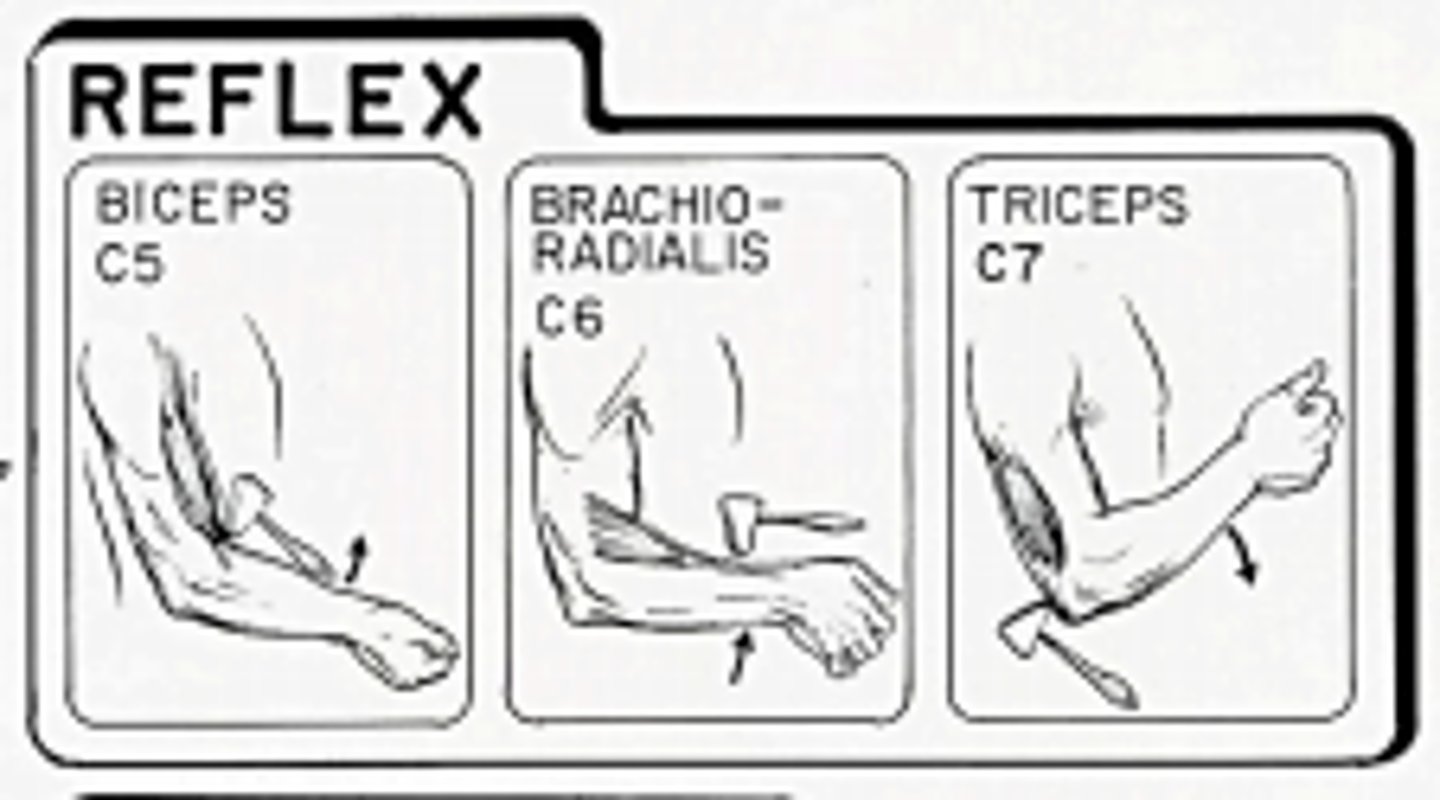
Deep tendon reflexes
Biceps
C5 + C6
Deep tendon reflexes
Triceps
C7, C8
Deep tendon reflexes
Brachio-radialis
C5 + C6 + RADIAL NERVE
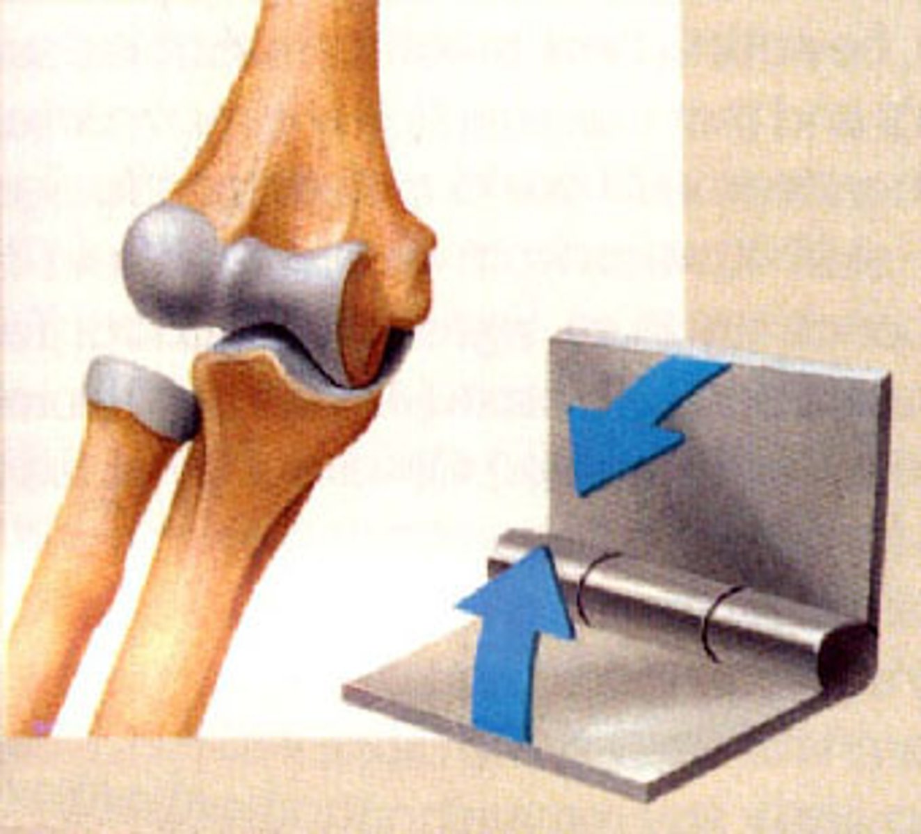
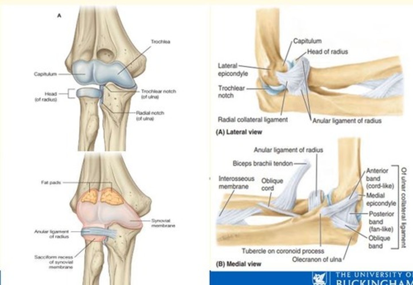
What type of joint is the elbow
Synovial hinge joint
What ligaments support the elbow?
Medial collateral ligament
Lateral collateral ligament
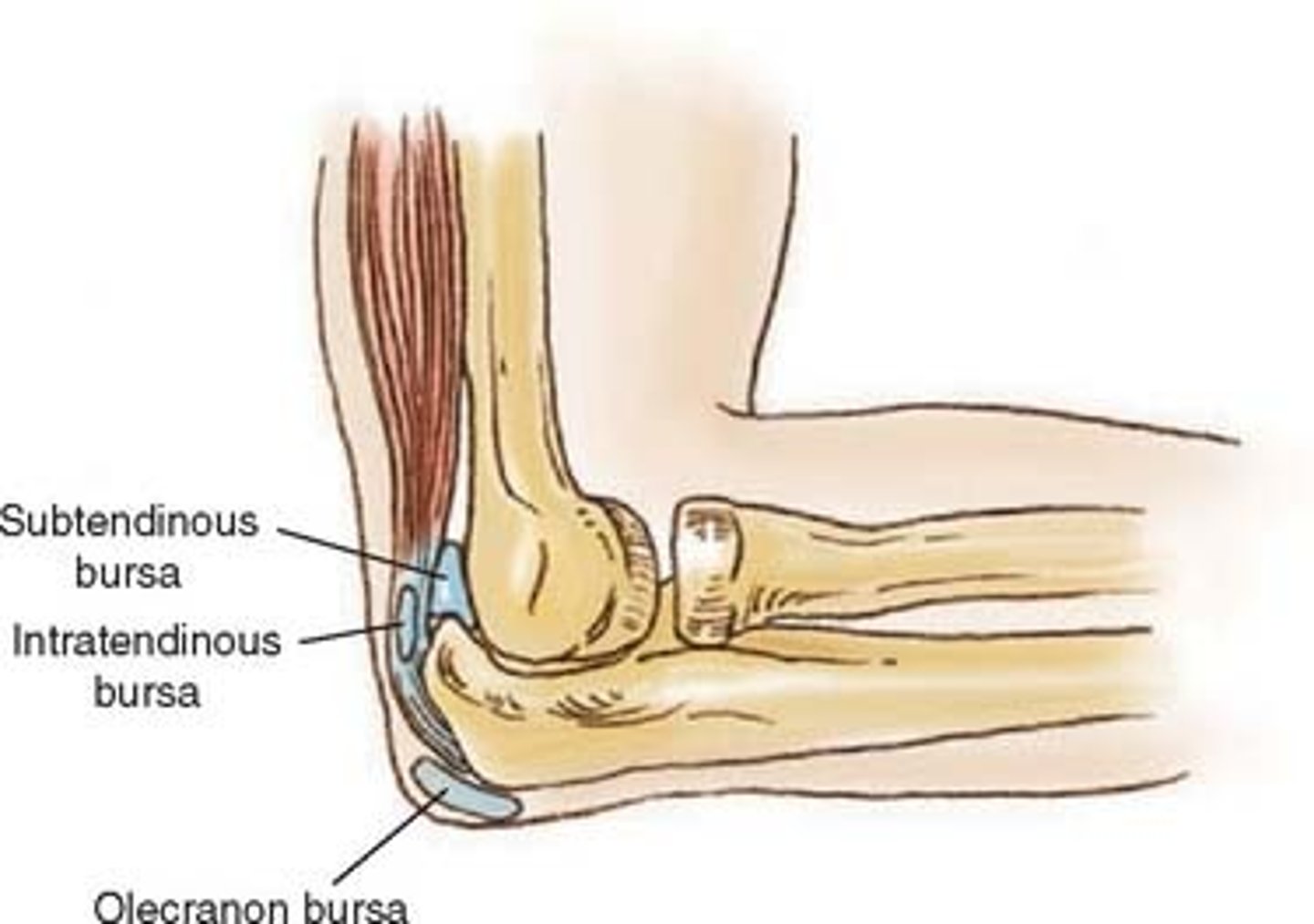
Bursae found around elbow joint
- Olecranon bursa (overlies olecranon posteriorly)
- Subtendinous bursa (between triceps and olecranon)

Olecranon bursa
Lies between olecranon process and skin
Subtendinous bursa
Bursa that prevents friction between bone and a muscle tendon
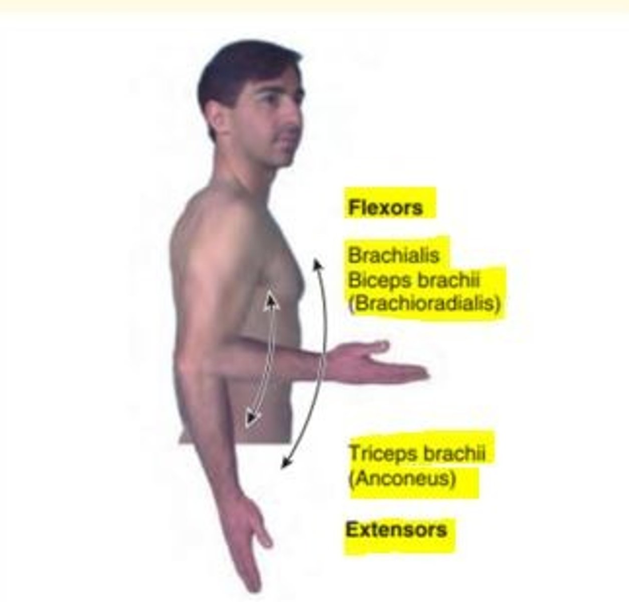
Movements at the elbow joint
Flexion and extension
CHIEF FLEXORS
- Brachialis
- Biceps brachii
MAIN EXTENSORS
- Triceps brachii
- Anconeus
Elbow injuries: avulsion fracture of medial epicondyle
This injury can occur after a fall caused by severe abduction of fully extended forearm
Elbow injuries: posterior elbow dislocation
This injury can occur after fall on outstretched hand (FOOSH)
___ nerve is vulnerable to damage with injury, fracture or dislocation of the elbow
Ulnar nerve is vulnerable to damage with injury, fracture or dislocation of the elbow
Cubital fossa
Elbow pit
Depression on the anterior aspect of the elbow

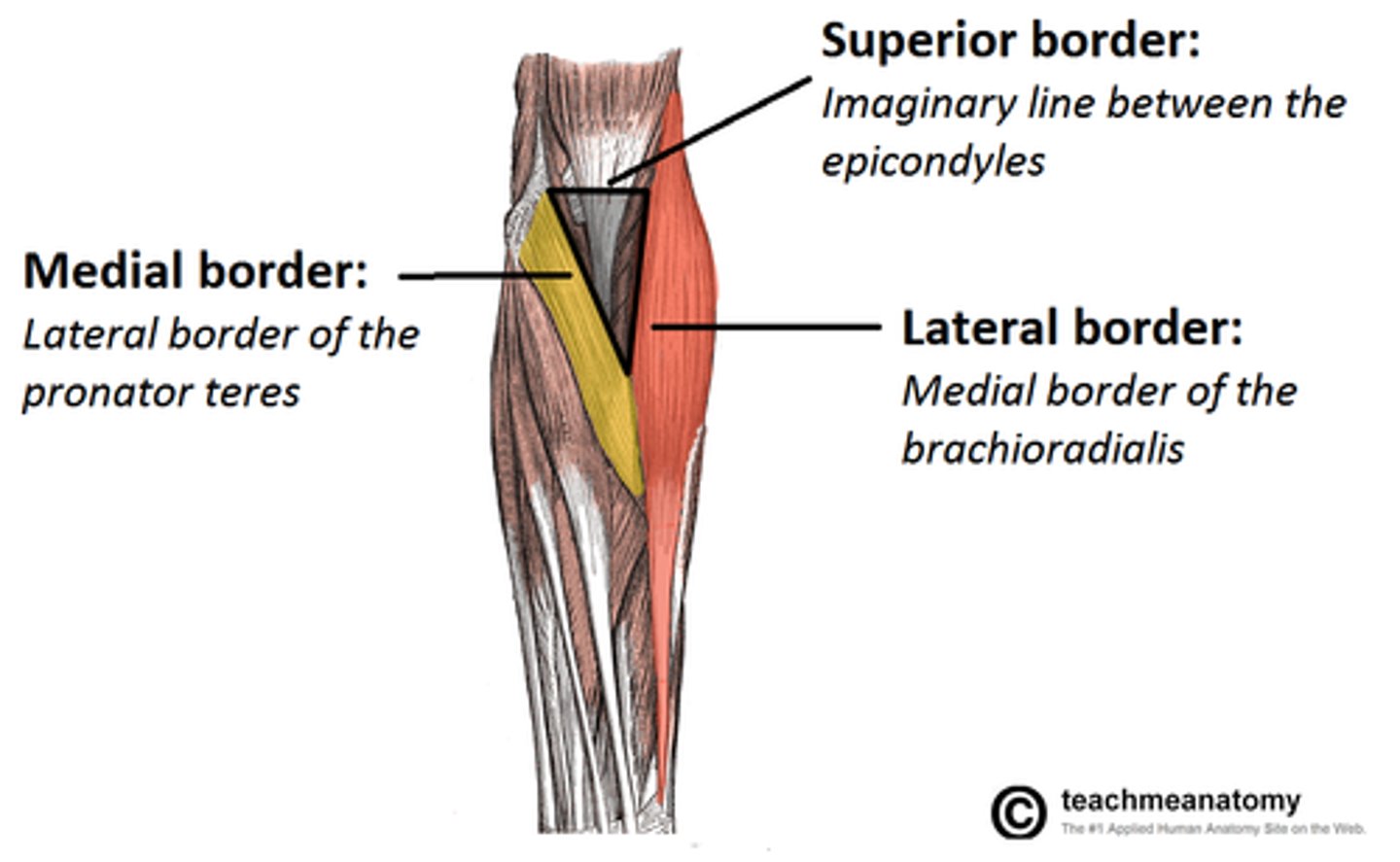
Borders of the cubital fossa
- Lateral = Brachioradialis (lateral)
- Medial = Pronator teres (medial)
- Superior = Imaginary line between two humeral epicondyles
Cubital fossa floor and roof
Floor = supinator muscle lateraly & brachialis muscle medially
Roof = skin & fascia, reinforced by bicipital aponeurosis
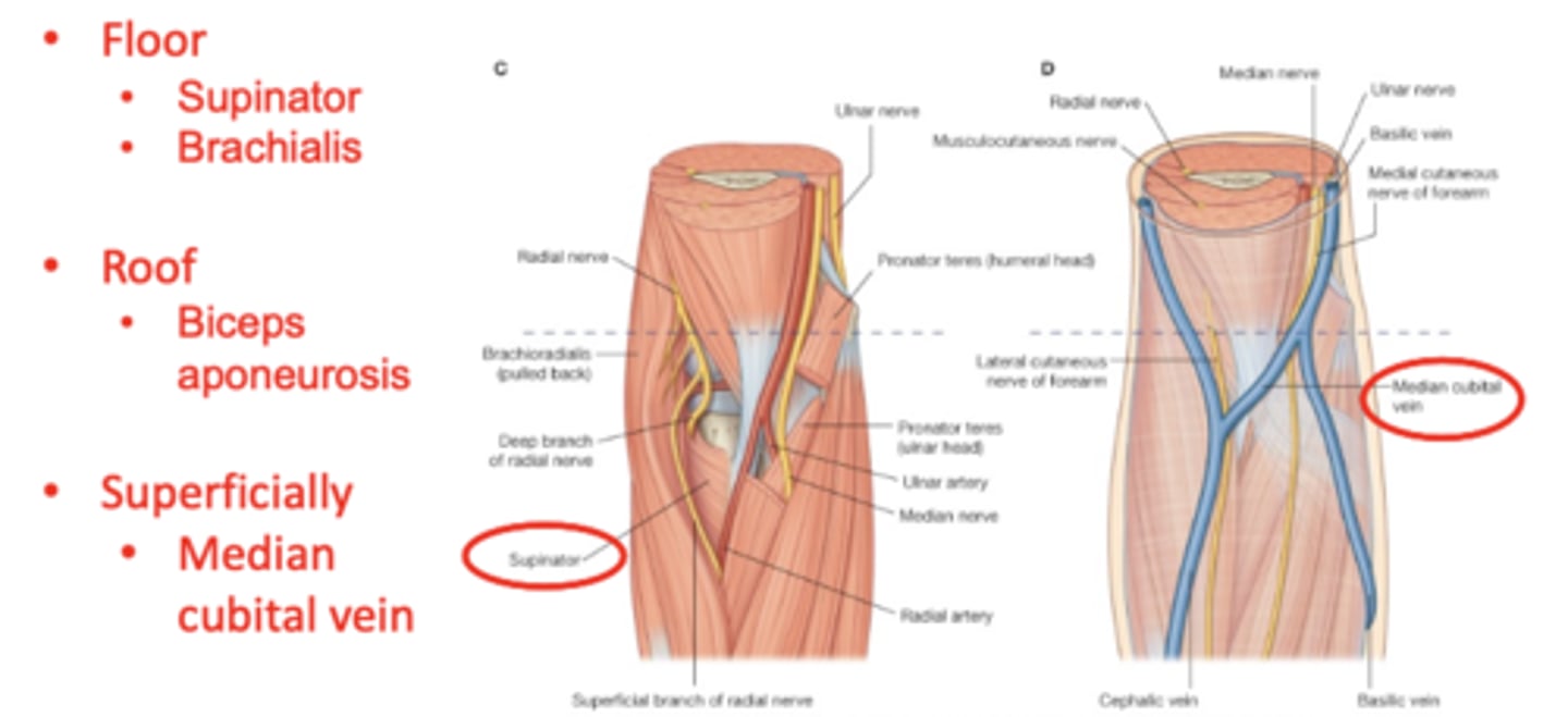
Contents of the cubital fossa
From medial to lateral:
- Median nerve
- Bifurcation of brachial artery into ulnar/radial
- Tendon of biceps muscle
- Radial nerve and its deep branch

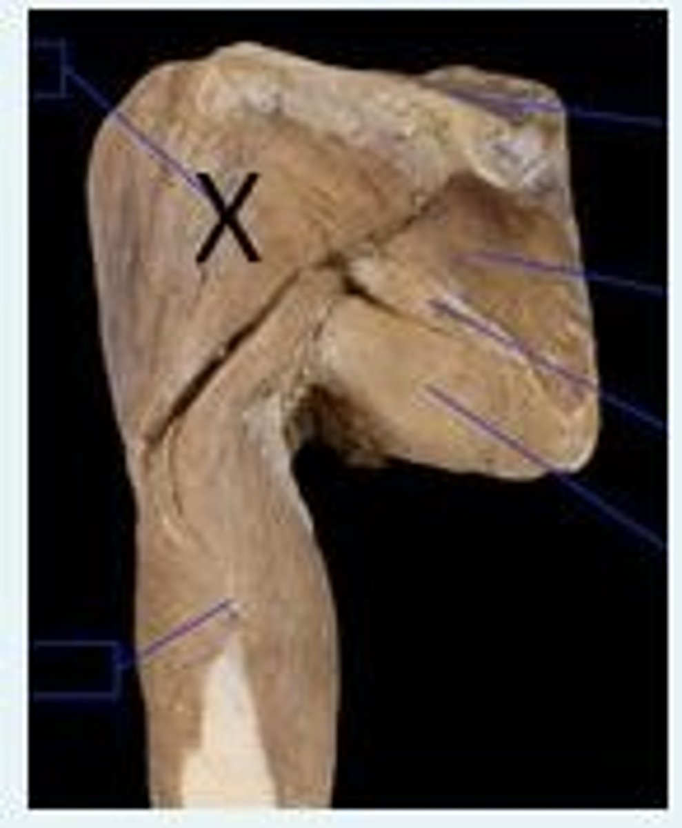
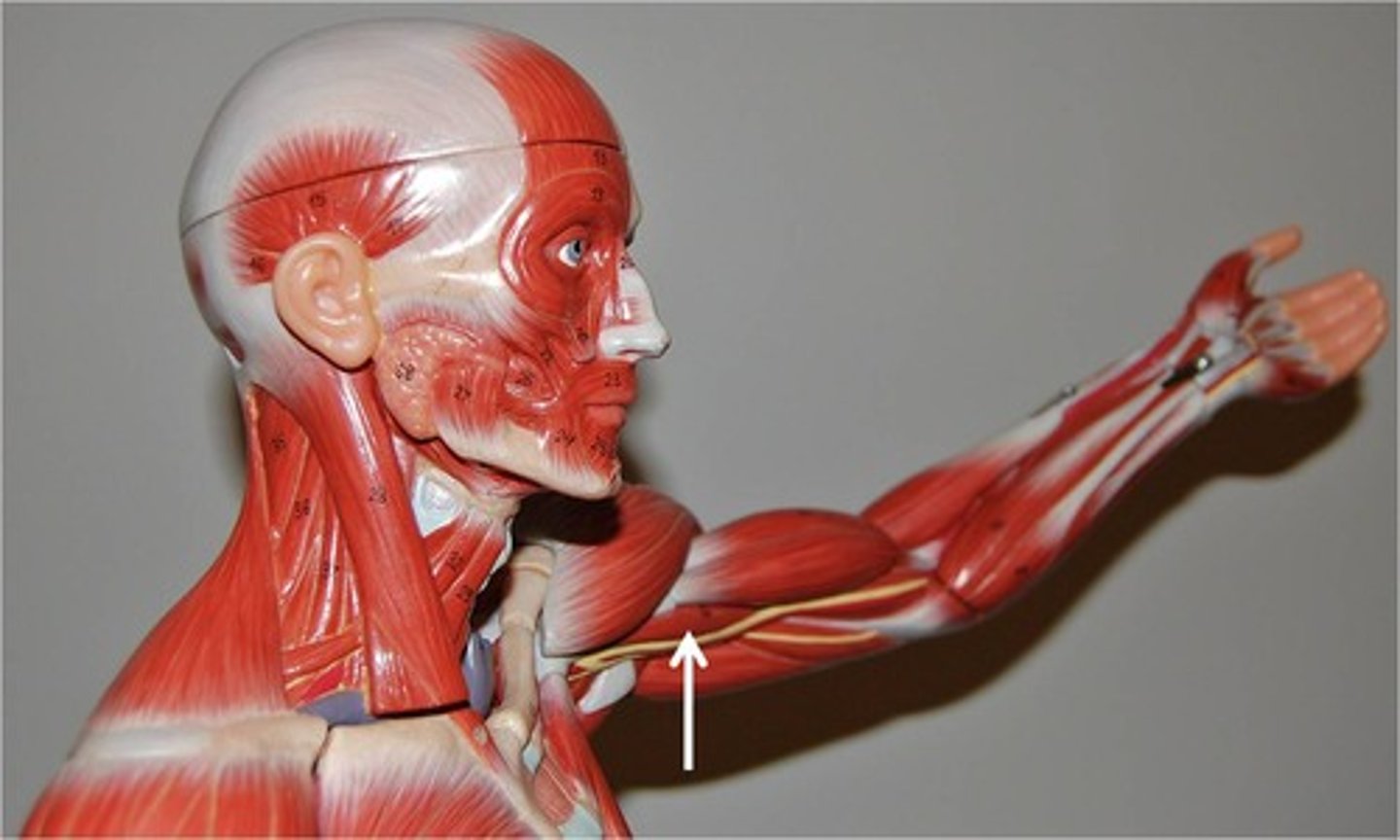
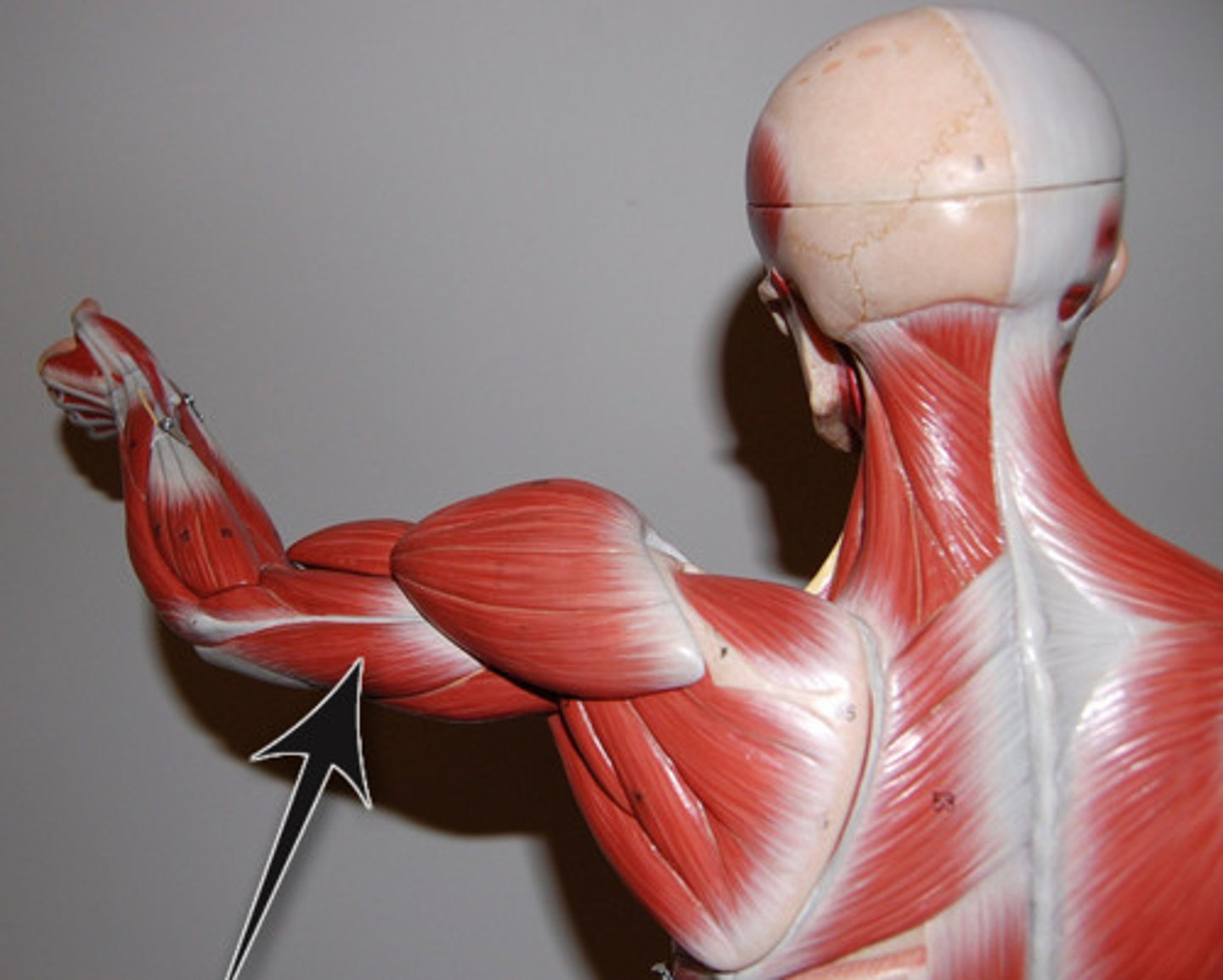
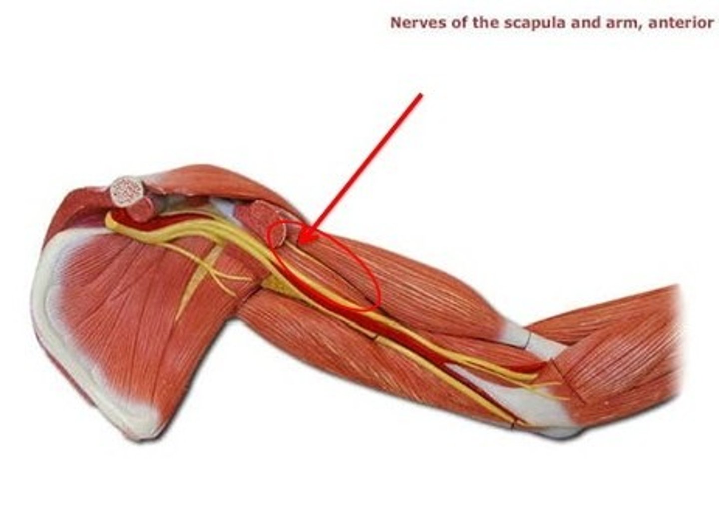
The muscle labelled X in the image above is the ___ muscle.
The nerve that supplies this muscle is the ___ nerve.
The muscle labelled X in the image above is the deltoid muscle.
The nerve that supplies this muscle is the axillary nerve.
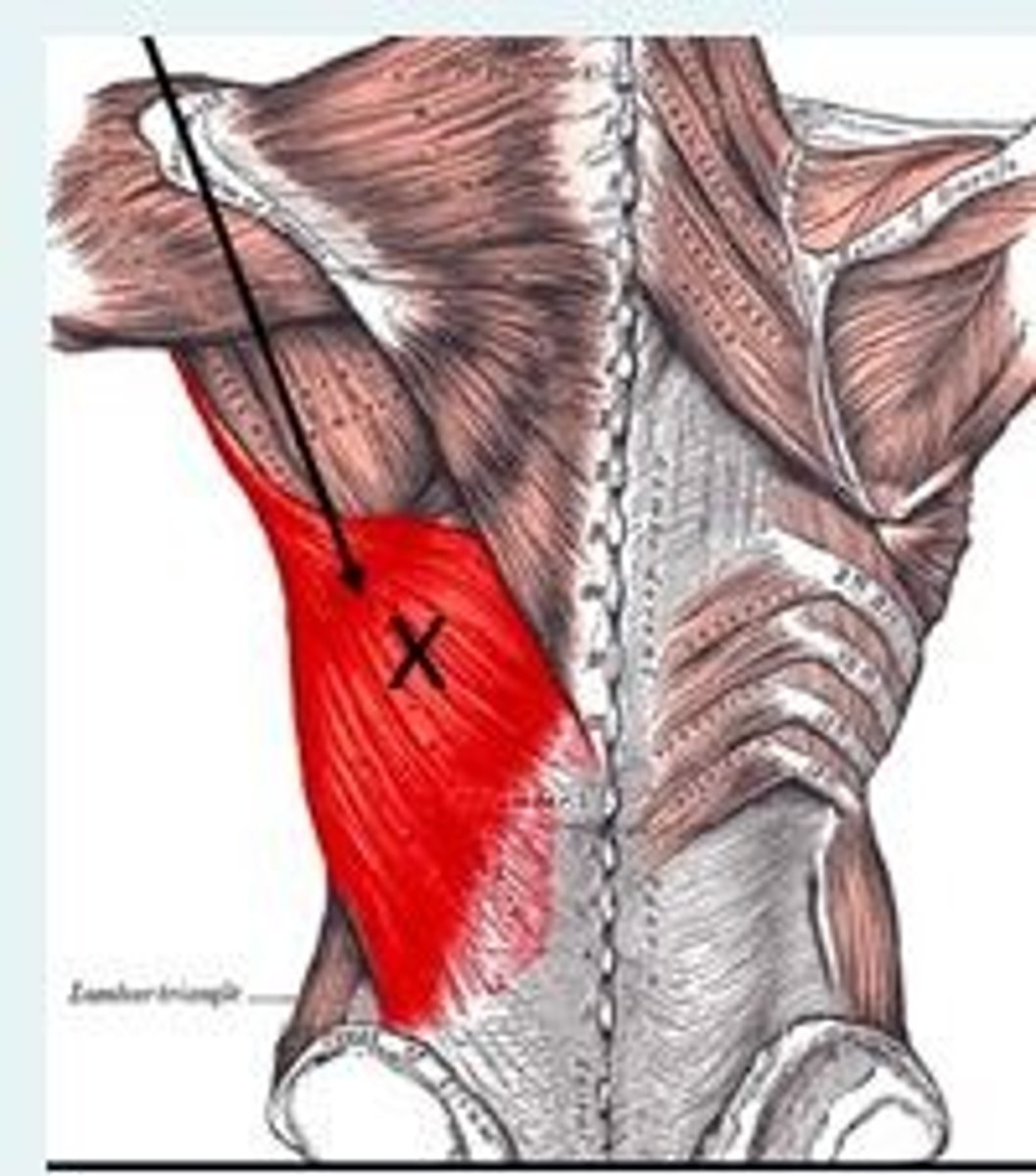
The muscle labelled X in the image above is the ___ muscle.
This muscle forms the ___ wall of the axilla.
The muscle labelled X in the image above is the latissimus dorsi muscle.
This muscle forms the posterior wall of the axilla.
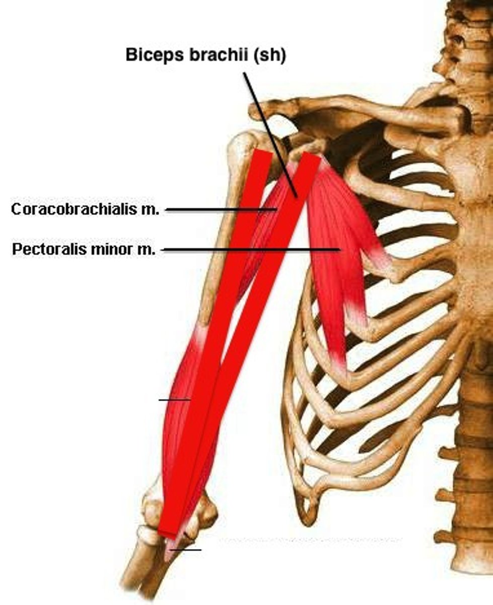
The biceps are supplied by the ___ nerve
Musculocutaneous
The three muscles have their origin at the coracoid process are...
a) Corachobrachialis, pec major, short head of biceps
b) Short head of biceps, long head of biceps, coracobrachialis
c) Long head of biceps, coracobrachialis, pec minor
d) Short head of biceps, pec minor, corachobrachialis
d) Short head of biceps, pec minor, corachobrachialis
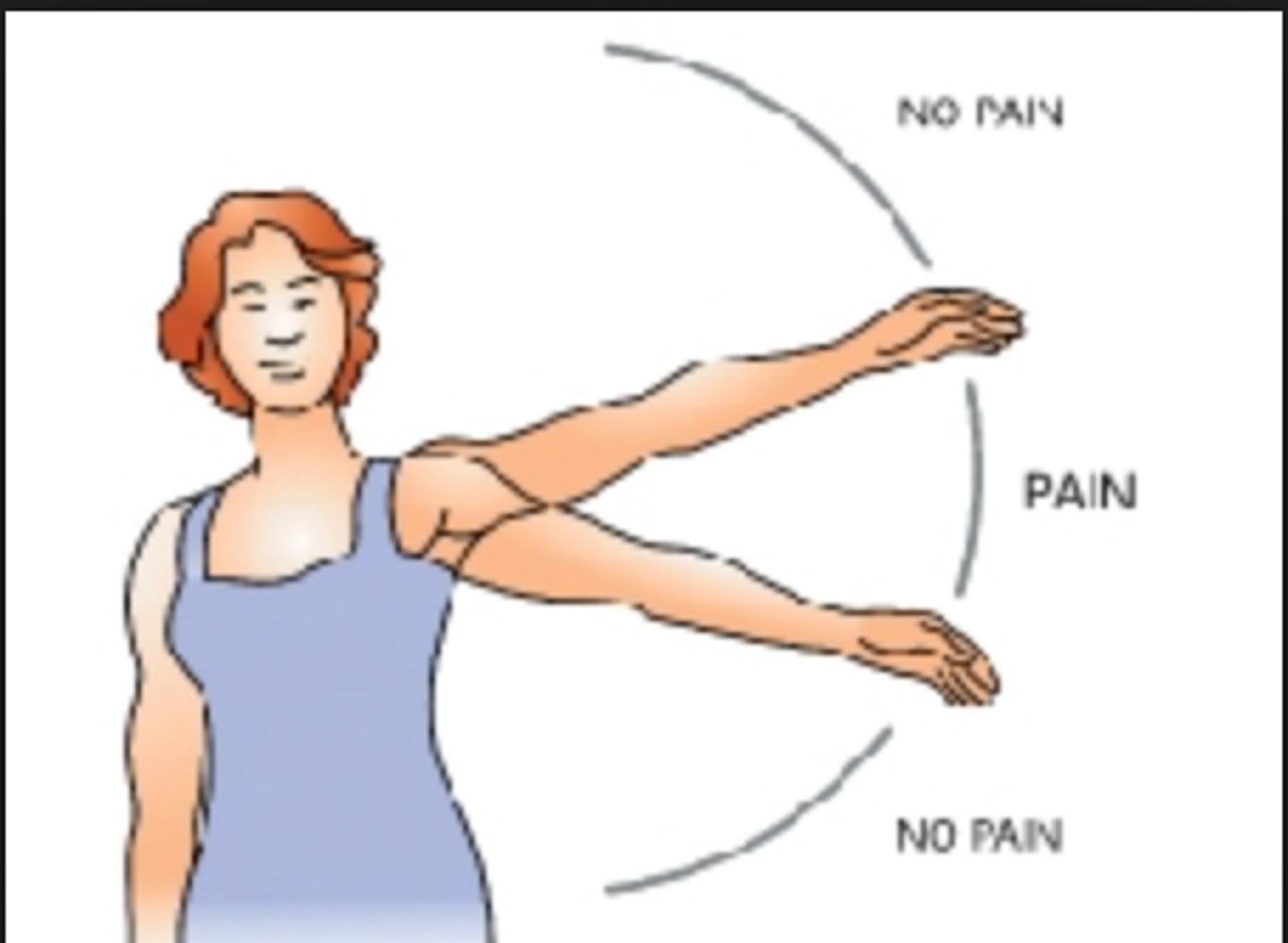
On examination, a patient experienced pain when abducting their shoulder between 60 and 120 degrees. The tendon for which one of the following muscles is most likely to be inflamed in this case?
a) Infraspinatus
b) Subscapularis
c) Supraspinatus
d) Teres major
e) Teres minor
c) Supraspinatus
Supraspinatus tendinitis is also known as impingement syndrome or painful arc syndrome. It results from the inflamed tendon pressing against the acromium.
A man suffers a penetrating wound through the posterior axillary fold, with resulting damage and weakness in shoulder adduction and medial rotation. Which one of the following muscles is most likely to have been damaged...
a) Latissimus dorsi
b) Teres minor
c) Infraspinatus
d) Trapezius
e) Serratus anterior
a) Latissimus dorsi
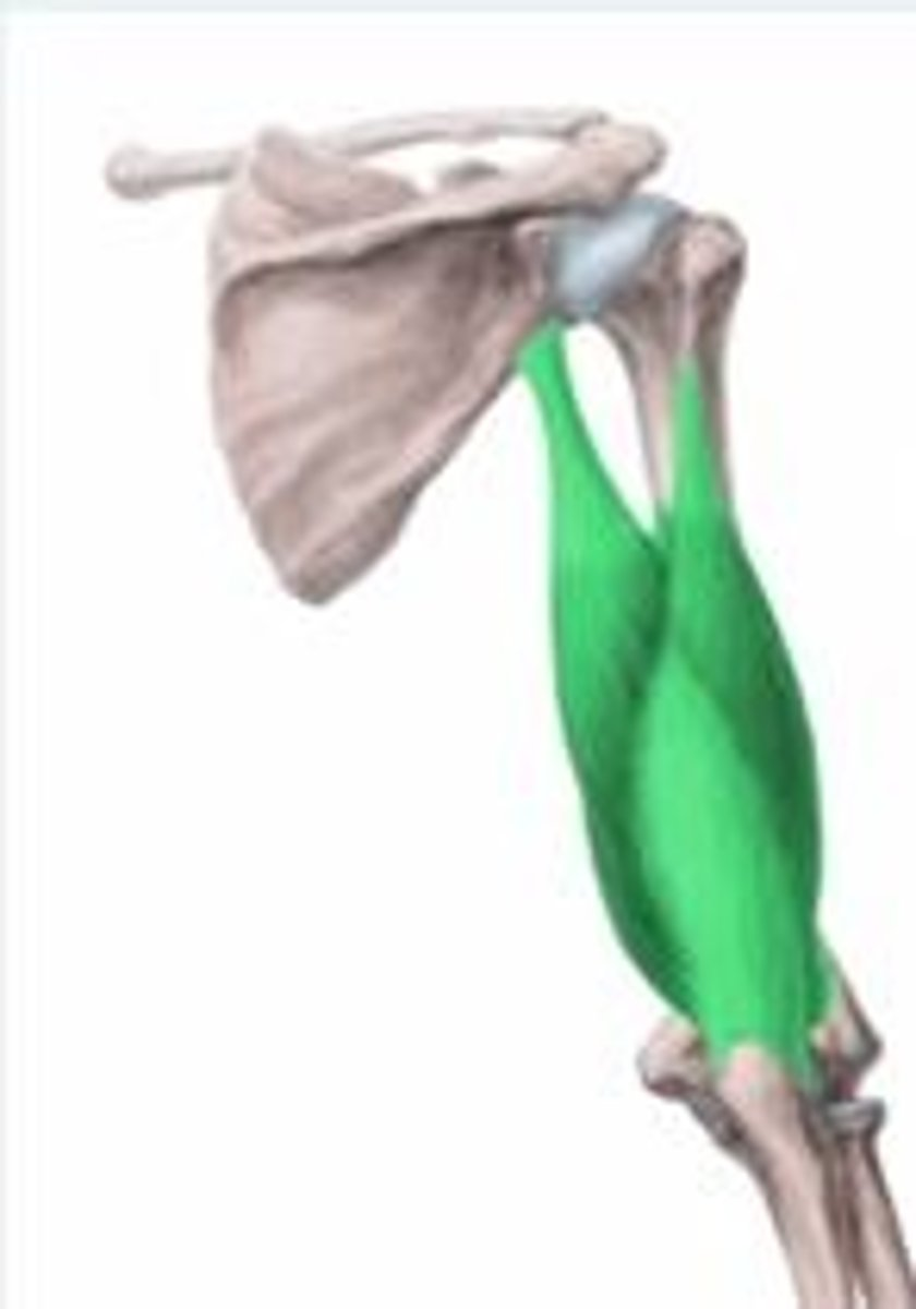
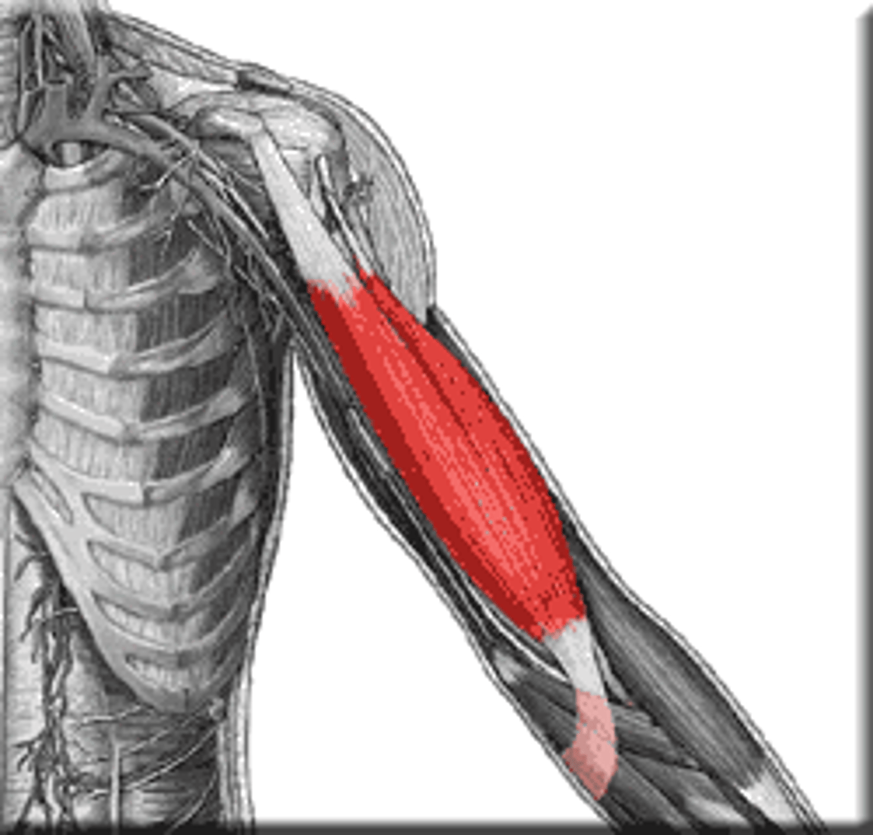
The structures highlighted in green in the following illustration form parts of which muscle?
Triceps
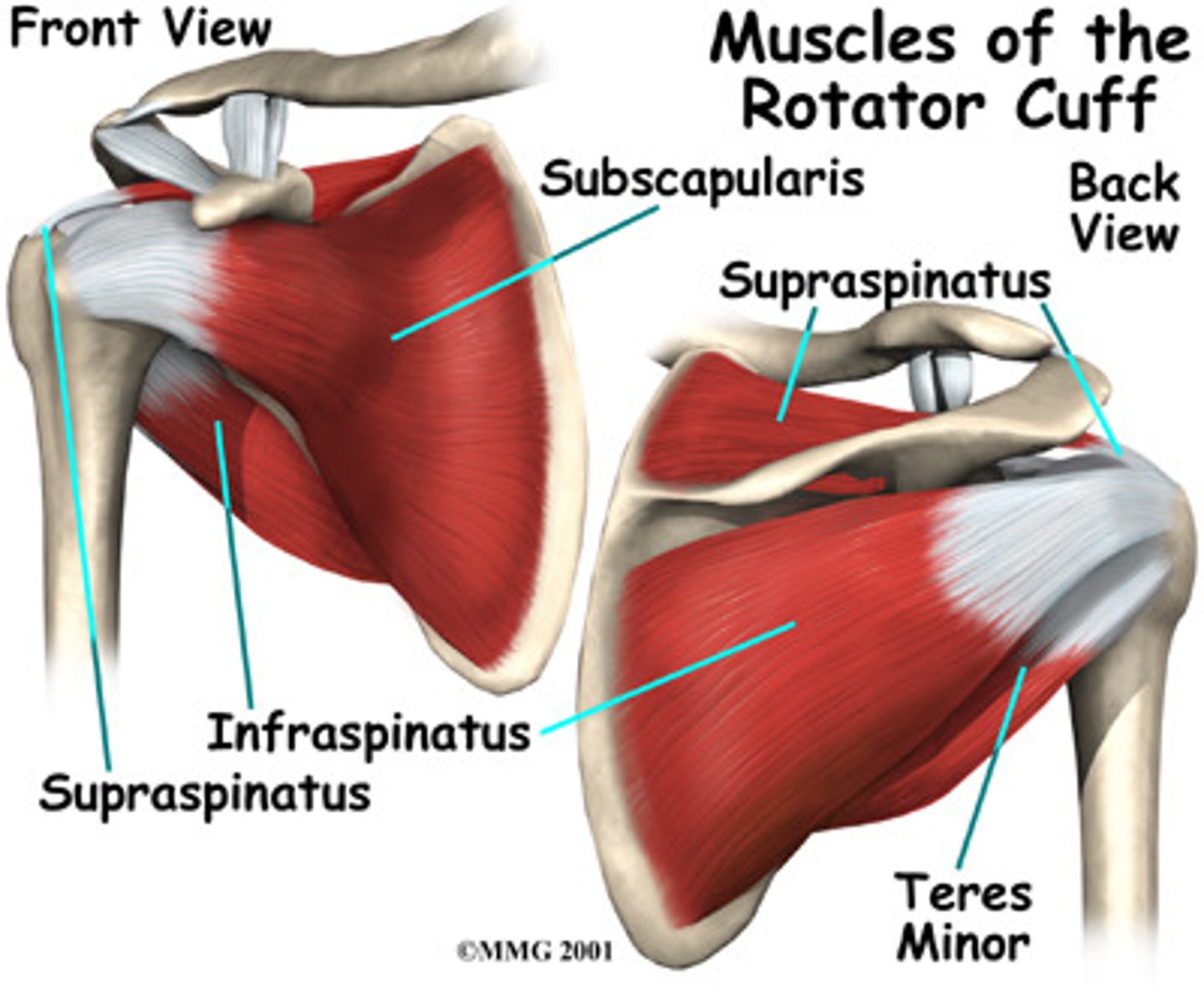
Which two of the rotator cuff muscles have opposing actions at the shoulder joint?
a) Subscapularis and supraspinatus
b) Teres minor and supraspinatus
c) Infraspinatus and subscapularis
d) Teres minor and infraspinatus
c) Infraspinatus and subscapularis
A) Which muscle is a major abductor of the arm, and can also be a flexor of the arm, or an extensor of the arm?
B) What nerve innervates this muscle?
A) Deltoid muscle
B) Axillary nerve
What medical term is given to the symptom of a tingling sensation?
Paraesthesia
What artery passes behind the middle of the clavicle and becomes named something different once crossing rib 1?
Subclavian artery