NVCC BIO 141 Chapter 8
 Call Kai
 Learn
Learn Practice Test
 Spaced Repetition
 Match
Match Flashcards
 Knowt Play
1/70

There's no tags or description
Looks like no tags are added yet.
Name | Mastery | Learn | Test | Matching | Spaced | Call with Kai | Chat |
|---|
No analytics yet
Send a link to your students to track their progress
71 Terms
Joint/Articulation
connects two bones
Synarthrosis
immovable joint
high stability
Amphiarthrosis
slightly movable joint
medium stability
Diarthrosis
freely movable joint
least amount of stability
Fibrous Joint
connect bones through fibers without allowing any movement (like sutures)
Cartilaginous Joints
allow only slight movement and consist of bones connected entirely by cartilage
synarthrosis/amphiarthrosis
Synovial Joints
freely movable joints with a joint cavity and fluid
diarthrosis
Fibrous Joint Subclasses
Sutures
Gomphoses
Syndesmoses
Sutures
Areas where the cranial bones have joined together
synarthrosis
Gomphoses
attachment of a tooth to its socket
synarthrosis
Syndesmoses
Joint in which two bones are bound only by ligaments
amphiarthrosis
radius/ulna
tibia/fibula
Cartilaginous Joint Subclasses
Synchondroses
Symphyses
Synchondroses
bones united by hyaline cartilage
synarthrosis (epiphyseal plate)
Symphyses
Bones united by fibrocartilage
amphiarthrosis
intervertebral joints
Parts of Joint Cavity
Articular Capsule
Synovial Fluid
Articular Cartilage
Adipose Tissue
Blood Vessels
Nerves
Articular Capsule
Outer Layer: dense irregular CT; keeps bones from being pulled a part; isolates internal environment from bloody supply
Inner Layer: Synovial membrane loose CT; secreting synovial fluid
Synovial Fluid
lubricates, supplies nutrients, shock absorption
circulates a small amount of fluid since too much or too little is bad
Articular Cartilage
Hyaline cartilage attached to articular bone surfaces
Ligaments
Connect bone to bone
Intrinsic: thickened articular capsules
Extrinsic: in or out of joint cavity
Tendons
Connect muscle to bone, crossing over/around joint when muscle contracts
stabilizes joints
Bursae
flattened fibrous sacs lined with synovial membrane and containing a thin film of synovial fluid
Tendon Sheaths
long bursae surrounding tendons in high stress regions
protecting long tendons as they cross
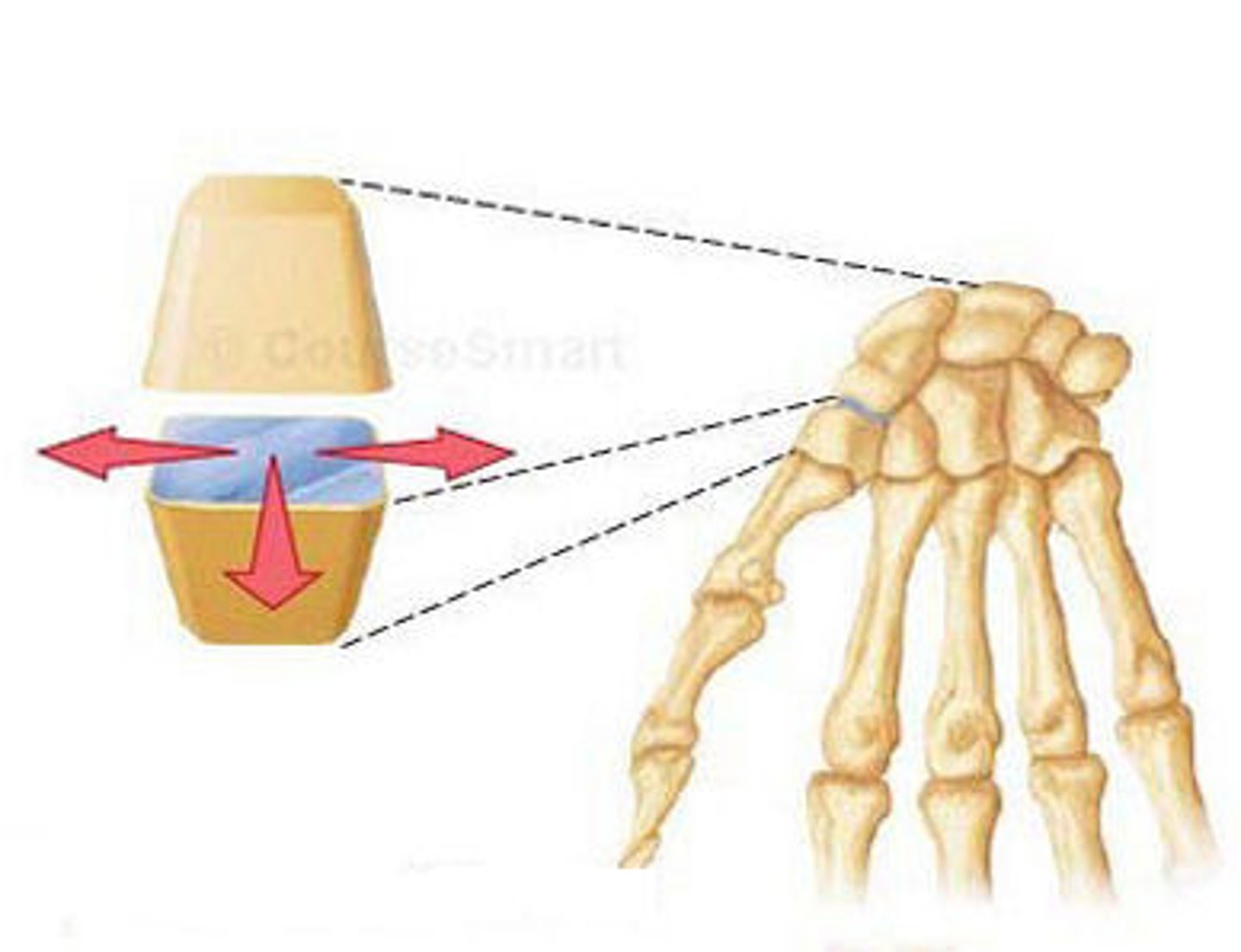
Nonaxial Joints
allow motion to occur in one or more planes without moving around an axis
wrists/ankles gliding
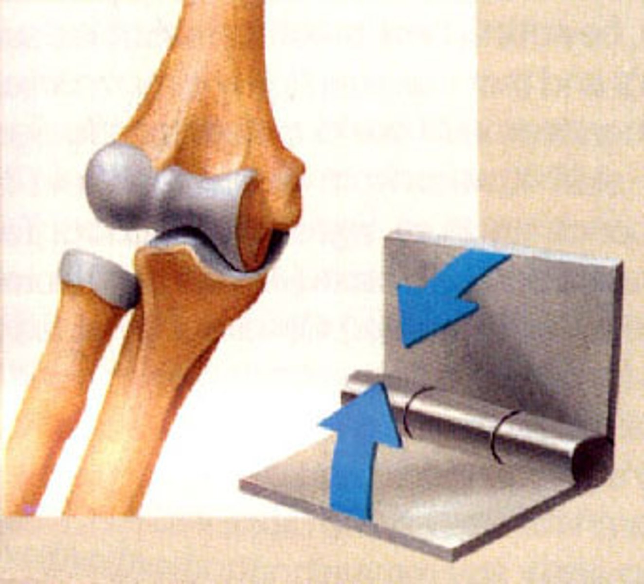
Uniaxial Joints
allow motion around only one axis (hinge)
elbow
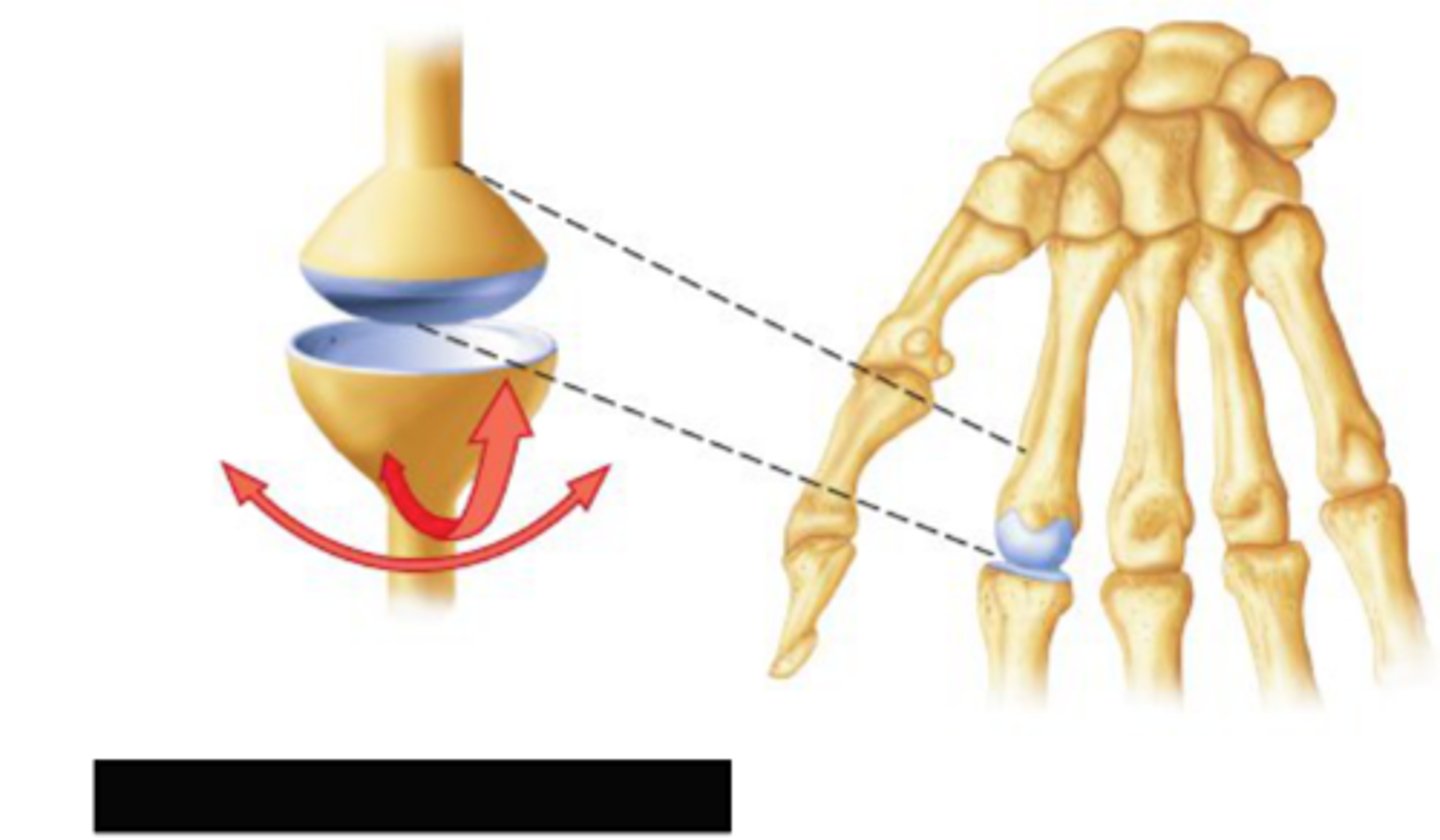
Biaxial Joints
movement in two planes
metacarpals
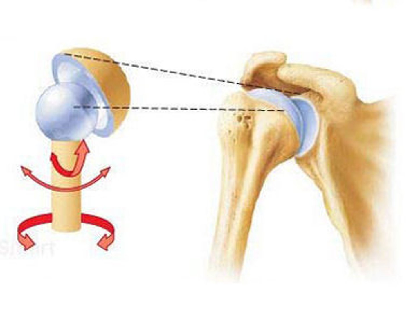
Multiaxial Joints
allow motion around three axes
shoulder
Flexion
Decreases the angle of a joint
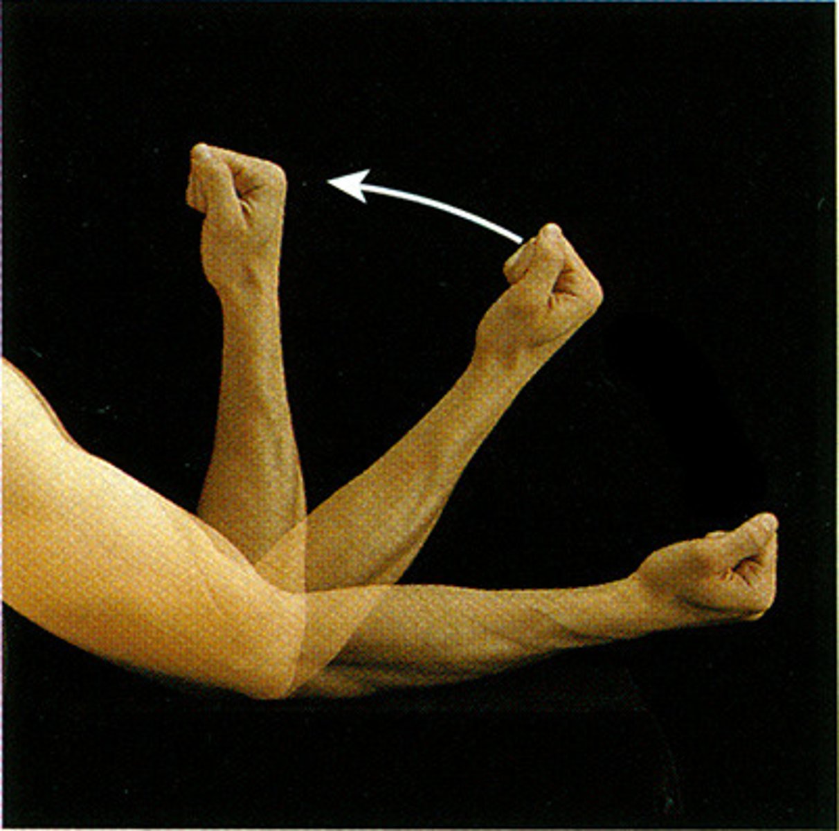
Extension
increases the angle of a joint
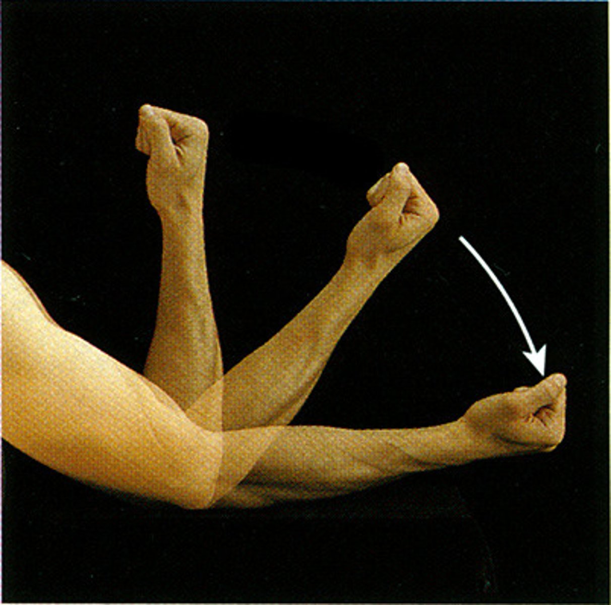
Hyperextension
the extreme or overextension of a limb or body part beyond its normal limit
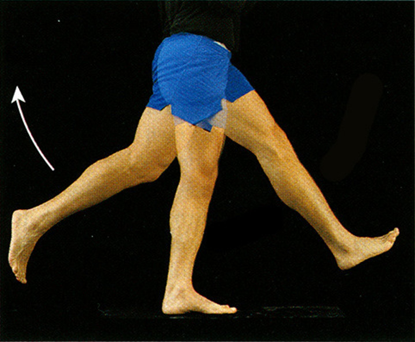
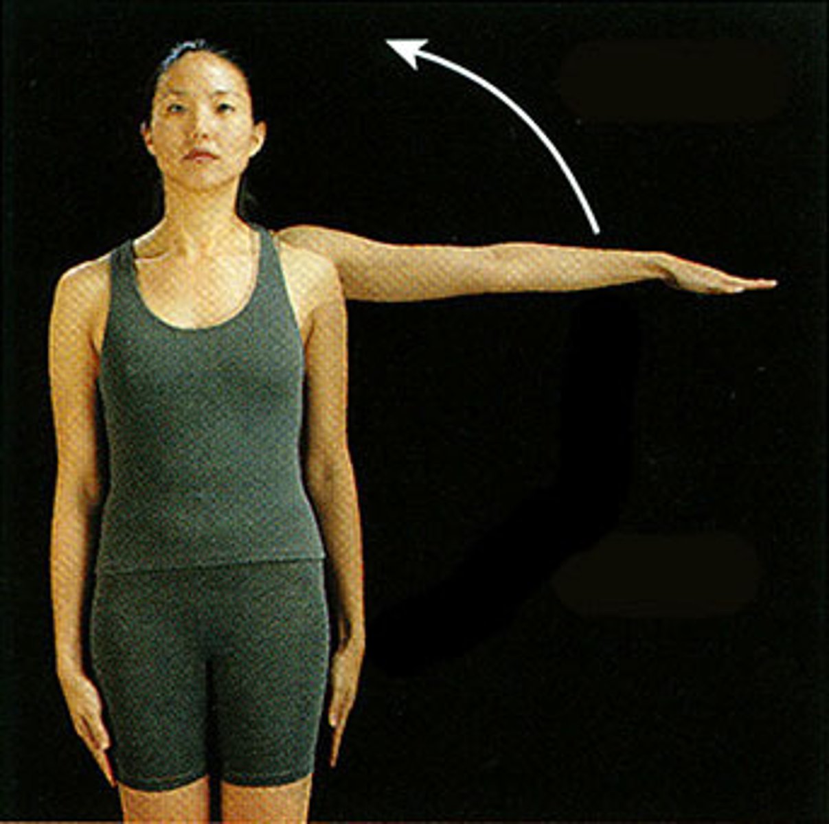
Abduction
Movement away from the midline of the body
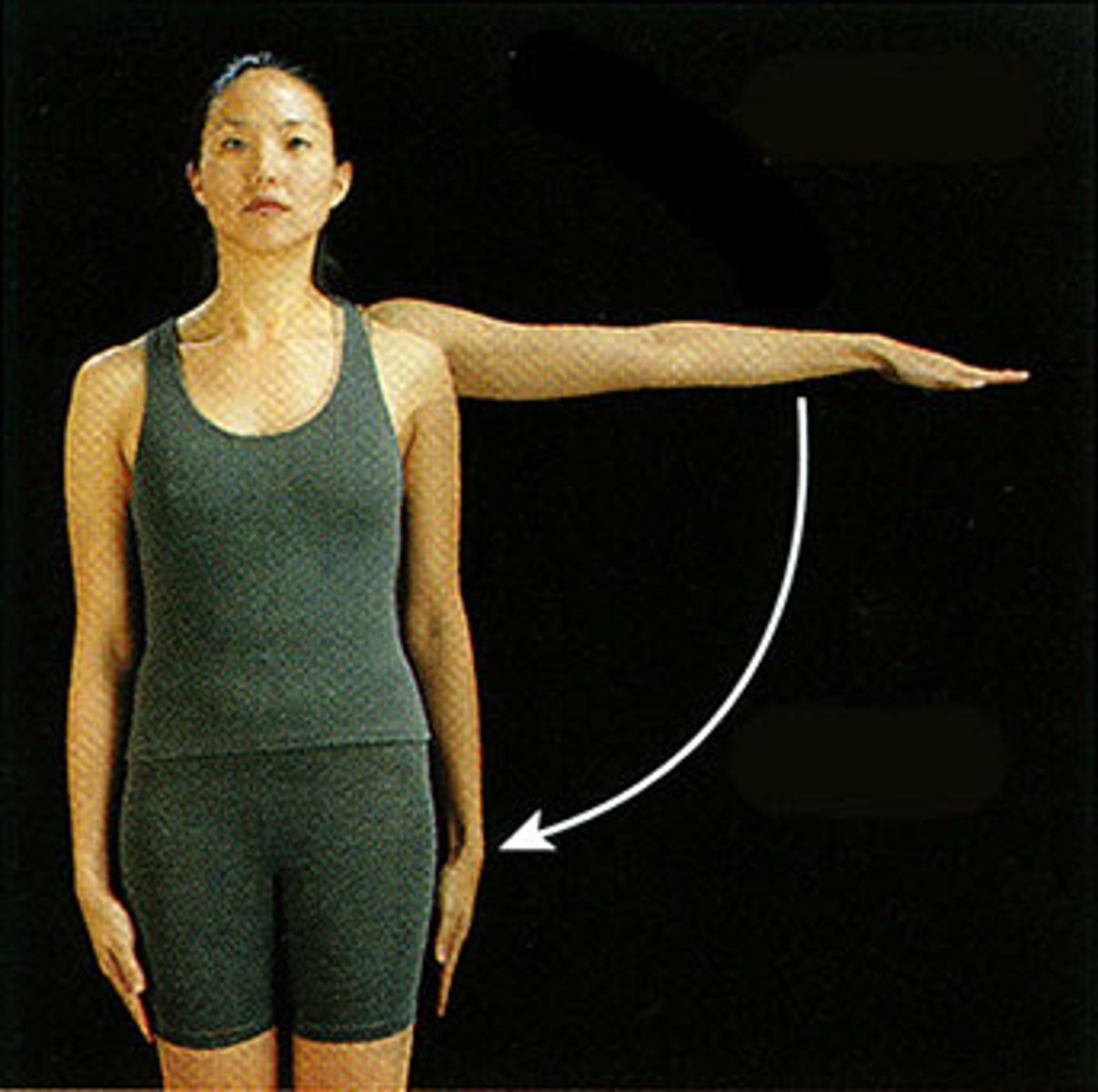
Adduction
Movement toward the midline of the body
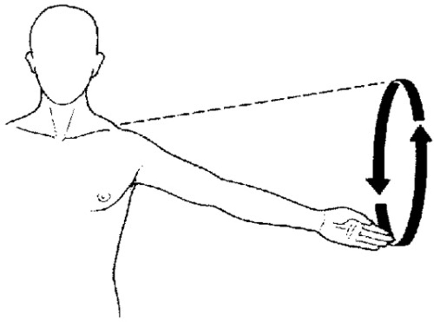
Circumduction
circular movement of a limb at the far end
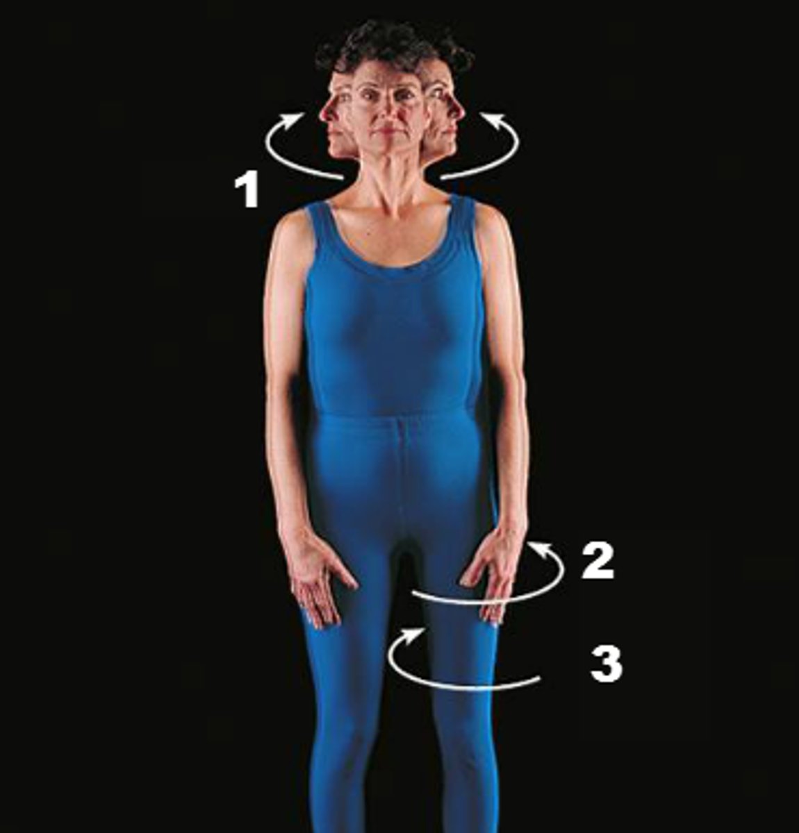
Rotation
twisting on an axis
Lateral: away from midline
Internal: towards midline
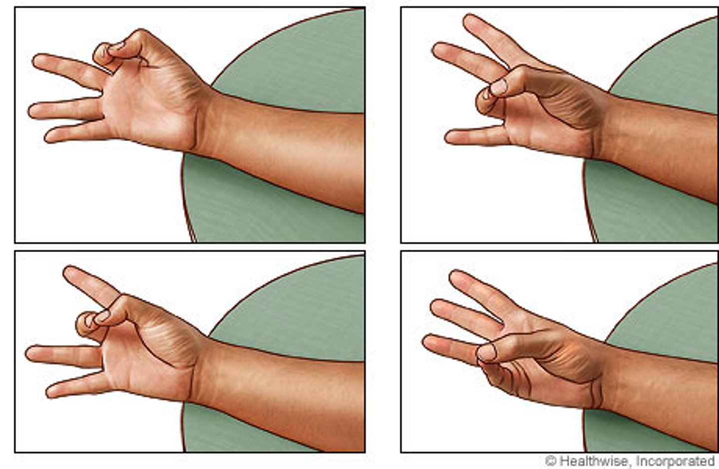
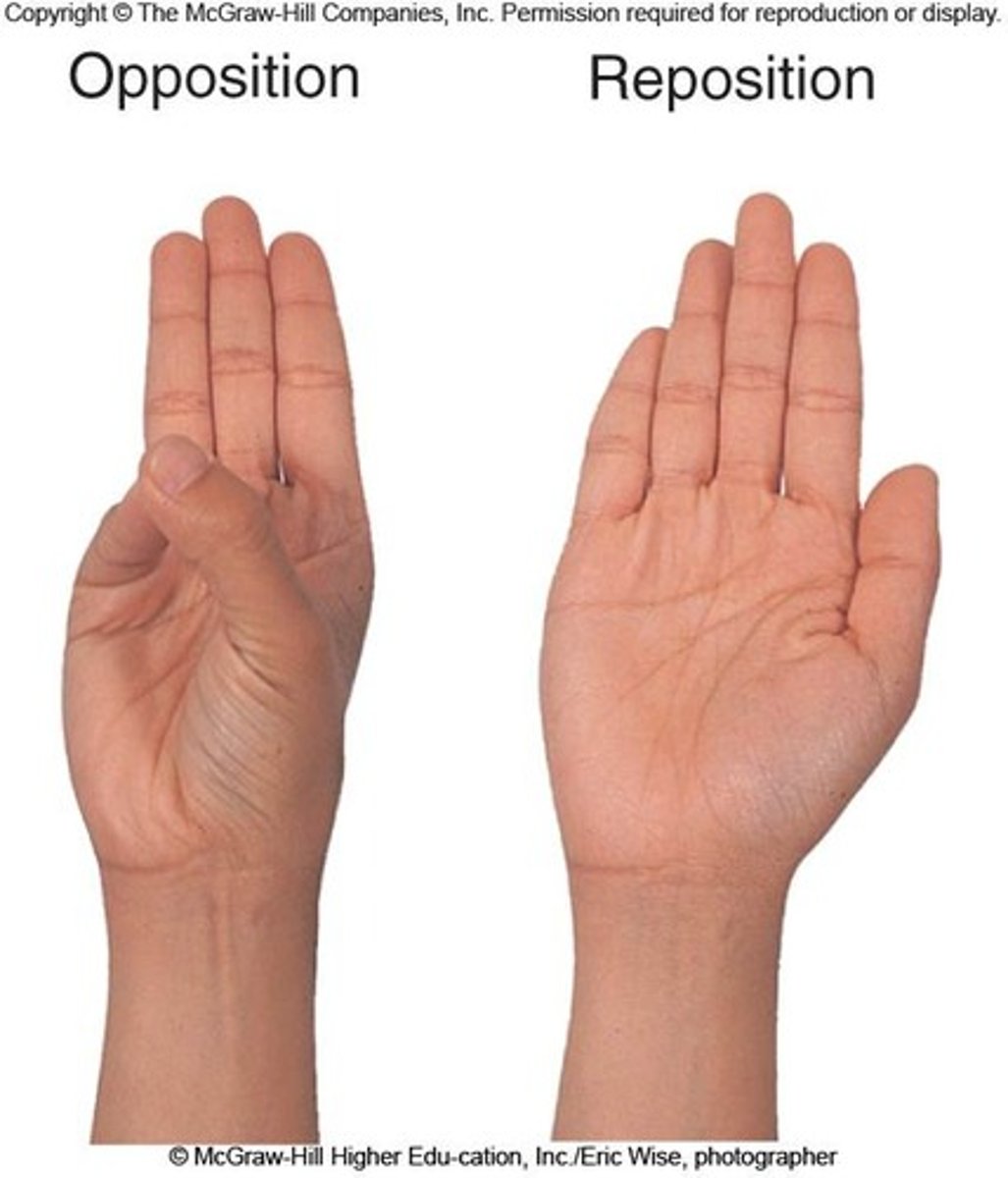
Opposition
thumb movement toward fingers or palm (grasping)
Reposition
return to anatomical position
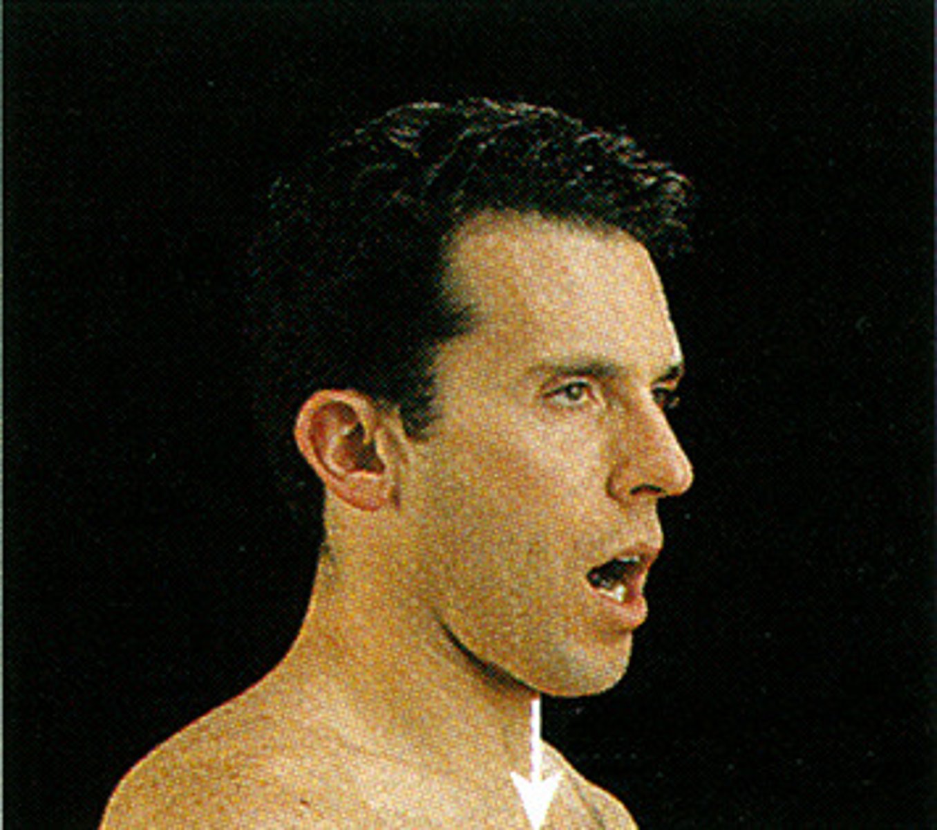
Depression
inferior movement of a body part
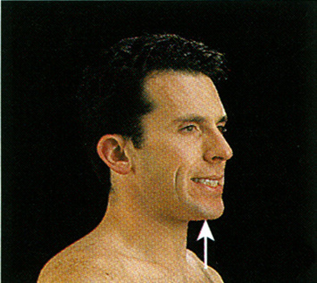
Elevation
raising a body part
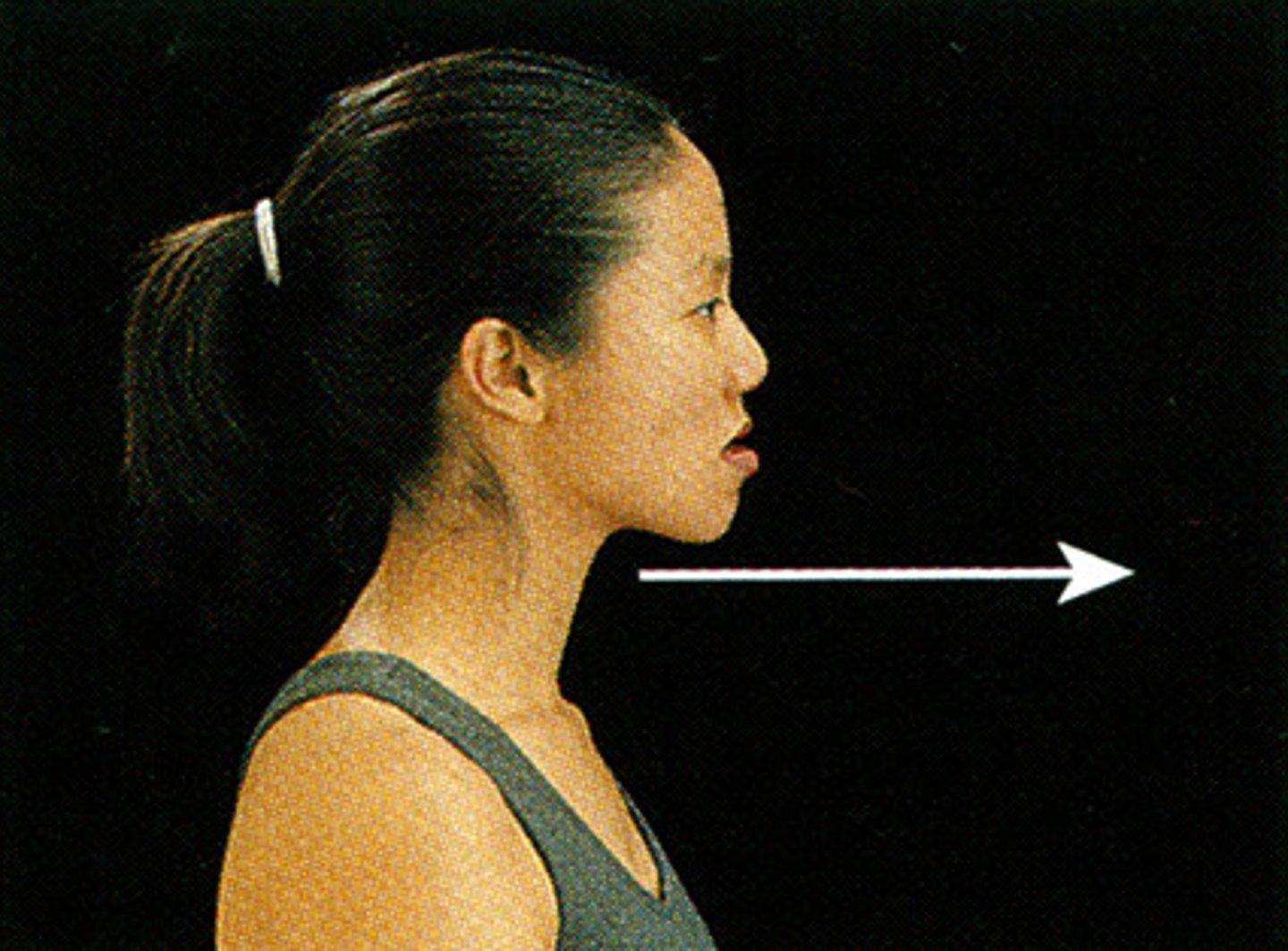
Protraction
anterior movement of a body part in the transverse plane
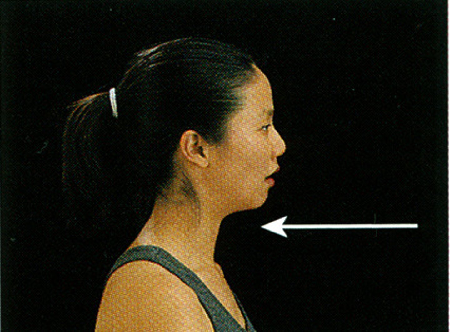
Retraction
moving a part backward
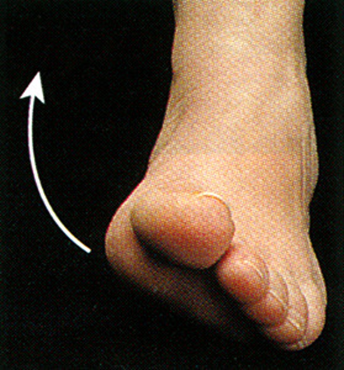
Inversion
rotation of sole of foot toward midline
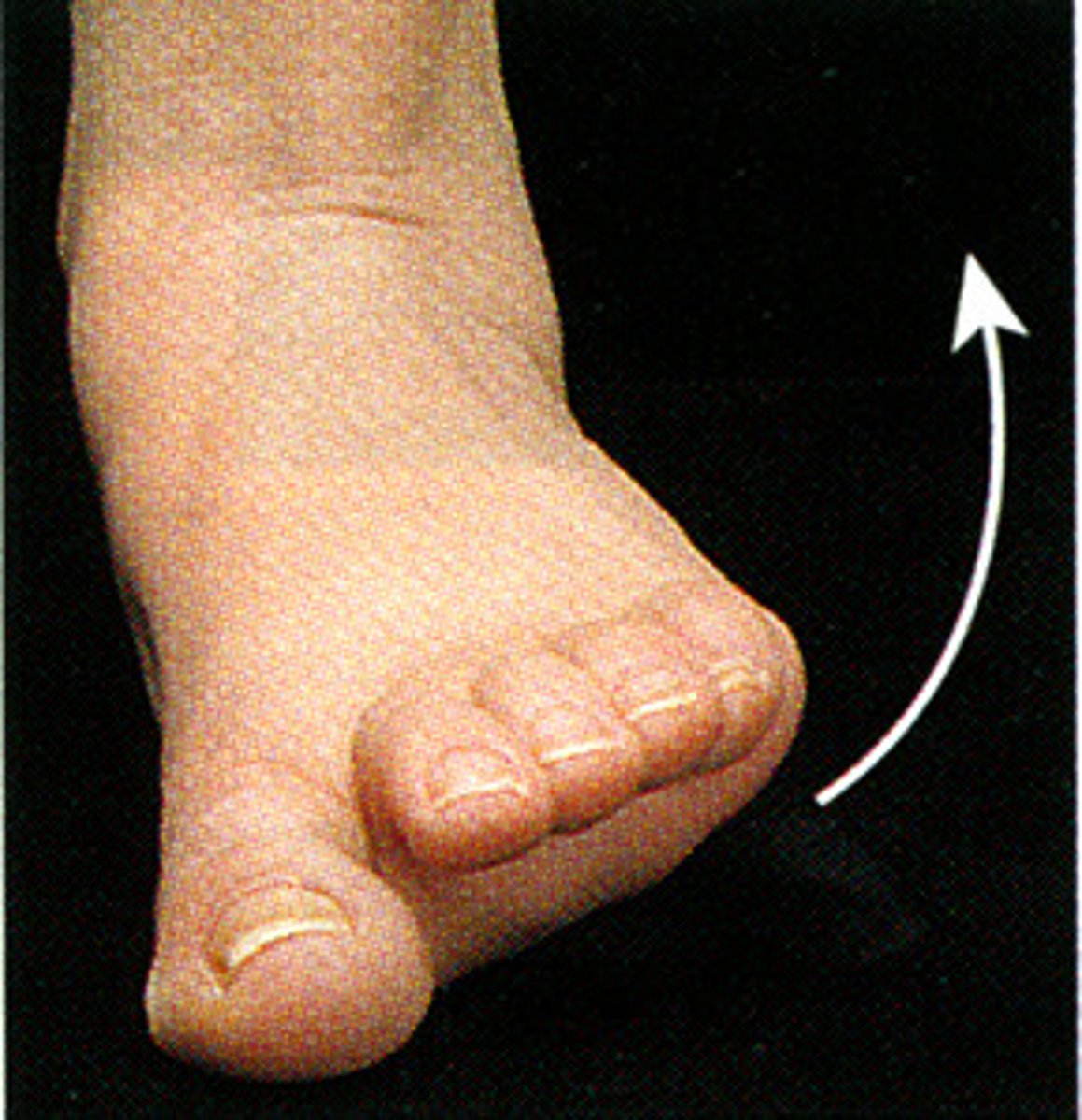
Eversion
rotation of sole of foot away from midline
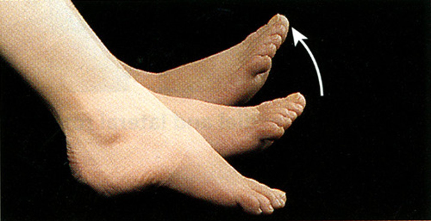
Dorsiflexion
bending of the foot or the toes upward
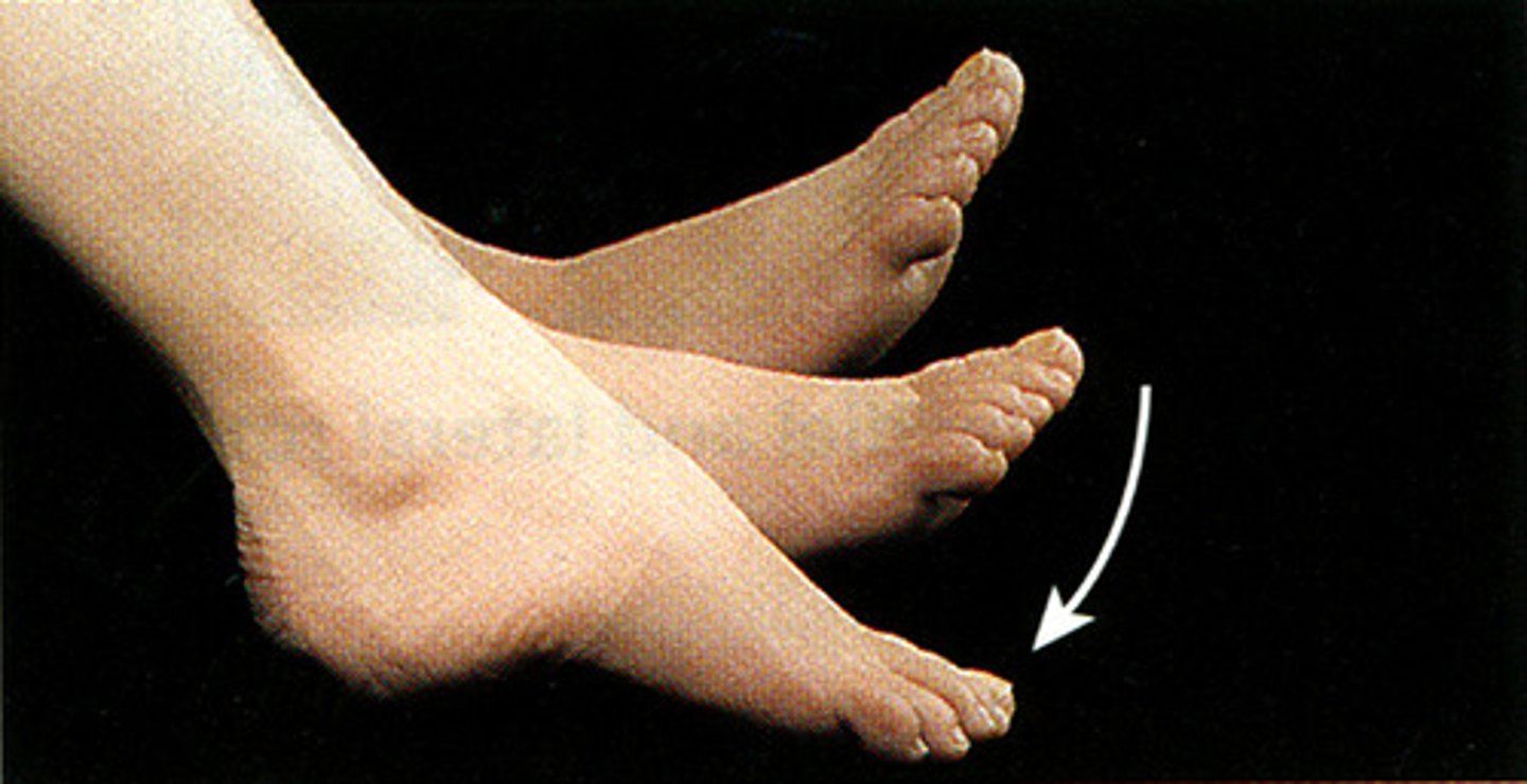
Plantarflexion
point toes down
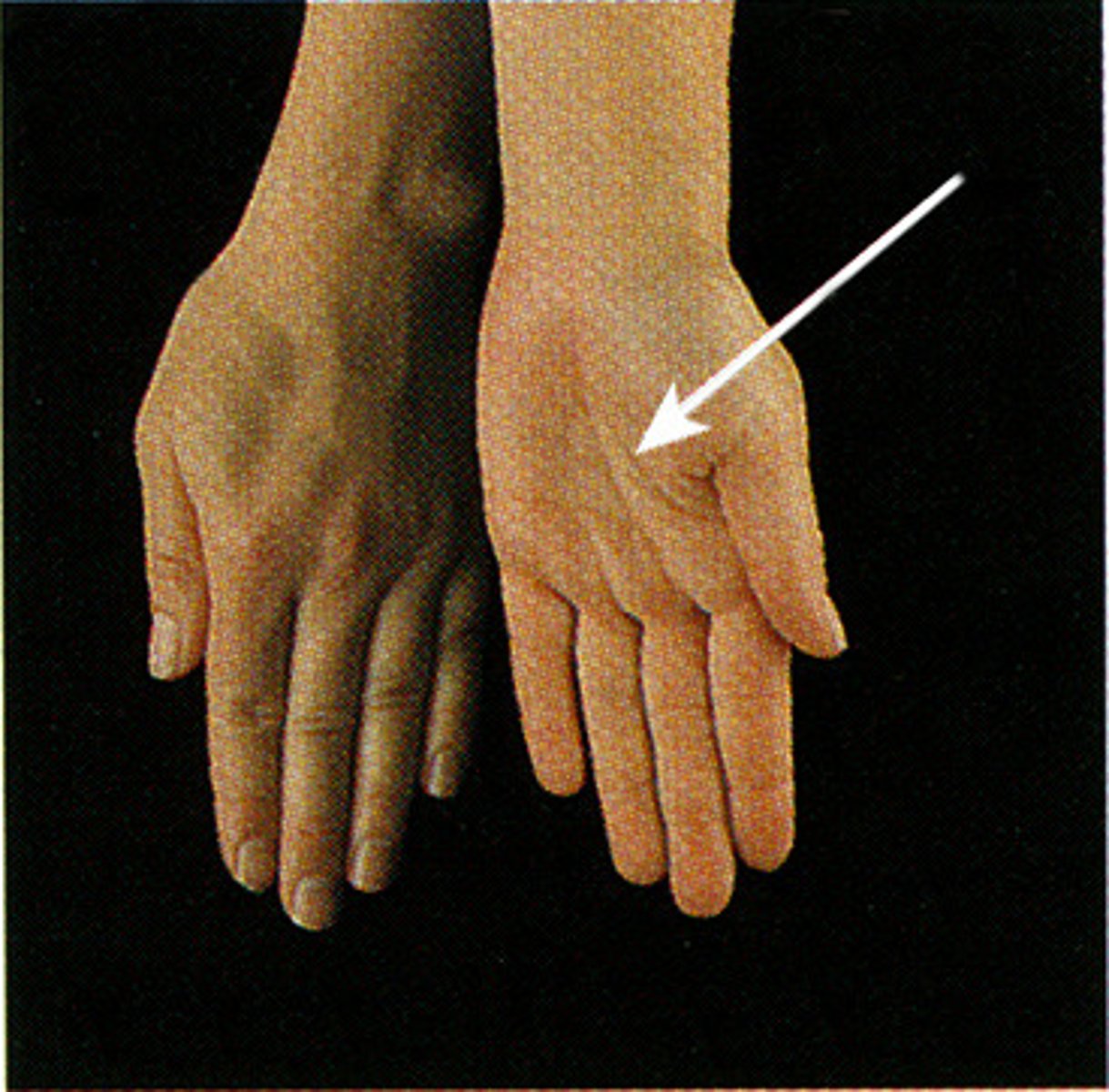
Supination
turning the palm upward
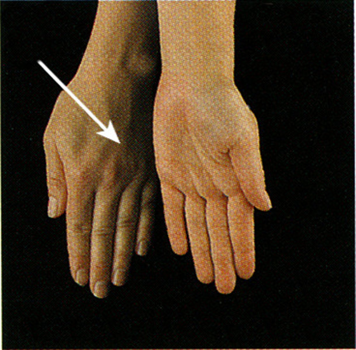
Pronation
turning the palm downward
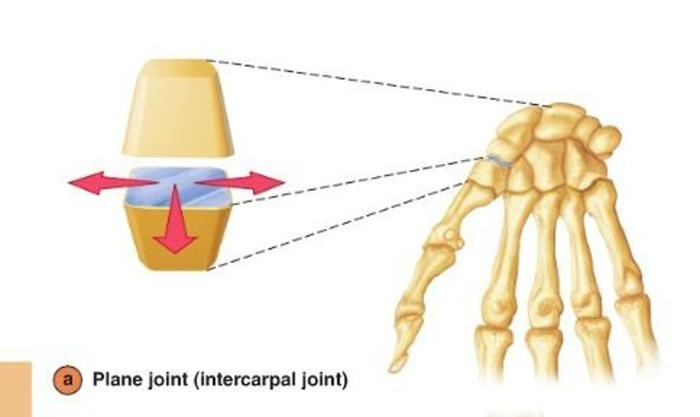
Plane Joint
simplest/least mobile
two flat surfaces
nonaxial (only gliding)
wrist and ankle
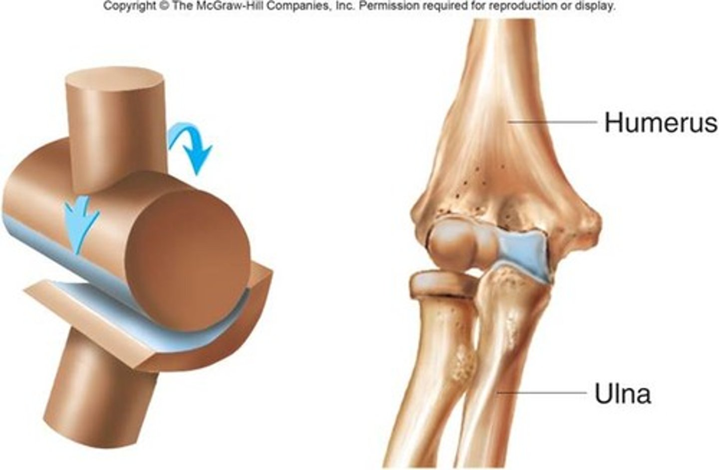
Hinge Joint
convex surface of one bone fits into the concave surface of another
uniaxial
elbow, knee, interphalangeal joints of fingers/toes
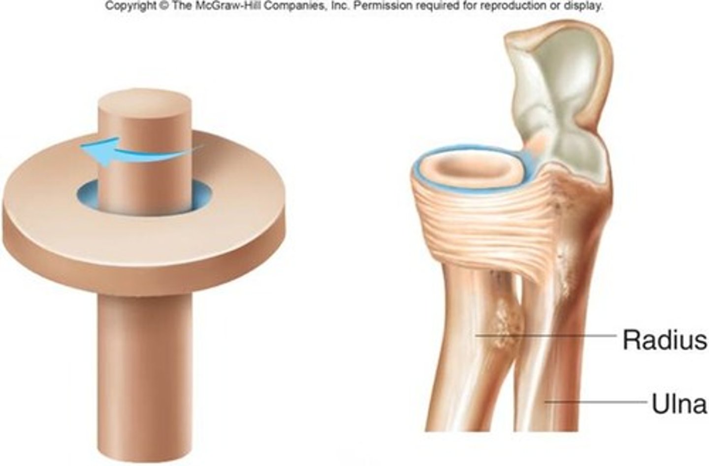
Pivot Joint
rotating bone turns around an axis; i.e. connection between radius/ulna and humerus
Condylar Joint
biaxial
oval convex regions fits into concave surface
flexion/extension
abduction/adduction
knuckles
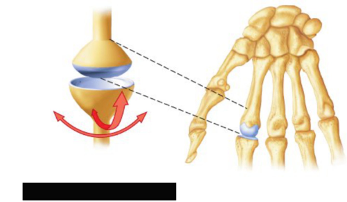
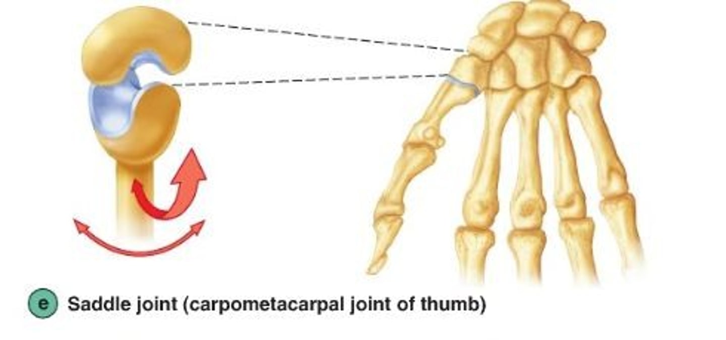
Saddle Joint
concave surface in one direction and convex in another; back and forth, side to side
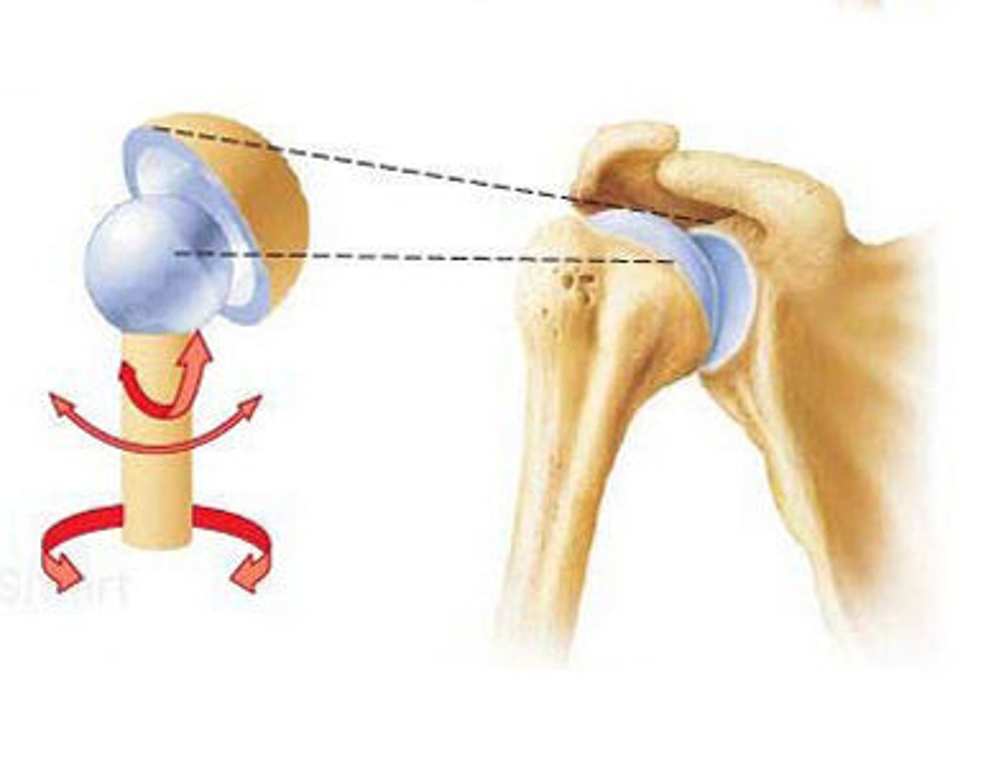
Ball-and-Socket Joint
multiaxial, shoulder and hip
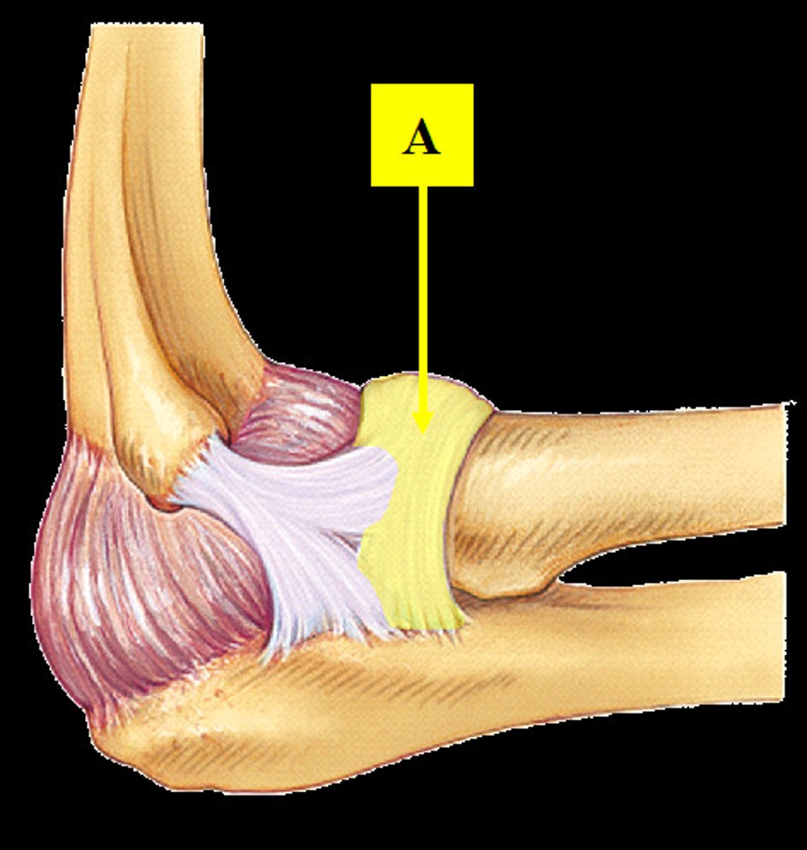
Radial Collateral Ligament
connects the lateral epicondyle of the humerus to the radius
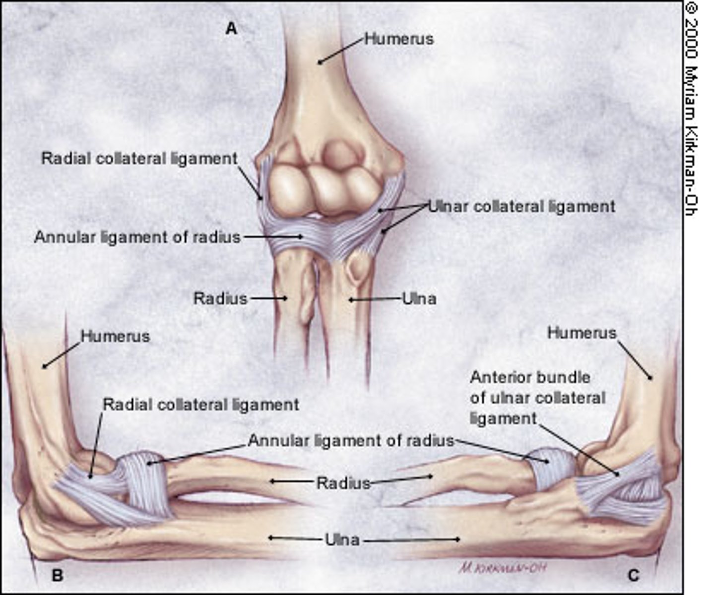
Ulnar Collateral Ligament
Connects the medial epicondyle of the humerus to the ulna
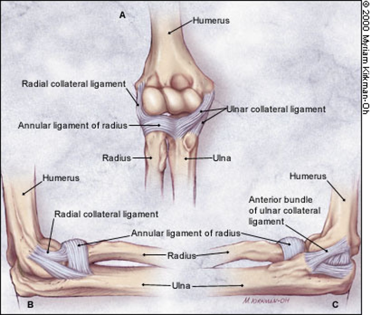
Anular Ligament
ring-shaped band of connective tissue below the elbow joint that encircles the head of the radius
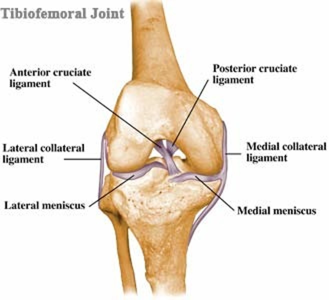
Tibiofemoral Joint
the point where the tibia meets with the femur
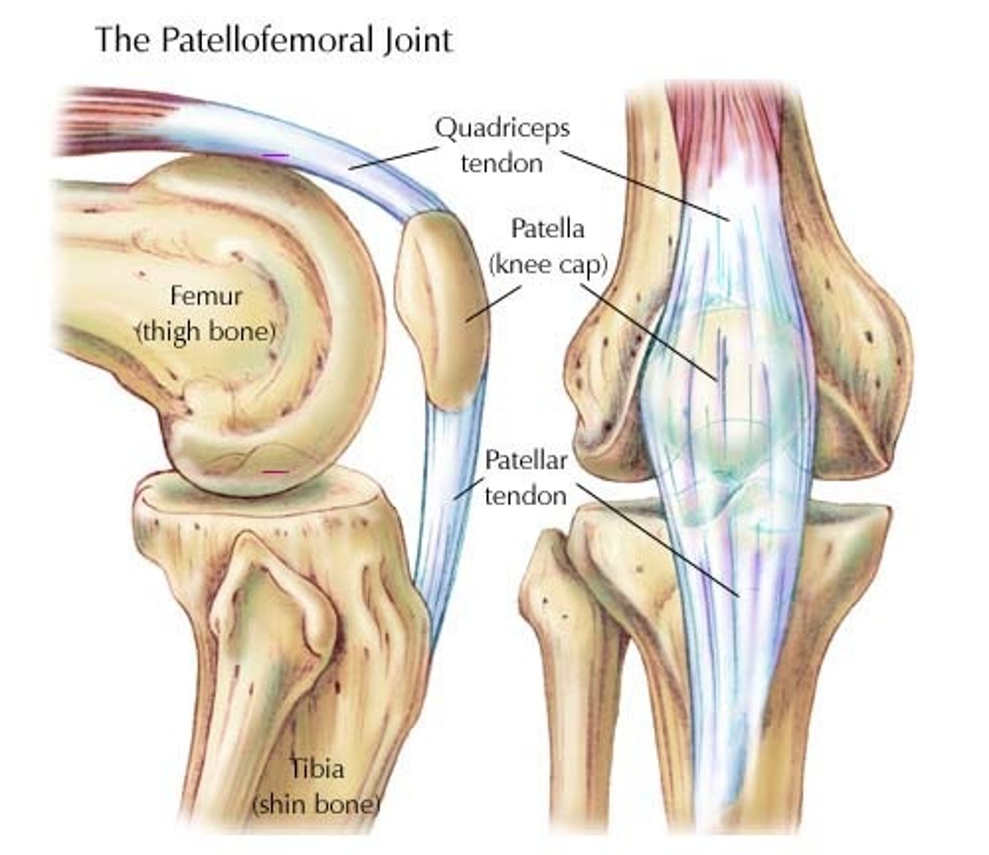
Patellofemoral Joint
the point where the kneecap and femur are connected in the trochlear groove
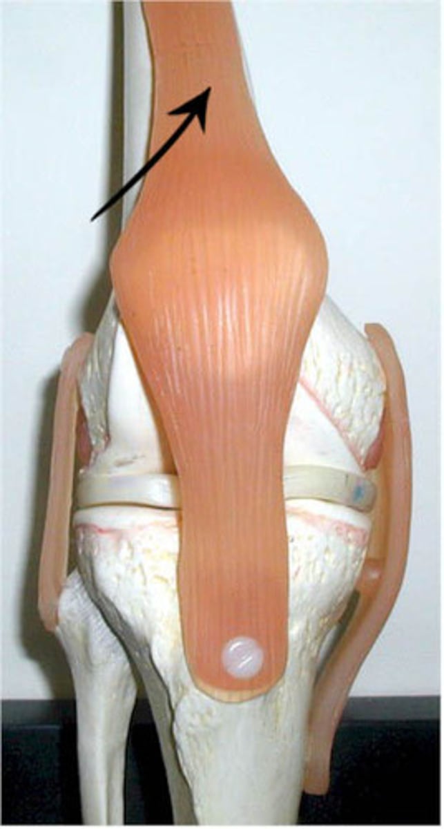
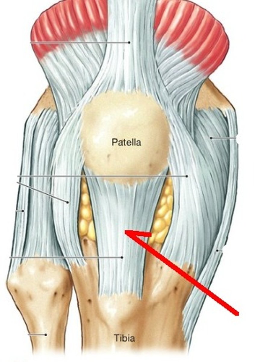
What surrounds the Patella?
Quadriceps Femoris Muscle Group
Patellar Ligament
connects patella to tibial tuberosity
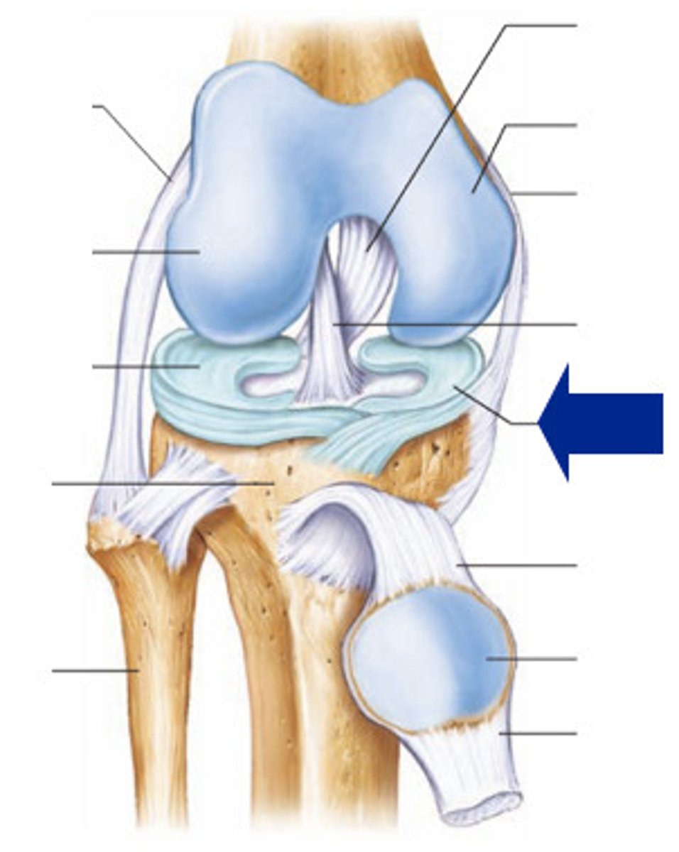
Medial/Lateral Menisci
fibrocartilage pads, at femur-tibia articulations, cushion and stabilize joint, give lateral support
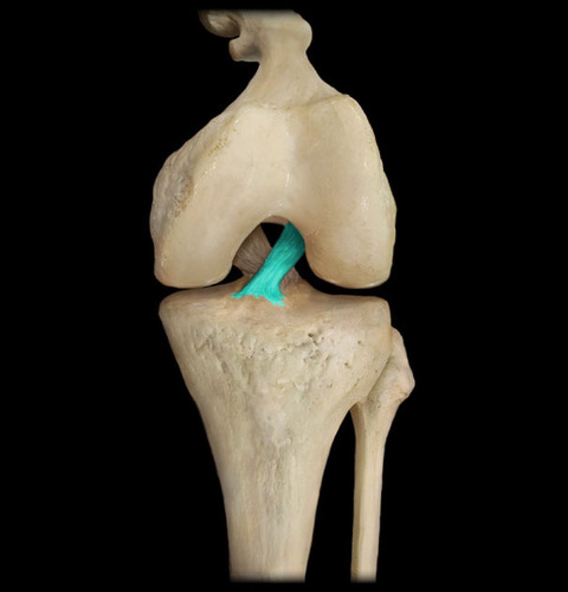
Anterior Cruciate Ligament
A ligament in the knee that attaches to the anterior aspect of the tibial plateau. restricting anterior movement of the tibia on the femur
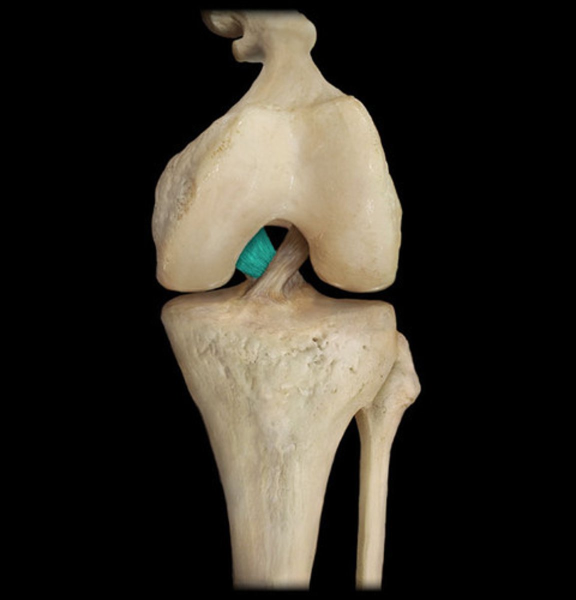
Posterior Cruciate Ligament
prevents posterior displacement of tibia
Glenohumeral Joint
The synovial ball-and-socket joint of the shoulder
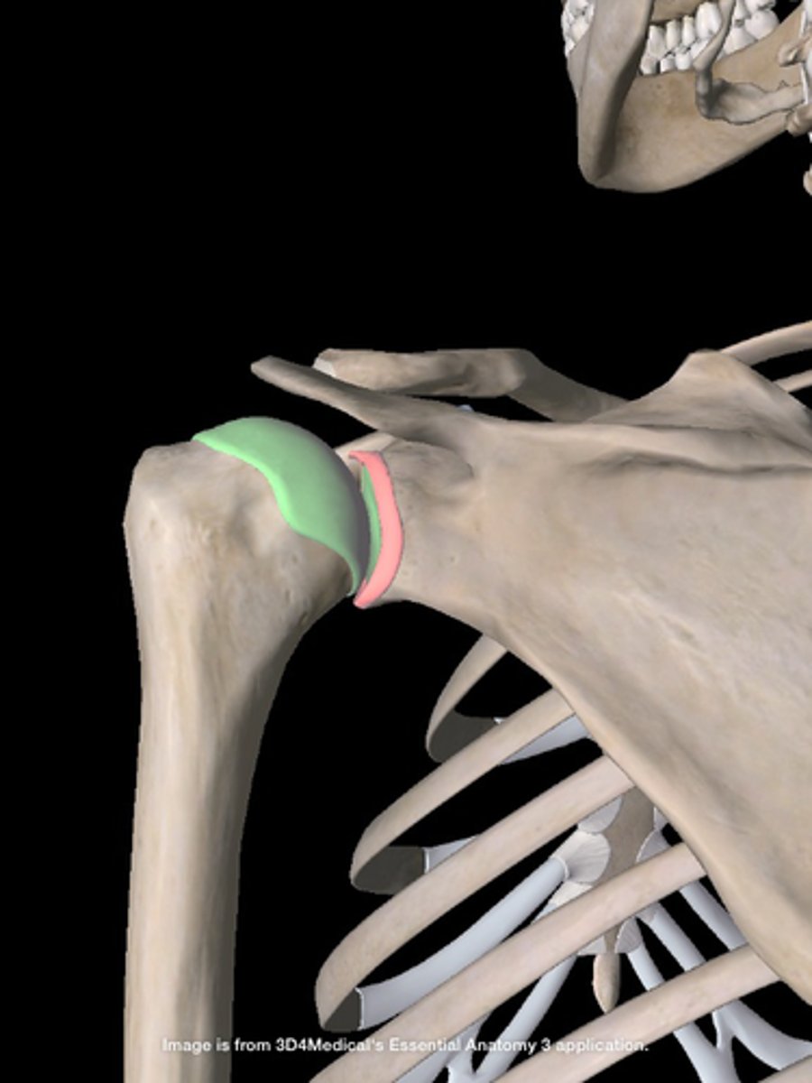
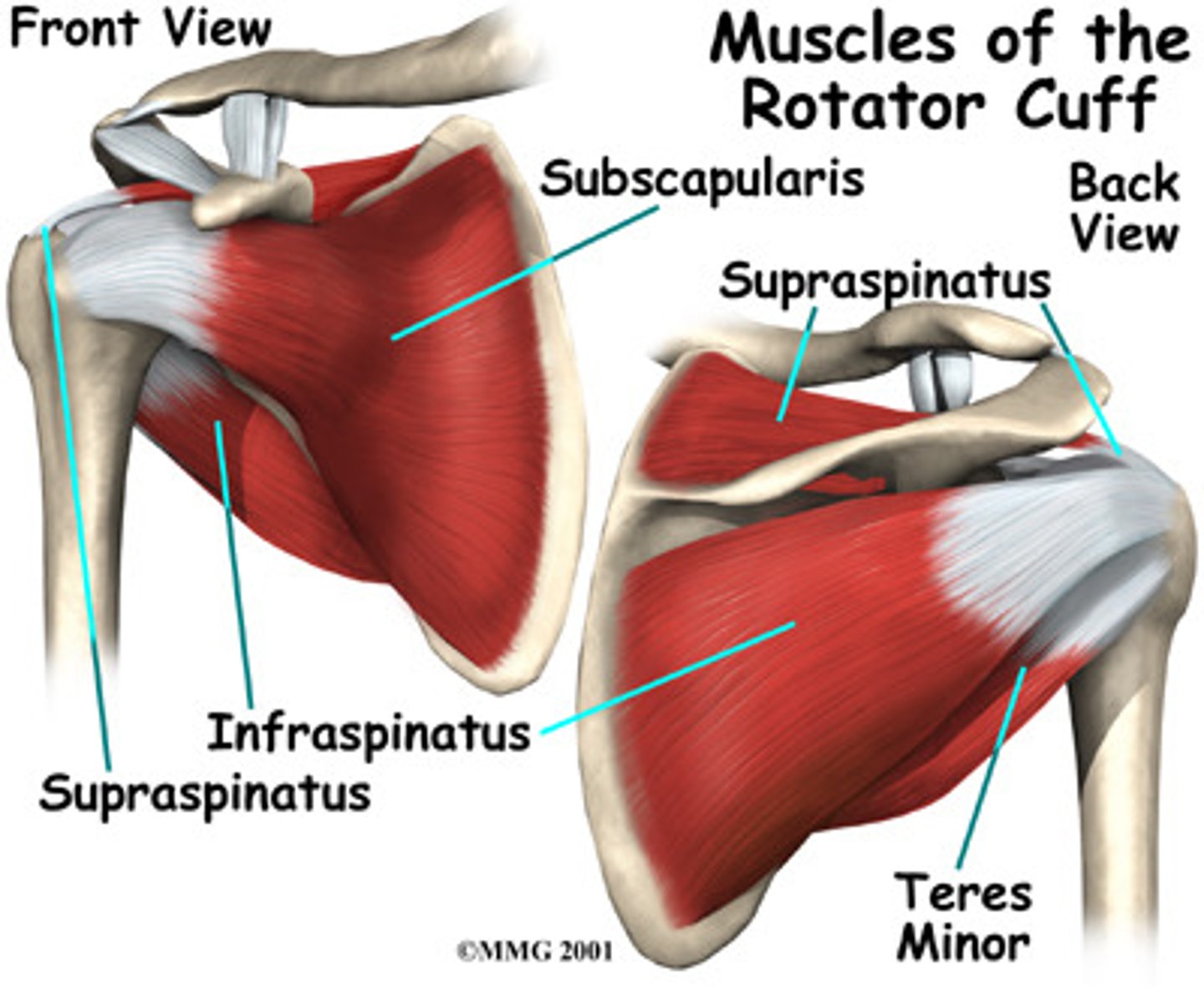
Rotator Cuff
supraspinatus, infraspinatus, teres minor, subscapularis
Why is the glenohumeral joint frequently injured?
shallowness of glenoid fossa
laxity of ligamentous structures
lack of strength and endurance in muscles
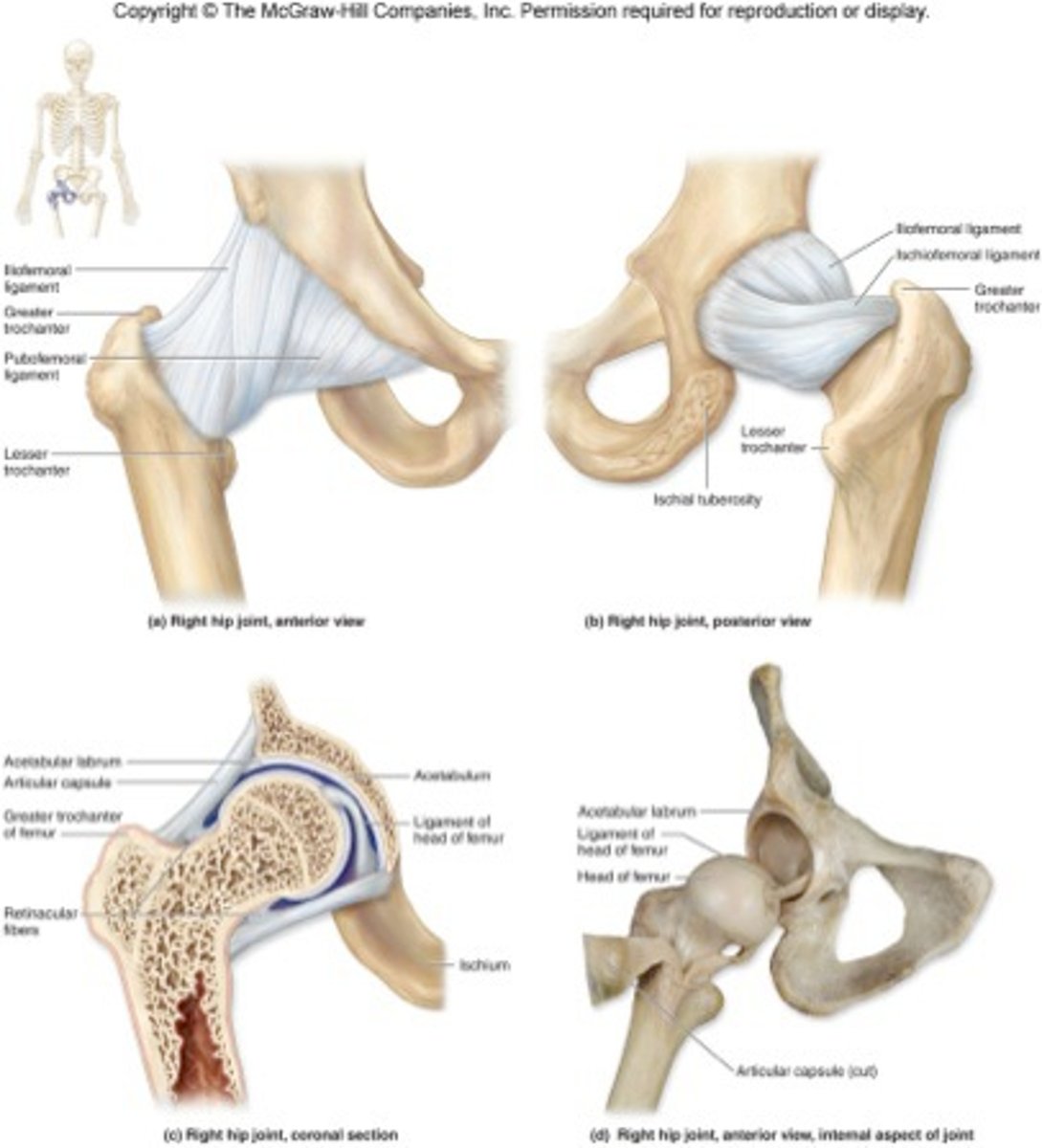
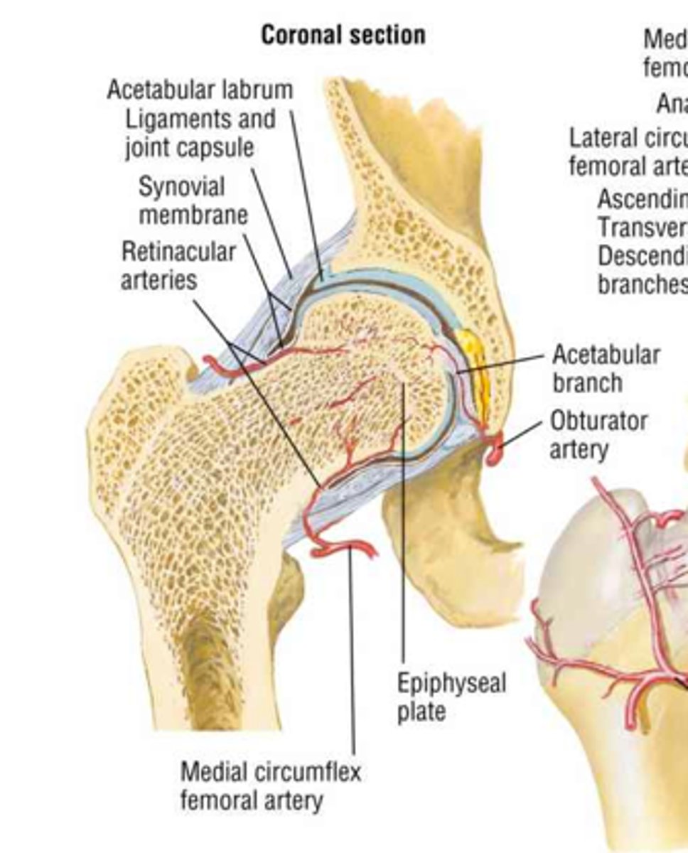
Coxal Joint
hip joint
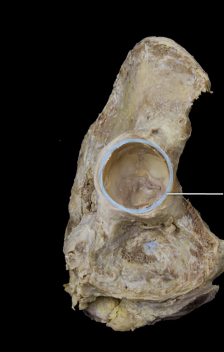
Acetabular Labrum
lip of fibrocartilage that surrounds outer margin of the acetabulum on the hip bone
Articular Capsule of Hip
very strong in hip joint
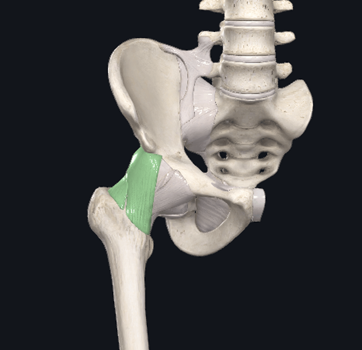
Iliofemroal Ligament
Y shaped
reinforced anterior side
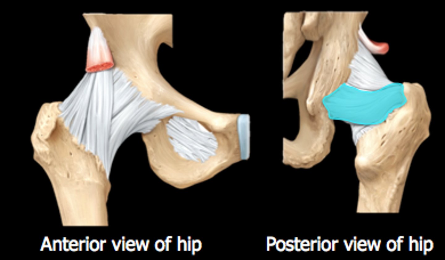
Ischiofemoral Ligament
spiral-shaped, posteriorly located ligament
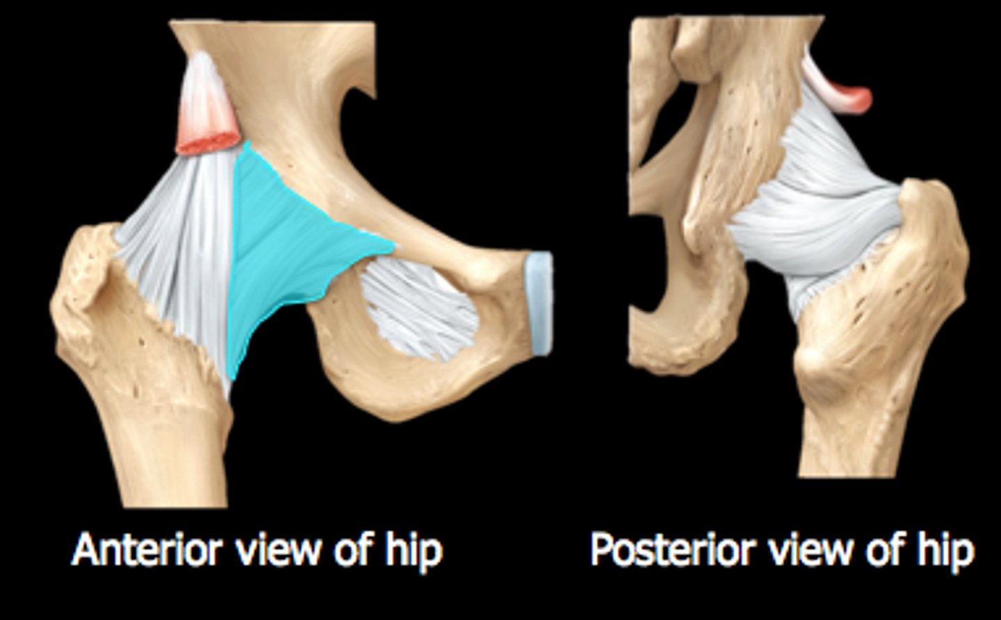
Pubofemoral Ligament
triangular thickening of capsule's inferior region
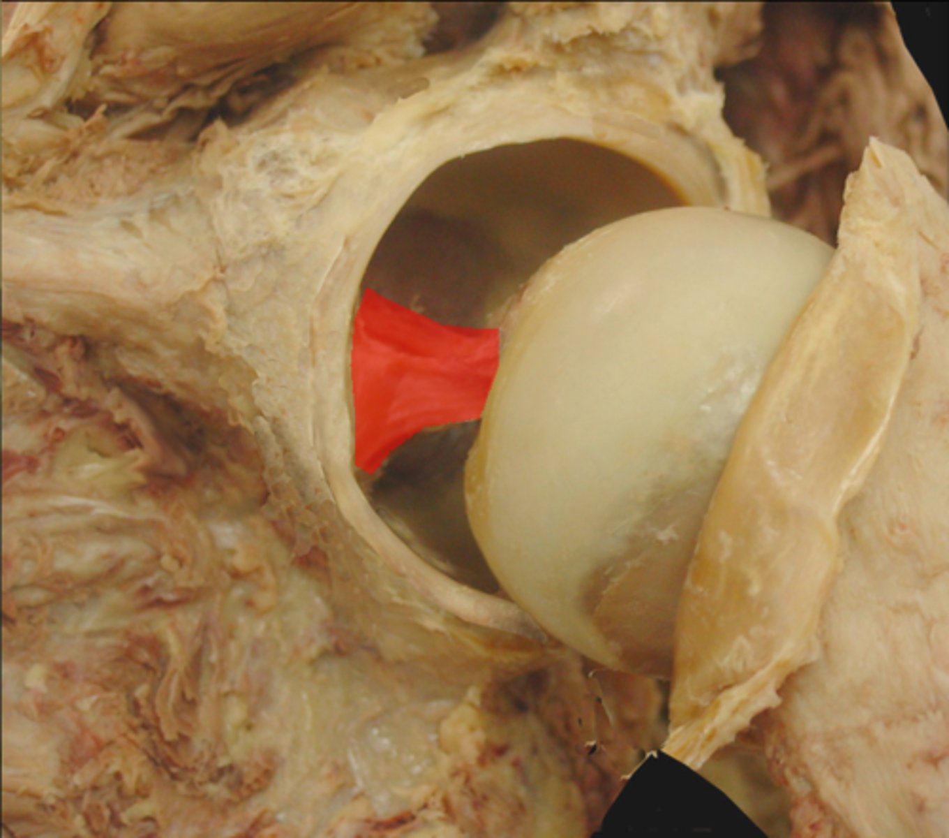
Ligament of the Head of Femur
ligament that spans the acetabulum of the hip bone and the fovea capitis of the femoral head
allows pathway for small artery