quiz 4 key terms - methods in neuroscience
 Call Kai
 Learn
Learn Practice Test
 Spaced Repetition
 Match
Match Flashcards
 Knowt Play
1/95

There's no tags or description
Looks like no tags are added yet.
Name | Mastery | Learn | Test | Matching | Spaced | Call with Kai | Chat |
|---|
No analytics yet
Send a link to your students to track their progress
96 Terms
immortalized cell lines
(ex: HEK293 - human embryonic kidney cells) cells that can be purchased and maintained in liquid nitrogen that are capable of endlessly replicating
advantages: unlimited growth/proliferation, can be maintained indefinitely, cheap, well-documented, suitable for high-throughput screenings/drug testing, don’t naturally have many neuronal genes —> good for testing sufficiency
disadvantages: accumulate genetic mutations, lack tissue context and complexity, not relevant to real in vivo results
primary dissociated cell culture
isolating cell bodies or groups of cells from living tissue
advantages: simplicity for studying cellular behavior, morphology, or interactions, lower cost than living or ex vivo tissue preparation, easy to prepare
disadvantages: takes about 5-6 hours to prepare mice samples (compared to HEK293 cells = 1 hour), limited physiological relevance/lacks tissue architecture, cells cannot survive for more than 2-3 days in media
stem cells
undifferentiated or partially differentiated cells that have the unique ability to divide and produce both identical stem cells (self-renewal) and differentiated cells (differentiation)
advantages: great for studying developmental differentiation/questions, better mimic in vivo environment due to not being uniform, regenerative medicine applications
disadvantages: difficult to culture/grow, costly, difficult to control what they differentiate into, tumor growth is common
ex-vivo tissue
tissue taken from a living organism and maintained or cultured outside the organism in a laboratory setting
advantages: maintain cellular diversity, tissue architecture, and original function —> making them more physiologically relevant, maintains circuitry, great for studying diseases
disadvantages: tissue degrades rapidly (1-2 days), very difficult technique to master, tissues are quite complex so you cannot attribute results to a particular gene or cell type
organoid
derived from stem cells, these 3D structures can mimic the 3D architecture and function of larger multicellular tissue (ex: brains)
limitations: very slow (70-180 days per organoid), variability in organoid morphology, do not faithfully reproduce mature circuitry and connectivity, necrotic core of dead cells
To sterilize instruments a __________ should be used. To sterilize the field / area of surgery __________ should be used.
(1) autoclave
(2) 70% ethanol
transfection
the process of introducing foreign DNA or RNA into a cell to study gene function or expression (usually used in immortalizaed cell lines)
sufficiency experiments:
1) Grow HEK 293 cells
2) Transfect 1 set of gene of interest and one with a control
3) Perform calcium imaging or electrophysiology to see if HEK293+GOI respond to a stimulus
electroporation
uses an electric field to temporarily permeabilize cell membranes, allowing the introduction of DNA, RNA, or other substances into the cell (usually used in primary cell culture/tissues)
cell sorting
the process of isolating specific cells from a mixed population based on their characteristics, such as size, surface markers, or fluorescence.
allows for the isolation of individual cells for downstream applications such as single-cell RNA sequencing, gene expression profiling, or protein analysis
limitations: expensive and technically challenging, causes some damage to cells, requires many cells with strong fluorescent tags, can be error prone and needs good analysis skills to sort out, requires marker/fluorescence on the cell-surface
calcium imaging
indirect measure of neuronal activity BUT allows you to observe/target specific cellular populations using Cre x GCaMP with great spatial resolution
can be performed in dissociated cells, ex vivo, or live/moving animals
very difficult technique to master even at the dissociated tissue culture level as subtle changes in how agonists are washed over the cells can affect their signal
expensive & time consuming
electrophysiology
direct measure of neuronal activation with higher temporal resolution
calcium imaging in vitro
1) if using Cre-ER x GCaMP mice give tamoxifen
2) perform primary cell culture
3) look at the change in fluorescence over time as you apply different agonists to cause activation
fiber photometry
one method for in-vivo calcium imaging is to attach a probe into the animal’s brain. this allows you to transmit optogenetic (blue light) to stimulate activation AND receive/record green light emissions (from GCaMP)
advantage: awake, moving animals
disadvantage: looking at overall fluorescence values, can’t differentiate patterns within particular neuronal population/cells
microendoscopy & GRIN lens
an in-vivo calcium imaging method where detailed recordings of individual neurons; high spatial and temporal resolution even deep within the brain
two photon microscope
another in-vivo calcium imaging method where you anesthetize the mouse, hold its head/body steady, and expose the spine/brain and directly apply agonists and/or stimulate senses while recording the green fluorescence
intraceullar recordings
a challenging electrophysiology technique that measures the activity of the entire neuron. it’s less common today than whole-cell patch clamp techniques that achieve higher signal-to-noise resolution
extracellular recordings
a less sensitive electrophysiology technique compared to intracellular, cannot measure graded potentials, sampling issues: data is biased toward the largest neurons and largest signals vs smaller ones
patch clamp recordings
an electrophysiology technique that allows you to record and manipulate single ion channels
whole-cell recording: allows researchers to measure the entire cell’s voltage/current; also good for injecting cells with dye for imaging
inside-out recording: allows researchers to study the influence of intracellular molecules on channel function
outside-out recording: allows researchers to study how ion channel activity is influences by neurotransmitters
physical lesion
very crude and difficult to control, challenging to tell if effect is from cells in area or axons running through (fibers of passage) area, irreversible
electrical lesion
spaital resolution very limited, microsimulation will impact all neurons in area, can affect passing axons/fibers as well as neuron bodies in area, very invasive
pharmacological lesion
reversible, some toxins are more specific than others AND some toxins are misunderstood (thought to be more specific than they are), can produce side-effects/inflammation
genetic lesion
time-consuming/expensive, best precision - can target specific cells/neurons, very good temporal control - you can choose when the lesion takes place (wait until adulthood)
chemogenetics
reversible (CNO will last 6 hours max), some off-target effects (you need to test your concentration of CNO with WT mice and compare to vehicle), can non-invasively activate deep brain regions
optogenetics
reversible within seconds (as soon as the light is removed), no off-target effects, either non-invasive (light on skin) or requires invasive surgery to deliver light deep into the brain
techniques possible in humans
pharmacological manipulation, external stimulation (transcranial magnetic stimulation and ultransonic neuromodulation)
cFos
an immediate early gene (IEG) - its expression is one of the first responses of neurons to stimulus or activity. a protein that is rapidly expressed in neurons in response to neuronal activity, particularly following synaptic stimulation or activation.
neuroimaging
allows us to non-invasively view the structure and function of the brain
structural brain imaging
produce images of the anatomical architecture of the brain, study changes over time, clinical use (tumors)
functional brain imaging
produce visual representations of the physiological processes that underscore neural activity, cannot demonstrate that a brain region causes a specific behavior/regulates a process, but can show correlations
white matter
axons/myelin
grey matter
cell bodies
black areas
ventricles
standard x-ray
mostly useless on brain, not enough contrast between structures, cerebral angiogram uses x-ray plus dues om arteries for circulatory system investigations
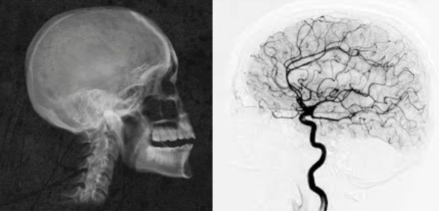
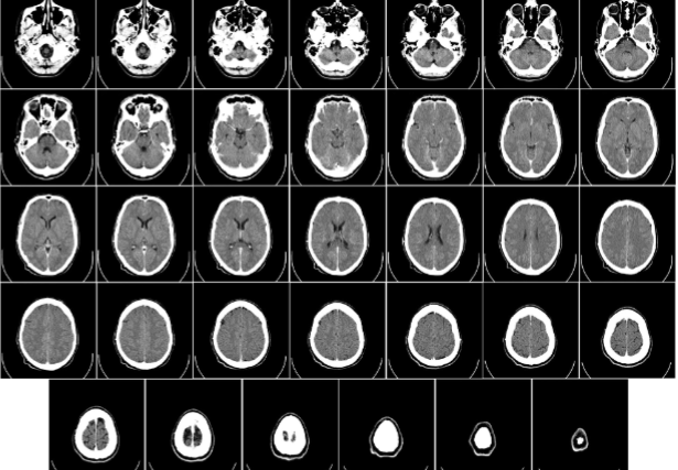
CT (computerized tomography) scan
thin x-ray beam with detector on opposite side of patient, rotates and translates to build a 3D image, distinguishes white matter, grey matter, and ventricles, good to distinguish injuries and tumors, cheaper and easier than MRI
limitations: contrast and resolution in brain is worse than MRI, only certain angles, significant radiation dose (~100x greater than standard x-ray)
MRI (magnetic resonance imaging)
takes advantage of magnetic properties of tissue to generate an image, protons (hydrogen atoms in water molecules) have spin, extremely strong magnets align all the proton’s spin in the body, radiofrequency pulses of the precession frequency excite protons/knock their spin out of the alignment, proton spins returning to alignment create their own radio frequency signal, measures by antennas in the MRI, strength measures in Telsa (T) and higher T = increase signal to noise ratio and provide better spatial resolution
limitations: expensive, time-consuming (30-60 min), noisy
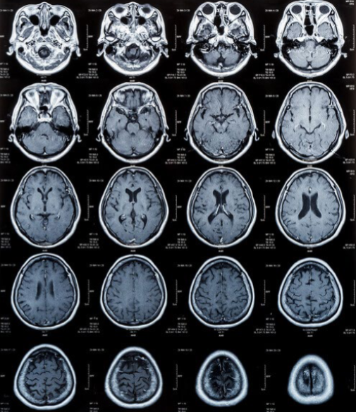
T1
longitudinal, CSF looks black
T2
transverse, CSF looks white
voxel
a 3D version of a pixel that represents a cubic volume of brain space
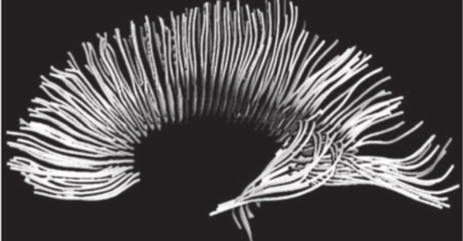
diffusion MRI
structures of axon fiber tracts via anisotropic diffusion of water molecules
functional MRI or fMRI
works similar to regular MRI but uses hemoglobin to look at oxygen metabolism over time as an indirect measure of neural activity, 4D image where the fourth direction is time (before and after stimulus is present), active neurons consume more oxygen than those at rest
limitations: slower temporal resolution (4-8s delay), limited spatial resolution, extreme sensitivity to subject motion, expensive
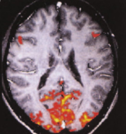
PET (positron emission tomography)
neural activity but no information on structure, inject a position-emitting isotope (fluorodeoxyglucose) into artery which will react with electrons to cause gamma photons to be released and detected by a device. one benefit over fMRI: you can get isotopes that bind to particular receptors or metabolism of neurotransmitter
limitations: some temporal resolution 4-8s as MRI (less than EEG, MEG), lower spatial resolution, no anatomical —> needs to be combined with MRI or CT
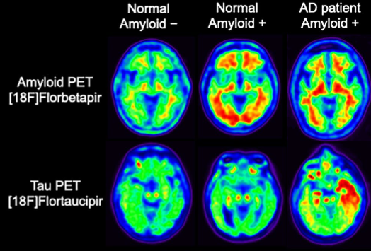
BOLD (blood oxygen level-dependent) effect
oxygen rich blood is sent to active brain regions as the active brain quickly depletes the oxygen
SPECT
a cheaper alternative that uses tracers with longer half-lives
EEG
measures gross electrical images of the brain and mostly surface level, noninvasive, amazing temporal resolution in ms, can be combined with fMRI
limitations: requires millions of active neurons to detect, cannot differentiate between individual neurons
MEG
measures magnetic field changes on the surface of the scalp that are produced by changes in the underlying pattern of neuronal activity (50,000 neurons need to be activated to detect); better spatial resolution than EEG
limitation: cannot differentiate between individual neurons, very expensive
event-related potentials (ERPs)
a stereotyped waveform that corresponds to a specific sensory/cognitive/motor event (both EEG and MEG)
IACUC
a committee you need approval from on your protocol and all personnel need training, surgeries need vet care/overseeing
replace, reduce, refine
three R’s
autoclaves
sterile supplies
isoflurane
keeps mice asleep during surgery (best recovery) and/or during short periods of handling (such as if you need to shave the mouse or inject dye easily, etc.)
ketamine/xylazine
good for procedures that will result in euthanasia at the end (CSF fluid collection)
buprenorphine
can be given 2x a day for pain management after surgery
stereotaxic surgery
a minimally invasive form of signal intervention that makes use of a 3D coordinate system to locate small targets (x, y, z is mapped)
brain atlas
defines the shape and location of brain regions in a common coordinate space (also used for DBS)
bregma
the place on the skull where the sagittal and coronal structures join the parietal and frontal lobe bones together (front)
lambda
meeting point of the lambdoid suture and sagittal suture
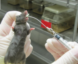
oral gavage
directly administer medications into the mouse’s stomach via mouth (tamoxifen)
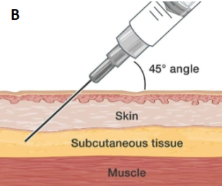
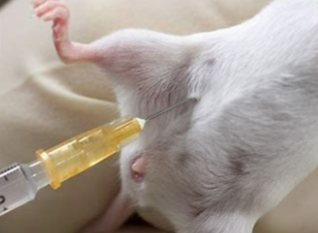
subQ
an injection into the subcutaneous tissue of the skin (itch/pain/dermal agonist tests); slower, sustained release
intraperitoneal injection (IP)
injecting into the body cavity (good for anesthetics, diphtheria toxin, CNO, other drugs); fast onset, short duration
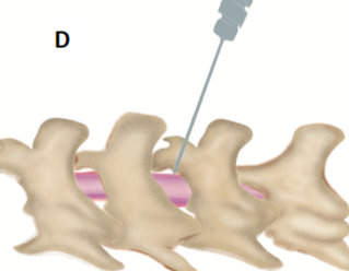
intrathecal (IT)
a way to deliver medication to the subarachnoid space of the spinal cord so that it reaches the CSF (very difficult!) direct, but not prolonged
blood
easiest to collect directly from the heart of an anesthetized mouse, can also be collected from the tail of a live mouse —> much more difficult
CSF (cerebral spinal fluid)
using a pulled capillary tube place in the base of the brain/spine of an anesthetized mouse; tissue collection usually involves euthanizing the mouse with CO2 and then immediately performing Perfusion or immediately on dry ice
ethology
study of non-human animal behavior
neuroethology
study of neural basis of an animal’s natural behavior; often used for the study of neural basis of specialized animal behavior
face validity
Does it look like the human disease? The model shows similar symptoms or characteristics.
construct validity
Does it have the same underlying cause or mechanism? The model reproduces the biology or genetics responsible for the disease.
predictive validity
Does it respond to treatments the same way humans do? Drugs or therapies that work (or don't work) in humans produce similar results in the model.
type I error
chance of false positives
type II error
chance of false negatives
effect size
magnitude of difference between conditions
power
ability to correctly detect a true positive result (reject the null hypothesis)
ethnogram
list of defined behavior before watching a behavior test
running wheel
rodent motor function model, measure time spent on wheel
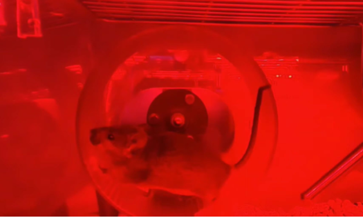
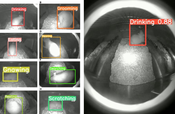
home cage activity
rodent motor function model, automatic monitoring of activity in the home cage, IR sensors that measure movement through the cage, AI-based video monitoring
rotarod
rodent strength and coordination model, mouse place on rotating cylindrical rod, slowly speed up, measure time until mouse falls, mice learn to do this better over time (can also be used as a memory assay)
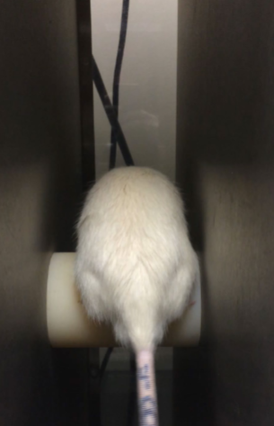
hanging wire
rodent strength and coordination model, measure time mouse can hang on suspended wire
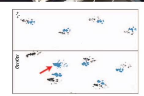
gait analysis
rodent strength and coordination model, dip feet in ink, allow to walk down a paper-lined tunnel, altered gaits can reveal ataxia (abnormal muscle coordination)
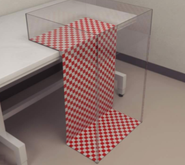
visual cliff
rodent sensory assay, “cliff” formed by high-contrast checkered surface, clear plexiglass extends from edge, mouse cannot actually fall, blind/vision impaired mice will continue forward - also can be conducted in human babies!
taste/flavor (choice assay)
rodent sensory assay, compare control vs experimental taste/flavor, quantify amount eaten/drank from each
olfaction (attraction/avoidance assays)
rodent sensory assay, compare time spent on multiple odorants
nociception
ability to detect noxious stimulus (pain), used to investigated neural basis of pain and therapeutic potential of drugs
tail flick
rodent nociception assay, tail placed in cold or hot water to produce a pain-provoking sensation, causes a reflex that moves the tail, measure latency before tail flick, slower response = nociception defect
hot plate
rodent nociception assay, place on calibrated hot plate, calibrated to generate withdrawal response in ~5-10 seconds, measure increases/decreases to this latency
formalin
measures response to noxious chemical stimuli, injection of itch-inducing formalin into paw, monitor total time spent itching the limb
morris water maze
spatial learning and memory, rodent is placed in the water at varied starting locations and must learn to search for the platform, multiple trials/days required to learn the task, pool is made opaque with milk/paint so rodent can no longer see the platform below the water
limitations: stressful, can be impacted by overall health of mutant animals, impacted by a wide variety of treatments
rodent arm maze
spatial learning and memory, rodent is placed in a maze and learns which arm to expect food in, relatively difficult task, multiple days/weeks of training required
key difference: not necessarily based on vision, less stressful
rodent VR
spatial learning and memory, virtual reality systems for more complex spatial learning tasks, can easily combine with electrophysiology, microscopy, optogenetics, etc.
novel object recognition
non-spatial learning and memory, rodents are curious and will spend time exploring novel objects, using non-spatial memory, old objects will be remembered and new will be explored, measure time exploring a novel vs old object
go/no-go task
attention and impulsivity assay, rodent trained to produce a response in the presence of a “go” cue, but not a “no-go” cue, generally paired with a reward/punishment, rodents are generally successful on “go” trials but have difficulty withholding responses on “no-go”
real-time place preference
a reward assay, inherent reward value of certain states are measured without conditioning, real-time neuromodulation when rodent is in certain place
resident-intruder assay
social behavior assay, measures territorial behavior in males, an “intruder” animal is added to the cage of a “resident” animal, aggressive behaviors are quantified: attacks, tail ratting, chasing/wrestling, useful to study drug impacts and neuronal control of aggression
social approach
a social behavior assay, rodent given choice to investigate another mouse or a novel object, most WT mice will spend more time socially, used as a model for autism-like social behaviors
open field test
rodents place in bare open chamber will initially stay near walls, and eventually explore the center, more anxious animals will continue to avoid center
marble burying assay
rodents tend to bury objects in their environment, more anxious animals tend to bury more objects
forced swim test
rodents place in large, graduated cylinder filled with water and forced to swim, initially swim vigorously trying to escape, eventually immobile and only maintains head above water, immobility is an indication of behavioral despair
limitations: conservation of energy or depression-like behavior? rats are better swimmers than mice
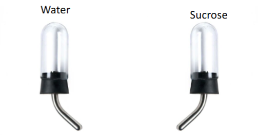
sucrose preference test
models anhedonia - loss of motivation to pursue normally rewarding activities, rodents allowed to choose between water and sucrose, models of depression will have reduced sucrose consumption