FETAL HEAD
 Call Kai
 Learn
Learn Practice Test
 Spaced Repetition
 Match
Match Flashcards
 Knowt Play
1/40

There's no tags or description
Looks like no tags are added yet.
Name | Mastery | Learn | Test | Matching | Spaced | Call with Kai | Chat |
|---|
No analytics yet
Send a link to your students to track their progress
41 Terms
macrosomia
birth weight of 4000g or greater
above 90th percentile
commonly result of poorly controlled maternal diabetes
what distinguishes microcephaly from anencephaly
intact calvarium
key sono of microcephaly
BPD or HC is 2-3 standard deviations below mean expected for GA
abnormal fetal biometric ratio
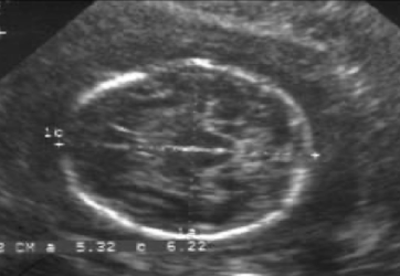
brachycephaly
head is rounder than usual
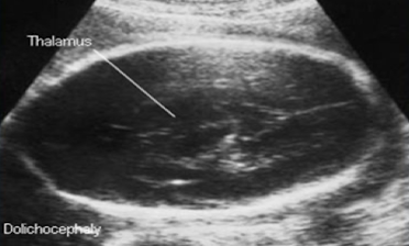
dolichocephaly
narrow BPD and long occipitofrontal diameter (OFD)
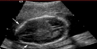
lemon sign
fetal head with bilateral denting of frontal bones
associated with spina bifida
cloverleaf skull
tri-lobed appearance of the head
associated with thanatophoric dysplasia and homozygous achondroplasia
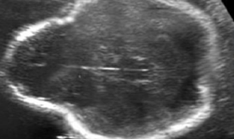
strawberry shaped skull
fetal head with normal BPD and narrow frontal diameter
associated with trisomy 18
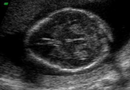
spalding sign
flattened and misshapen fetal head with overlapping of cranial bones
associated with fetal demise
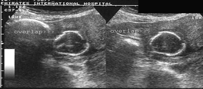
scalp edema
fetal hydrops and scalp thickening (>/= 3mm)
presence of the _______ excludes agenesis or corpus callosum
CSP
what is agenesis of corpus callosum m/c associated with
dandy walker malformation
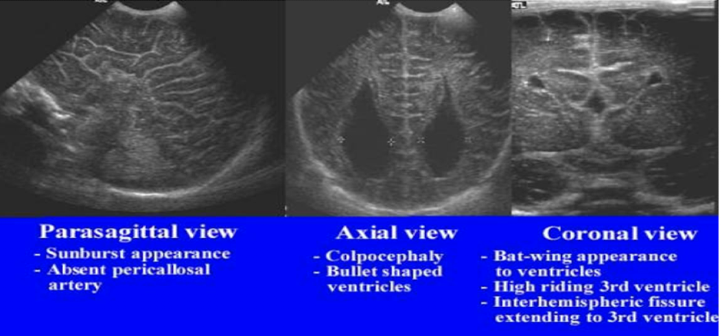
sono of ACC
absence of CSP
colpocephaly (disproportionate prominence of occipital horns of lateral ventricles) bullet shaped
sunburst appearance
intracranial calcifications are most commonly the result of
fetal viral infection (cytomegalovirus m/c)
what is the most common intracranial tumour
teratoma
aqueductal stenosis
results from obstruction, atresia, stenosis of aqueduct of sylvius
causes ventroculomegaly
sono of aqueductal stenosis
ventriculomegaly
3rd ventricle dilaltion
flexion and adduction of thumb
vein of galen aneurysm
fusiform median cyst in the region of the transverse fissure immediately superior to the cerebellum
abnormal turbulent Doppler signals within the aneurysm
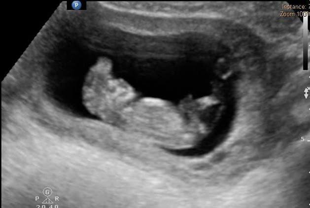
acrania
absence of normal cranial vault with a normal appearing or disorganized brain above orbits
increased maternal serum AFP is associated with
open neural tube defects
anencephaly
absence of cranial vault, complete or partial absence of forebrain,
presence of brainstem, midbrain, skull base, facial features
sono of anencephaly
no normal cranial morphology and brain tissue above orbits
exophthalmos (bulging of eyes)
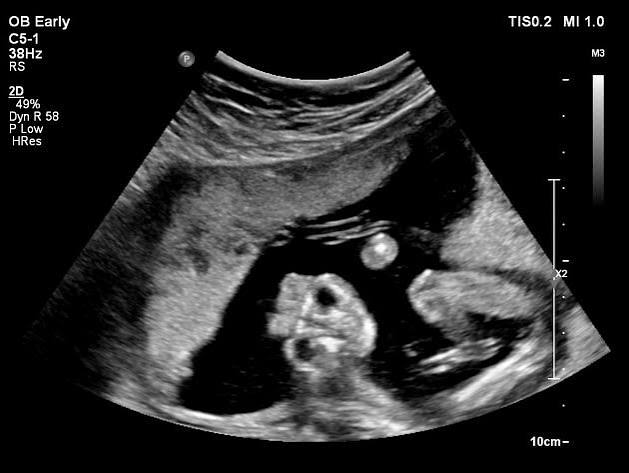
cephalocele
neural tube defect in which meninges alone or meninges and brain herniate through defect in calvarium (no increase in AFP)
meningocele: meninges only
encephalocele: brain tissue only
encephalomeningocele: brain tissue and meninges
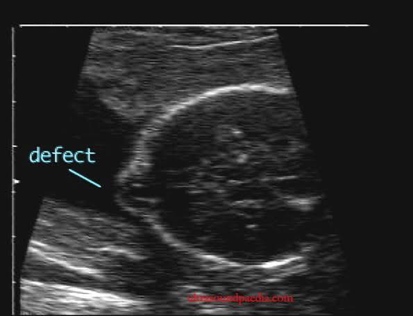
encephalocele
ventriculomegaly
dilation of cerebral ventricles without defining the cause
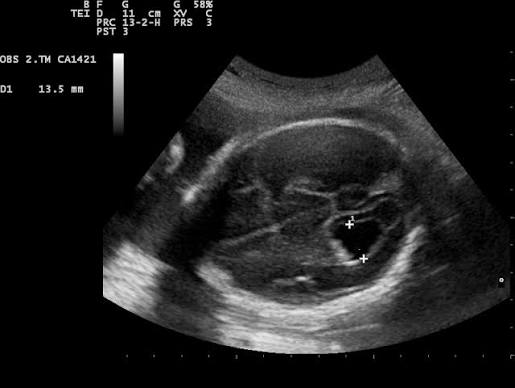
m/c causes of ventriculomegaly
arnold chiari (3rd, 4th, lateral ventricles dilated)
open NTDs
double aqueduct stenosis
dandy walker
hydrocephaly
ventriculomegaly most commonly associated with increased intracranial pressure
usually due to lesion causing obstruction
aqueduct of sylvius obstruction causes enlargement
of lateral ventricles and 3rd ventricle
what is holoprosencephaly
incomplete separation of the two hemispheres
what is holoprosencephaly m/c associated with
Trisomy 13
also 18
alobar holoprosencephaly
mono ventricle
fused thalamus
no fissure, CSP, CC
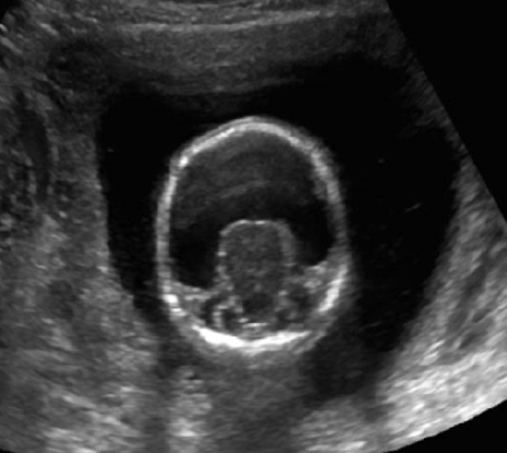
semilobar holoprosencephaly
single ventricle cavity
partial/complete fused thalami
rudimentary flax
no CSP, CC
lobar holoprosencephaly
almost complete division of ventricles
CC may be present
CSP still absent
hydranencephaly
absence of cerebral hemispheres with an incomplete or absent falx and sac-like structure contain CSF surrounding brainstem and basal ganglia
sono of hydranencephaly
brainstem, cerebellum, cisterna magna unaffected
lacking a mantle or rim of cerebral brain tissue
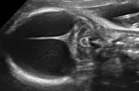
schizencephaly
clefts in the cerebral mantle with communication between the ventricles and subarachnoid space
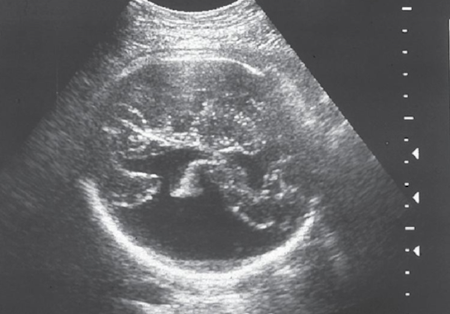
dandy-walker malformation
agenesis or hypoplasia of cerebellar vermis
dilated 4th ventricle
enlarged posterior fossa (cisterna magna)
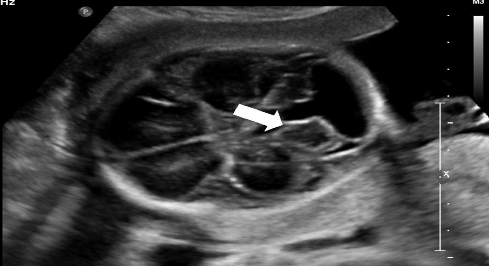
arachnoid cyst
benign cyst in the cerebrospinal axis
do not communicate with ventricular system
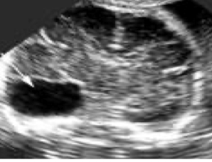
porencephaly
cysts filled with cerebrospinal fluid that communicate with ventricular system or subarachnoid space
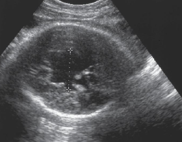
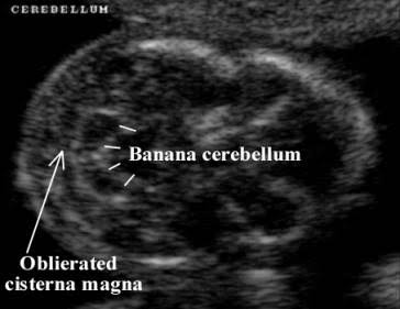
arnold chiari type II
displacement of cerebellar tonsils, part of cerebellum, 4th ventricle, pons and medulla oblongata
accompanied by hydrocephalus and myelomeningocele
what is the main sono finding in chiari type 2
banana sign
(curved cerebellum)