BIOL2230 - Neurophysiology
1/151
There's no tags or description
Looks like no tags are added yet.
Name | Mastery | Learn | Test | Matching | Spaced | Call with Kai |
|---|
No analytics yet
Send a link to your students to track their progress
152 Terms
Describe the major divisions of the nervous system
CNS and PNS
PNS: sensory and motor
Sensory: somatic, visceral and special receptors
Motor: somatic, autonomic and enteric
Describe the functional organisation of the spinal nerves
Motor and sensory nerves leave the spinal cord and bundle together into fascicles
Fascicles contain multiple axons and are surrounded by connective tissue
Each spinal nerve contains multiple fascicles
Describe the structure of the cerebral cortex
It contains 6 cortical columns
The left brain processes the right side of the body and vice versa
It contains pyramidal (large dendritic trees, long axon for communication outside cerebral cortex) and stellate neurons (short dendritic trees, short axon for communication inside cerebral cortex)
What are the three types of white matter in the brain?
Commissural: connects the left and right hemispheres
Association: connects areas of one hemisphere
Projection: travel to and from the cortex via the brainstem
Describe the anatomy and function of the thalamus
Anatomy: symmetrical, made of multiple nuclei
Function: relays motor information, regulates consciousness, sleep and alertness
What are the structures that make up the limbic system?
Hippocampus, amygdala, cingulate gyrus, parahippocampal gyrus
Thalamus, hypothalamus
What are the structures that make up the brainstem?
Midbrain
Pons
Medulla oblongata
Describe the function of the pons
Sleep, bladder control, respiration, hearing, balance, posture, eye movements, taste, swallowing, facial expression/sensation
Also contains pontine respiratory centre for voluntary control and adjustment of breathing
Describe the function of the medulla oblongata
Receives information from chemoreceptors, interacts with motor nerves and pontine respiratory centre to control breathing
Also controls cardiovascular system
What are the principles of equimolality and electrical neutrality?
Equimolality: the concentrations of ions in the ICF and ECF must be equal
Electrical neutrality: the ICF and ECF anions must balance, and the ICF and ECF cations must balance
What does the Nernst equation find?
The equilibrium potential of a certain ion, given its valence and concentration within and without the cell
Describe the characteristics of a graded potential
Small, rapid VM changes
Irregular size
Spreads electrotonically
Fast
Dissipates at an exponential rate
Strength and duration is proportional to stimulus strength
Susceptible to noise
Depolarising or hyperpolarising
Describe the characteristics of an action potential
Large VM changes
Regular
Spread slowly
Long distance, can regenerate
Frequency codes stimulus strength
Not affected by noise
Only depolarising
Frequency: ~5-100Hz
Pulse length: 1-400ms
Conduction velocity: 1-120m/s
What are the advantages and disadvantages of each electrophysiological recording type?
Extracellular
+ Easy to do
- Small signal which dissipates
- Signal is local field potential
Intracellular
+ Measures one cell
- Invasive, harder to do
Patch clamp
+ Measures one ion channel
+ Can compare activity of different neuron parts
- Very hard to do
What are the major categories of neurotransmitters?
Classical: single amino acids, biogenic monoamines, ACh
Neuropeptides
Purines
Others
Compare electrical and chemical synapses
Electrical:
Rapid
Unidirectional or bidirectional
Excitatory or inhibitory
Sign-conserving
Chemical:
Slower
Unidirectional
Excitatory (Type I) or inhibitory (Type II)
Not sign-conserving
Describe direct coupling
NT binds to GPCR, which activates alpha-subunit that acts as a ligand for an ion channel
Describe indirect coupling
NT binds to GCPR, which activates alpha-subunit which activates/inhibits an enzyme. The enzyme activates a secondary messenger that opens/closes an ion channel.
What are post-synaptic potentials?
Small, temporary changes in membrane potential as a result of a neurotransmitter binding
What is modality of a sensory receptor?
The form of energy of a stimulus; the adequate stimulus is the modality a receptor responds best to
What are the functional categories of sensory neurons?
Exteroceptors (external): mechanoreceptors, photoreceptors, chemoreceptors, thermoreceptors
Interoceptors (internal): chemoreceptors, baroreceptors, osmoreceptors
Proprioceptors (posture, position): a type of mechanoreceptors
What are the structural categories of sensory neurons?
Simple: dendrites are sensory receptors
Complex: connective tissue surrounds dendrites
Special senses: receptor cell synapses onto dendrites
Where is the trigger zone in pseudo-unipolar cells?
Where the axon meets the dendrites, so that GPs generated here can be converted to an AP
Distinguish between phasic and tonic receptors
Phasic: quickly adapt
Tonic: slowly adapt
What factors determine acuity?
Receptive field of sensory units
Density
Overlap of receptive fields
Convergence
Lateral inhibition
What are the overall properties of cutaneous mechanoreceptors?
Detect touch, pressure, vibration and stretch
Low threshold (high sensitivity)
Signal with glutamate
Mostly A-beta (large, myelinated)
Describe hair follicle receptors
RA1: rapidly adapting, small receptive field
Detect bending of hairs in response to air movement or touch
Only present in hairy skin
Wrapped around base of hair follicle
Free nerve endings
Describe the properties of thermoreceptors
Free nerve endings in superficial layers of skin
RA2
Population coding (activity of receptors with different ranges compared)
Signal when temperature changes
Which neurons use the spinothalamic vs. DCML tracts?
Spinothalamic: nociceptors, thermoreceptors, mechanoreceptors
DCML: mechanoreceptors, proprioceptors
Describe the trigeminothalamic pathway
1. Trigeminal nerve from face to nuclei in brainstem and spinal cord
2. To Thalamus
3. To somatosensory cortex
Describe the primary somatosensory cortex
Postcentral gyrus
Determines stimulus type, intensity and location
Describe the secondary somatosensory cortex
Upper lip of lateral sulcus
Higher order processing
Integrates sensory and motor information
Integrates information from both body sides
Integrates with limbic system for learning and memory
Describe the somatosensory association area
Posterior parietal cortex
Receives input from both somatosensory cortices
Integrates visual and sensory input
Coordinates motor response
What are some causes of neuropathic pain?
Lesion
Disease/infection
Compression/pinching/trauma of nerves
Inflammation
Damage to ion channels
Describe the properties of nociceptors
Free nerve endings
Mechanoreceptors, thermoreceptors, chemoreceptors or polymodal
No background firing
Large receptive fields
High threshold
Tonic
Signal using glutamate and substance P
Define and describe inflammatory soup
Tissue damage causes the release of:
ATP, ions, prostaglandin, Substance P
Bradykinin
Serotonin
Histamine
This impacts pain perception by nociceptors
Describe the pathway of nociceptive perception to the brain
1: nociceptor activated and synapses onto dorsal horn using glutamate and Substance P
2: decussate at spinal cord, then to thalamus
3: to somatosensory cortex
Signals also sent to limbic system for emotional/behavioural response
How does the limbic system cause descending pain pathways?
It sends efferent pathways via the spinal cord to the dorsal horn
Either excitatory (serotonin onto nociceptor) or inhibitory (noradrenaline, steroids or opioids released onto other receptors)
How do interneuron suppress pain?
Interneurons release endogenous opioids onto first-order neurons to inhibit Substance P release
Or onto secondary neurons to cause inhibitory post-synaptic potentials
Define pain sensitisation and its types
Sensitisation: increased sensitivity to pain
Hyperalgesia: more pain than normal
Allodynia: pain from a non-painful stimulus
What is the gate-control theory of pain?
Mechanical stimulation of A-beta fibres (cutaneous mechanoreceptors) activate inhibitory interneurons (using GABA or glycine) to inhibit C-fibres
Describe the structure of the olfactory epithelium
2 layers
Olfactory mucosa: receptor cells, basal replacement cells
Lamina propria: connective tissue, mucus-secreting cells
Describe the structure of olfactory chemoreceptors
Bipolar cells
Dendrites in olfactory mucosa for reception
Dendrites are olfactory knobs containing cilia
High sensitivity
Receptive to one chemical each
Explain olfactory transduction
Chemical binds to G-protein coupled receptor
G(olf) protein activates adenylyl cyclase
ATP converted into cAMP
cAMP opens cation channels (depolarisation)
Ca2+ opens Cl- channels (more depolarisation)
GP generated, generating AP
Glutamate released onto 2ndary neurons
Explain how olfactory information is processed in the brain
Mitral and tufted cells project to olfactory/piriform cortex located in the temporal lobe
Activation of different receptor types compared (population coding)
Entorhinal cortex then travels to thalamus for relay elsewhere
Explain how sweet signalling occurs
Sweet tastant opens G-protein coupled receptor
G-protein activates adenylate cyclase
ATP converted to cAMP
cAMP activates a protein kinase that closes K+ channels (depolarisation and NT release)
Explain how gustatory information is processed in the brain
Three cranial nerves involved: 7, 9 and 10
1: cranial nerves travel to gustatory nucleus in medulla
2: to thalamus
3: to primary gustatory cortex in insula lobe
Compare Type II and Type III cells in taste buds
Type II: bitter, umami and sweet, signals using ATP with no clear synapse (may activate Type III)
Type III: sour and salty, signals using serotonin (may inhibit Type II)
Describe the structure of the organ of corti
Contains inner and outer hair cells
Outer hair cells have stereocilia embedded in the tectorial membrane, which is connected to the basilar membrane
When endolymph moves, this moves the tectorial membrane and activates OHCs
When the endolymph moves, this directly activates IHCs
No kinocilium in the organ of corti stereocilia!!!
Compare the ion concentrations of the endolymph and perilymph
Perilymph: low K+, high Na+
Endolymph: high K+, low Na+ (electrochemical force driving K+ in when channels open)
Describe the pathway of auditory information to the brain
Travels via CN 8 to brainstem, then thalamus, then primary auditory cortex
What is the structural organisation of the primary auditory cortex?
Arranged by frequency
Name each of the cranial nerves and their function
1: olfactory
2: optic
3, 4, 6: eye movement
5: trigeminal
8: vestibular, auditory
10: vagus
Describe the pathway of vestibular information to the brain
Vestibular nerve (CNVIII) synapses onto vestibular nucleus
To cerebellum, vestibular cortex and eye muscle cranial nerve nuclei
Describe the vestibuloocular reflex
Receives information from semicircular canals (endolymph movement) and proprioception
CNVIII signals to nuclei of cranial nerves to cause eye movement in opposite direction to head
Describe the vestibulospinal reflex
Receives information from otolith organs
CNVIII signals to vestibular nuclei in brainstem
To spinal cord, then motor neurons
Describe the vestibular cortices in the brain
Parietoinsular vestibular cortex processes spatial orientation and movement
Multimodal association area integrates vestibular information with vision and proprioception
Describe the structure of the photoreceptor outer segment
A stack of discs
Visual pigment molecules (VPMs) are embedded in the membrane of discs
Contain opsin and chromaphore
Compare rods and cones
Rods:
More common
Night vision
High sensitivity
Saturate quickly
Long, conical
Cones:
Less common
Colour vision
Low sensitivity
Saturate slowly
Short, tapered
Detect movement well
Describe the general structure of a photoreceptor
Outer segment for transduction
Inner segment containing nucleus
Synaptic terminals
Explain the process of photoisomerisation and phototransduction
Light isomerises chromophore
Conformational change of opsin
Transducin activated
PDE activated by transducin alpha-subunit
cGMP broken down by PDE
Describe photoreceptor activity during dark
High cGMP levels
cGMP Na+ channels opened
Cell depolarised
VGCCs open
Glutamate release
Describe photoreceptor activity during light
Low cGMP levels
cGMP Na+ channels close
Cell hyperpolarised
No glutamate release
Describe the visual processing function of retinal bipolar cells
Temporal processing
Describe the visual processing function of retinal ganglion cells
Determine basic features of the image (brightness, colour, motion, uniformity, edge detection)
Describe the pathway of visual information in the brain
Optic nerves cross over at optic chiasm
Synapse at lateral geniculate nucleus
Neurons project to primary visual cortex
What sites does the myosin globular head contain?
Actin-binding and myosin ATPase
What properties make an NMJ different from a chemical synapse?
Only excitatory
ACh is used
Motor endplate is folded
Motor endplate contains nicotinic ACh receptors
1 AP causes 1 muscle twitch
AChE removes ACh from synaptic cleft
Describe the structure of voltage-gated calcium channels in myocytes
DHP receptor (conformationally changed by depolarisation)
Ryanodine receptor (opened by change in DHP)
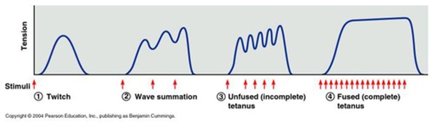
Describe the four stages of increasing muscle tension
Treppe: full relaxation, increasing tension until plateau
Wave summation: partial relaxation, overlap of twitches, Ca2+ released faster than removed
Unfused tetanus: brief, partial relaxation, troponin-tropomyosin saturated by Ca2+, all myosin-binding sites on actin exposed
Fused tetanus: no relaxation, maximal contraction, continual exposure of actin
Describe the factors affecting muscle tension
Stimulation frequency
Fibre diameter
Fibre length at start of contraction
Fatigue
Compare ATP hydrolysis of fast and slow-twitch muscle
Fast: glycolysis
Slow: oxidative phosphorylation
Compare skeletal, cardiac and smooth muscle
Skeletal: striated, voluntary, multi-nucleated, only excitatory, Ca2+ from SR, troponin-tropomyosin, NMJ
Cardiac: striated, involuntary, 1-2 nuclei, longer APs for no overlap of contraction, Ca2+ from ECF and SR, troponin-tropomyosin, NEJ
Smooth: non-striated, involuntary, 1 nucleus, excitatory or inhibitory, Ca2+ from ECF and SR, calmodulin, NEJ
What are the three ways of classifying smooth muscle?
Neurogenic (requires neural stimulation) vs. myogenic (capable of initiating contraction)
Phasic (bursts of contraction) vs. tonic (continuous contraction, regulated by [Ca2+])
Multi-unit (neurogenic, tonic) vs. single-unit (myogenic, phasic)
What causes Mysathenia Gravis and Lambert-Eaton Syndrome?
Autoimmune diseases, where immune cells target parts of the neurons
What receptors does ACh bind to on the motor end plate?
Nicotinic acetylcholine receptors
Compare upper and lower motor neurons
UMNs: connect motor cortex (voluntary) or brainstem (involuntary) to LMNs directly/indirectly
LMNs: originate in cranial nerve nuclei (involuntary) or ventral horn (voluntary) and innervate muscle
Compare the two types of LMNs
Alpha: large diameter, myelinated, innervate extrafusal fibre (for movement)
Gamma: smaller diameter, myelinated, innervate intrafusal fibre (for proprioception)
Describe the Renshaw loop
LMN excites muscle and also releases ACh onto Renshaw cells
Renshaw cells are inhibitory interneurons next to alpha LMNs and provide negative feedback to prevent overactivation
Describe how LMNs integrate sensory and motor information to form the final common pathway
LMNs receive +/- input from UMNs, sensory feedback from spindle fibres and interneurons
The effects of these are summed and may/may not cause an AP
This is important since NMJs are only excitatory, so if an AP is generated, a twitch will occur
Compare small and large motor units
Small: in small muscles (e.g., eyes), small increase in force, precise, smaller diameter fibres, slower fatigue
Large: in large muscles (e.g., quads), large increases in force, less precise, larger diameter fibres, faster fatigue
Large motor units have larger cell bodies, more leak channels and are harder to depolarise
What proprioceptors are present in muscle?
Muscle spindles: muscle length
Golgi tendon receptors: muscle tension
Describe how muscle spindles detect muscle length changes
Part of intrafusal fibre
Innervated by afferent signals (stretch receptors; Type 1a and 2) and efferent signals (gamma LMN)
Always some depolarisation for low-level muscle activation
Muscle stretch causes depolarisation and increased firing
This causes alpha LMN to counteract it and contract (and opposite occurs when muscle contracts and afferents are hyperpolarised)
What is the difference between Type 1a and 2 stretch receptors?
1a: primary endings wrapped around muscle to detect muscle stretch
2: secondary/flower spray endings to detect muscle length
What are the four categories used to classify reflexes?
Level of processing: cranial/spinal
Somatic/autonomic
Innate/conditioned (e.g., Pavlov's dog)
Monosynaptic/polysynaptic
What are the properties of stretch reflexes?
Stimulus: spindle fibre stretch
Response: contraction to counteract stretch
Somatic reflex
Describe the Golgi tendon reflex
Stimulus: stretch of Golgi tendon
Integrator: spinal cord (disynaptic), excitatory to hamstring, inhibitory to quads
Response: hamstring contraction, quads relaxation
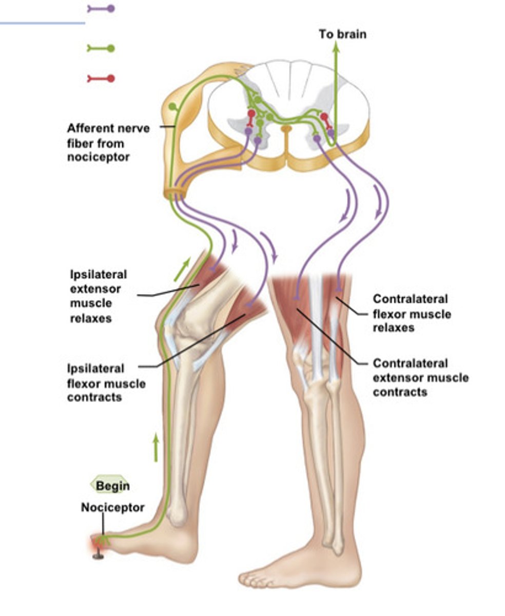
Describe the withdrawal and crossed-extensor reflex
Stimulus: nociceptor activation in leg
Response: afferent neuron synapses onto 5 other neurons: one to excite the hamstring, one to inhibit the quads (this withdraws the leg), one to excite the quads and one to inhibit the hamstring (to stabilise the other leg), and one to decussate and travel to the brain
Describe the main areas of the motor cortex
Primary: pre-central sulcus, UMNs originate
Premotor: in front of M1, coordinates voluntary movement
Prefrontal association areas: front of brain, plans voluntary movement, thoughts and personality
Describe the structure and neurons of the motor cortex
Arranged somatotropically
Contain pyramidal neurons (UMNs) which synapse onto interneurons
Also contain Betz cells, pyramidal neurons which synapse directly onto LMNs for precise movements
Compare the pyramidal and extrapyramidal tracts
Pyramidal: voluntary, originate in motor cortex, either corticospinal or corticobulbar, decussate at medulla
Extrapyramidal: voluntary and involuntary, originate in brainstem nuclei, simple movement
Describe the corticospinal tract
UMNs originate in cerebral cortex (either frontal or parietal lobe)
Fibres travel through brainstem to medullary pyramids
Some decussate here, forming the lateral tract for arm, hand movements
Others decussate at the end of the spinal cord, forming the anterior tract for balance, posture
Describe the corticobulbar tract
UMNs travel from motor cortex to synapse onto:
LMNs in cranial nerve nuclei
Or interneurons that project to LMNs
Describe the inputs and output of the cerebellum
Inputs:
Mossy fibres from brainstem and spinal cord, synapse onto granule cells
Climbing fibres from inferior olive, synapse onto Purkinje cells
Both provide sensory and motor information
Purkinje cells then synapse from cerebellar cortex to deep nuclei
Output: deep nuclei which affect movement
Describe the inputs and outputs of the basal ganglia
Inputs: motor information from cerebral cortex to striatum first, then other basal ganglia
Outputs: return to cerebral cortex via thalamus, then travel via pyramidal pathways
Describe how the basal ganglia inhibits/excites movement
Globus pallidus interna normally inhibits the thalamus
Direct pathway: for movement, the striatum inhibits GPi to excite movement
Indirect pathway: for no movement, the striatum excites GPi to inhibit movement
What neurotransmitters control the direct and indirect pathways?
Substantia nigra: dopamine excites direct and inhibits indirect pathway
Striatum interneurons: ACh inhibits direct and excites indirect pathway
Describe the main symptoms of dementia
Memory impairment, executive dysfunction, impaired communication, impaired recognition, personality
Describe the causes and symptoms of Alzheimer's disease
Cause:
Beta amyloid plaques (EC)
Neurofibrillary tangles of tau protein (IC)
Neurophysiology: frontal lobes and hippocampus
Symptoms: dementia
Describe the causes and symptoms of Parkinson's disease
Cause: Lewy bodies (aggregates of alpha-synuclein)
Neurophysiology: substantia nigra (excites movement)
Symptoms: hypokinetic movment disorder (loss of movement)