Study guide lab micro
 Call Kai
 Learn
Learn Practice Test
 Spaced Repetition
 Match
Match Flashcards
 Knowt Play
1/34

There's no tags or description
Looks like no tags are added yet.
Name | Mastery | Learn | Test | Matching | Spaced | Call with Kai | Chat |
|---|
No analytics yet
Send a link to your students to track their progress
35 Terms
What are the main differences between the four biosafety levels of containment with respect to type of microorganisms that are handled at each of these levels?
The containment levels range from the lowest
biosafety level 1 (BSL-1), to the highest at level 4
(BSL-4)
What is the Biosafety Level 1 (BSL-1)?
- Low-pathogenicity microorganisms
-Precautions and personal protective equipment
(PPE) required are minimal
-Laboratory access is not restricted, and work can
be conducted on regular open bench tops
-An autoclave or a licensed agency is used for
decontamination of biohazard waste
-Personal protection and decontamination
procedures for BSL-1 include hand washing and
disinfection
What is Biosafety Level 2 (BSL-2)?
-Suitable for working with microorganisms
of moderate pathogenicity
-The same precautionary measures as BSL-1
-A BSL-2 laboratory facility requires the following:
1) A competent scientist: supervisor / professor
2) Adequate training: in handling pathogenic agents
3) Biosafety cabinets: for handling liquid cultures
4) Access to the laboratory should be restricted to
authorized personnel only.
What is Biosafety level 3 (BSL-3)?
-Higher pathogenicity than levels 1 and 2
-Exposure to these agents may lead to serious or
potentially fatal diseases
-The degree of training and protective measures
for supervisors and personnel should be adequate
for handling such agents.
What is Biosafety level 4 (BSL-4)?
- Facilities work with the infectious agents of
the highest degree of pathogenicity
-Requires the maximum level of training,
protection and security. There are no vaccines or
treatment for such infectious agents, and their
diseases are frequently fatal.
According to the classification of biosafety levels of containment as in point 1 above,to which biosafety level would our lab belong?
Biosafety Level 2 (BSL-2)
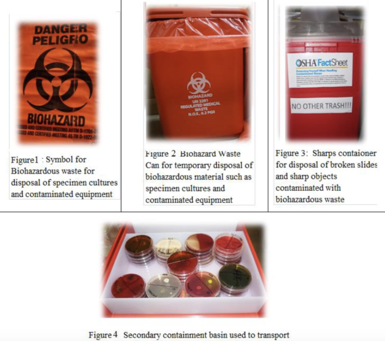
Briefly describe what the biohazard sign indicates and the proper disposal of biohazard waste.
- The Biohazard sign: it indicates that infectious
agents may be present in a container, a room, or a
building, and that PPE should be worn when entering
those areas or handling those containers
-The proper disposal of biohazard waste requires strict segregation, specialized containment, and authorized treatment methods like autoclaving or incineration before final disposal
Describe the proper technique of handwashing according to the CDC.
-20 seconds of lather time
-It is important that you wash your hands both before and after working with your material. You will be trained in proper handwashing and will be responsible for maintain these procedures for the duration of the semester. Proper handwashing is a skill that is required in may workplaces such as food handling and healthcare.
Describe the proper way to store a microscope.(part 1)
1. Always use both hands when carrying a microscope. Use one hand to hold the arm and the other to support the microscope under the base.
2. Place microscopes gently over tables. If you need to move it to a different place, lift them up and move them gently; please don’t try to slide them over the tables.
3. Be careful not to damage any parts when you remove the cover and uncoil the electric cord
Describe the proper way to store a microscope.(part 2)
4. After every use of the microscope, you should:
5. Remove your slide from the stage.
6. Wipe the oil from the stage using regular paper towels or kim-wipes.
7. Wipe the oil from the oil immersion lens and clean it with lens paper and lens cleaning solution.
8. Clean the oculars with lens paper and lens cleaning solution
List the optical parts and components of the light compound microscope in the order in which light travels through them. These are components 10-15 (marked with a
star) in the list above.
1. Illuminator (light source):
2. Condenser:
3. Diaphragm lever:
4. Specimen:
5. Objective lens:
6. Prism:
7. Ocular lens
What is the first order for in which light travels through a microscope?
-Illuminator (light source): this is the source of light in the microscope
What is the second order for in which light travels through a microscope?
Condenser: this component contains lenses that collect the light from the illuminator, concentrate it, and direct it upwards towards the specimen
What is the third order for in which light travels through a microscope?
Diaphragm lever: the iris diaphragm is an internal part of the condenser. It regulates the intensity of light admitted by the condenser and adds contrast to the image.
What is the fourth order for in which light travels through a microscope?
Specimen: the specimen is obviously not a component of the microscope, but it would be at this point that light would be moving through it in the pathway of light through a microscope
What is the fifth order for in which light travels through a microscope?
Objective lens: the ocular lens provides the initial magnification of the specimen and projects it on a mirror like component known as the prism. There are 4 or 5 different objective lenses (4X, 10X, 40X,
and 100X) attached to the nosepiece of a microscope. A more detailed description of these lenses is given below.
What is the sixth order for in which light travels through a microscope?
Prism: reflects the image provided by the objective lens and projects it towards the ocular lens.
What is the last order for in which light travels through a microscope?
Ocular lens: magnifies image reflected from the prism and magnifies it a second time (10X or 20X) leading to total magnification of an image.
What is the function of the optical parts of the microscope.
They play a direct role in illuminating and producing a magnified image of the specimen observed under the microscope.
What are the functional components of the Microscope?
Stage adjustment knobs
Coarse focusing knob
Fine focusing knob
Light intensity control knob
Stage adjustment knobs:
used to control the movement of the mechanical stage.
Coarse focusing knob:
this knob is used in the initial stages of focusing and it should only be used with the screening and low power objective lenses.
Fine focusing knob:
this knob is used to achieve precise focus and is the only knob that should be used when using the high power or oil immersion lenses.
Light intensity control knob:
controls the amount of light from the illuminator
What is the magnifying power of the four objective lenses ?
Ocular (eyepiece) (standard) 10x
Screening lens 4X
Low power objective 10x
High power objective 40x
Oil immersion objective 100x
Define or explain what total magnification is.
-It is the size of the image of an
object being observed under the microscope. It
results from the combined action of the objective
and ocular lenses
Given a combination of lenses in a microscope, calculate the total magnification
Magnifying Power of Microscope Lenses
-Ocular Lens (Eyepiece): 10x (standard)
-Objective Lenses: Scanning Objective → 4x =40x
Low-Power Objective → 10x = 100x
High-Power Objective → 40x =400x
Oil Immersion Objective → 100x =1000x
🔹 Total Magnification = Ocular Lens × Objective Lens Example: High-Power (40x) × Ocular (10x) = 400x total magnification
Explain what is meant by saying that the lenses in a microscope are parcentered
The specimen stays centered when switching between objective lenses.
Explain what is meant by saying that the lenses in a microscope are parfocal
The specimen stays in focus when switching between objective lenses, needing only minor adjustments.
List the four major groups of bacteria as differentiated by the gram stain results based on the gram reaction and the cellular
morphology of bacteria:
1) Gram positive cocci
2) Gram positive bacilli
3) Gram negative cocci
4) Gram negative bacilli
In terms of clinical significance, give the two main reasons while the gram stain is considered an extremely important test.
1) The gram stain may be the first fundamental
test in the identification of bacteria
2)The gram stain may allow the doctor to start a
more accurate treatment
List the gram stain reagents in the order in which they are applied during a gram stain procedure.
1) Crystal Violet: the primary stain
2) Iodine (mordant): intensifies the color of the primary stain
3) Acetone–Alcohol (decolorizer): removes the primary stain
4) Safranin (counterstain): re-stains an organism that has lost the primary color to the decolorizer
List the main causes of erroneous results when performing a gram stain procedure.
1. Making the smear too thick: which doesn’t allow
proper decolorization leading to inconclusive
results showing both gram reactions in the
same organism
2. Improper application of decolorizer and (or)
improper rinsing of slides: this also yields to
both gram stain reactions in the same organism